ABSTRACT
Objective: To investigate the correlation between respiratory syncytial viral load and length of hospitalization in infants with acute wheezing episodes. Methods: This was a two-year, cross-sectional study of infants ≤ 12 months of age with bronchiolitis at the time of admission to a tertiary hospital. For the identiication of respiratory viruses, nasopharyngeal secretions were collected. Samples were analyzed (throughout the study period) by direct immunoluorescence and (in the second year of the study) by quantitative real-time PCR. We screened for three human viruses: rhinovirus, respiratory syncytial virus, and metapneumovirus. Results: Of 110 samples evaluated by direct immunoluorescence, 56 (50.9%) were positive for a single virus, and 16 (14.5%) were positive for two or more viruses. Among those 72 samples, the most prevalent virus was respiratory syncytial virus, followed by inluenza. Of 56 samples evaluated by quantitative real-time PCR, 24 (42.8%) were positive for a single virus, and 1 (1.7%) was positive for two viruses. Among those 25 samples, the most prevalent virus was again respiratory syncytial virus, followed by human rhinovirus. Coinfection did not inluence the length of the hospital stay or other outcome s. In addition, there was no association between respiratory syncytial virus load and the length of hospitalization. Conclusions: Neither coinfection nor respiratory syncytial viral load appears to inluence the outcomes of acute bronchiolitis in infants.
Keywords: Bronchiolitis; Coinfection; Viral load; Hospitalization; Respiratory syncytial virus, human.
Lack of association between viral load and
severity of acute bronchiolitis in infants
Ana Paula Duarte de Souza1, Lidiane Alves de Azeredo Leitão2, Fernanda Luisi2, Rodrigo Godinho Souza2, Sandra Eugênia Coutinho2, Jaqueline Ramos da Silva2, Rita Mattiello2, Paulo Márcio Condessa Pitrez2, Renato Tetelbom Stein2, Leonardo Araújo Pinto2Correspondence to:
Leonardo A. Pinto, Instituto de Pesquisas Biomédicas, Hospital São Lucas da PUCRS, Avenida Ipiranga, 6690, 2º andar, Instituto de Pesquisas Biomédicas, CEP 90.610-000, Porto Alegre, RS, Brasil.
Tel.: 55 51 3320-2313. Fax: 55 51 3320-3312. E-mail: leonardo.pinto@pucrs.br
Financial support: This study received inancial support from the Fundação de Amparo à Pesquisa do Estado do Rio Grande do Sul(FAPERGS, Foundation for the Support of Research in the State of Rio Grande do Sul) and the Brazilian Coordenação de Aperfeiçoamento de Pessoal de Nível Superior (CAPES, Ofice for the Advancement of Higher Education).
INTRODUCTION
Respiratory distress and wheezing are very common respiratory symptoms in children and may be the clinical expression of a wide variety of problems in the respiratory tract. Regardless of the cause, wheezing is a frequent reason for seeking medical care in the emergency room,
especially during the irst years of life.(1) The main cause
of wheezing in infants is acute viral bronchiolitis, which is often accompanied by other risk factors, such as maternal smoking and premature birth.
One study reported that almost half of a population under one year of age seen in an emergency department presented with respiratory symptoms.(2) In another study,
it was reported that 17% of children with wheezing had
been hospitalized at least once during the irst year of
life.(3) The leading cause of hospitalization among those
infants was acute bronchiolitis caused by infection with viruses of the family Paramyxoviridae, which includes the human respiratory syncytial virus (RSV), of the genus Pneumovirus. Other agents that have been often linked to bronchiolitis and recurrent wheezing: viruses of the family Adenoviridae, including the various human adenoviruses within the genus Mastadenovirus; viruses
of the family Picornaviridae, such as those of the genus
Enterovirus, which comprises several human rhinoviruses (HRVs), including the species Humanrhinovirus A, B, and
C; other viruses of the Paramyxoviridae family, especially the various Human parainluenza virus species of the
Respirovirus genus and the Human metapneumovirus
(HMPV) species of the genus Metapneumovirus; and viruses of the family Orthomyxoviridae, which includes the genera
Inluenza virus A, B, and C. The rate of coinfection is also high. In one study, RSV occurred as a single infection in 68.8% of children with wheezing, whereas nearly a third were coinfected with another respiratory virus.(4) The
viruses most frequently associated with RSV are HMPV and HRVs.(5) Such infections result in high costs to the
health system, as well as impairing the quality of life of the infants and the family. One major question that is still open to debate is the role that the viral load, especially that of RSV, plays in determining the severity of acute wheezing episodes.(6) In the present study, we evaluated
the occurrence of infection and coinfection with respiratory viruses in infants with wheezing at the time of hospital admission, as well as the association between viral load and outcomes related to disease severity.
1. Laboratório de Imunologia Clínica e
Experimental, Instituto de Pesquisas
Biomédicas, Centro Infant, Pontifícia
Universidade Católica do Rio Grande do
Sul – PUCRS – Porto Alegre (RS) Brasil. 2 Laboratório de Respirologia Pediátrica,
Instituto de Pesquisas Biomédicas, Infant Center, Pontifícia Universidade
Católica do Rio Grande do Sul – PUCRS
– Porto Alegre (RS) Brasil.
Submitted: 22 September 2015. Accepted: 25 February 2016.
METHODS
This was a cross-sectional study of infants up to 12 months of age with acute bronchiolitis who were admitted to a tertiary hospital—Hospital São Lucas, operated by the Pontiical Catholic University of Rio Grande do Sul—in the city of Porto Alegre, Brazil, between September of 2009 and August of 2011. The inclusion criteria were being ≤ 12 months of age; having been admitted with a clinical diagnosis of acute bronchiolitis (based on prodromal symptoms, with wheezing, crackles, and tachypnea); having been recruited within the irst 48 h of hospitalization; and having a ≤ 72-h history of clinical manifestations of lower respiratory tract infection (wheeze or respiratory distress). Patients with a history of lung disease related to prematurity (such as bronchopulmonary dysplasia) were excluded, as were those with congenital heart disease, chronic lung disease (cystic ibrosis or bronchiolitis obliterans), or clinical symptoms suggestive of Bordetella pertussis infection, as well as those who had used macrolides previously.
Data regarding clinical conditions at the time of hospital admission, vital signs, and signs of respiratory distress were obtained from medical charts. Medical histories were collected from parents or guardians with a standardized questionnaire. Information regarding the clinical course of the disease until discharge, such as the length of hospital stay, duration of oxygen therapy, and wheezing, was collected by the study physicians and researchers, as was information related to demographic variables at admission.
On the irst day of hospitalization, all patients underwent nasopharyngeal lavage for the identiication of respiratory viruses. To avoid the inclusion of infants in the convalescence period, we recruited and collected samples only from patients who had a ≤ 72-h history of clinical manifestations of lower respiratory tract infection (wheeze or respiratory distress). Sample collection and immunoluorescence are routine in the assessment of infants with bronchiolitis at Hospital São Lucas., although PCR is not. All samples collected during the second year of the study were frozen at −80°C and stored for subsequent PCR testing.
Direct immunoluorescence (DIF) for RSV, adenovirus, parainluenza, and inluenza was performed for the detection of antigens in nasopharyngeal secretions. For DIF, a speciic antibody labeled with luorochrome conjugate (Biotrin, Dublin, Ireland) was used for the monoclonal antibody group-speciic and type-speciic detections and culture conirmation. This test was performed in all patients evaluated during the study period (September of 2009 to August of 2011) and was used in order to investigate the role that the number of different viruses plays in determining the severity of acute bronchiolitis. We deined two groups of patients: those infected with a single virus; and those infected with two or three viruses.
Samples collected in the second year of the study were submitted to real-time PCR for RSV, HRV, and HMPV. Total RNA was extracted by the TRIzol method (Life Technologies, Carlsbad, CA, USA) according to the manufacturer’s instructions. cDNA was synthesized with a Superscript III kit (Invitrogen, Karlsruhe, Germany) and quantiied with the Qubit assay (DNA HS; Invitrogen). The quality of the cDNA for each patient was tested by ampliication of the endogenous β-actin gene with a real-time PCR system (StepOne™; Applied Biosystems,
Foster City, CA, USA), including TaqMan Master Mix (Applied Biosystems) and speciic primers (Applied Biosystems). Samples that did not amplify β-actin were excluded from the analysis. Quantitative real-time PCR reactions were performed to amplify HRV-, RSV-, HMPV-speciic genes using 4 ng of cDNA in triplicate for each patient. Primer sequences, synthesized and cloned into pUC57 plasmids (GenScript, Piscataway, NJ, USA), were used in order to perform a 10-fold dilution and generate a standard curve starting at 4 ng. This test was performed in all patients during the second year of the study (September 2010 to August 2011) and was used in order to investigate the effect that viral load has on the markers of bronchiolitis severity.
The viral load (in copies/mL) was calculated from the amount of cDNA used in the PCR. Statistical analysis of the viral load was performed using GraphPad Prism, version 5.02 (Graphpad Software, San Diego, CA, USA).
The study was approved by the Research Ethics Committee of the Pontiical Catholic University of Rio Grande do Sul (Protocol no. 09/04 678). The parents or legal guardians of all participants gave written informed consent.
Data were summarized as mean ± standard deviation or as or median and interquartile range, depending on their distribution. Characteristics were compared between the two groups. The variables presented nonparametric distribution. The nonparametric Kruskal-Wallis test was used in order to compare continuous variables between the groups. To correlate continuous variables (e.g., viral load and length of hospitalization), we used Pearson’s or Spearman’s correlation tests. The level of statistical signiicance was set at p ≤ 0.05. Data analysis was performed with the Statistical Package for the Social Sciences, version 17.0 (SPSS Inc., Chicago, IL, USA).
RESULTS
Between September of 2009 and August of 2011, we recruited 127 patients, 110 of whom met all of inclusion
criteria (irst wheezing episode). Clinical information
was collected for all of the patients included. In all 110 patients, the nasal samples collected were adequate for analysis by DIF, and 56 samples were submitted to
quantitative real-time PCR in order to identify speciic
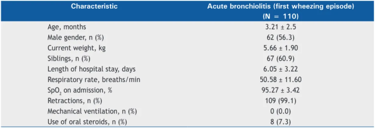
respiratory viruses (HRV, RSV, and HMPV). The mean age of the patients was 3.21 months, and 62 (56.3%) of the 110 patients were male (Table 1). Of the 110 infants evaluated, 109 (99.1%) had subcostal or intercostal retractions and required oxygen therapy. The mean hospital stay was 6 days, and none of these infants were admitted to the intensive care unit or needed mechanical ventilation (Table 1).
Of the 110 samples analyzed by DIF, 72 (65.4%) tested positive for a virus and 38 (34.6%) tested negative. The most common virus was RSV, which was
identiied in 65 (90.2%) of the 72 positive samples, followed by inluenza, identiied in 15 (20.8%); parainluenza, identiied in 10 (13.8%); and adenovirus, identiied in 3 (4.1%). In the sample as a whole, 56
(50.9%) of the 110 patients tested positive for one virus and 16 (14.5%) tested positive for two or three
length of the hospital stay or any other variable (Table 2). Among the 56 samples analyzed with quantitative
real-time PCR (during the second year of the study), speciically to evaluate the impact of viral load, RSV was again the virus most often detected, being identiied
in 19 (76%) of the 25 positive samples, followed by
HRV, which was identiied in 4 (16%), and HMPV, which was identiied in 2 (8%). The quantitative real-time PCR analysis showed that, in the second year of the
study, 31 (55.3%) of the 56 patients tested negative, 24 (42.8%) tested positive for one virus, and 1 (1.7%) tested positive for two viruses.
The mean viral loads for RSV, HRV, and HMPV were 1,340,000 copies/mL, 614,000 copies/mL, and 175,000 copies/mL, respectively. As can be seen in Figure 1, the RSV viral load in the nasal secretions, as
determined by real-time PCR, showed no signiicant
correlation with markers of clinical severity in our study sample (p > 0.05). We found that viral loads did not
inluence the length of hospitalization or the duration
of wheezing episodes.
DISCUSSION
In our study, RSV viral loads in infants with wheezing
did not inluence the length of the hospital stay,
which was used as a marker of the severity of acute bronchiolitis or wheezing episodes. Given the role of viral infections in wheezing, the hypothesis that higher viral loads or coinfection with different types of viruses
could inluence the natural history of acute wheezing
is reasonable and logical.(7) However, the association
between viral loads and severity remains unclear and controversial in the literature, as does that between coinfection and severity.(8-11) In clinical practice, the
size of the viral load and infection with more than one virus may generate uncertainty about the prognosis of such infections.(4) In the present study, the severity
of acute wheezing was not affected by the RSV viral loads in nasopharyngeal secretions.
When we used DIF in order to diagnose infection
with respiratory viruses (inluenza, parainluenza,
adenovirus, or RSV), we found that approximately 65% of the samples were positive for at least one such pathogen. Our DIF results are similar to those obtained in previous studies in the literature, in which the reported rate of infection with respiratory viruses among children with respiratory symptoms ranges from 45% to 70%.(12) In the present study, RSV was the
most prevalent pathogen, whether alone or together with another virus.
Real-time PCR was positive for RSV, HRV, and HMPV in
44.6% of the analyzed samples. That rate of positivity is in contrast with the 93.5% reported in a study that
used conventional and real-time PCR for 12 different
respiratory viruses.(13) However, it is similar to the rates reported in studies using only real-time PCR,
which ranged from 44% to 64%.(14,15) Our study was
underpowered to analyze the impact of coinfection
on disease severity. Currently, the reported effects of
Table 1. Characteristics of patients according to the questionnaire completed at admission and variables associated with clinical severity.a
Characteristic Acute bronchiolitis (first wheezing episode)
(N = 110)
Age, months 3.21 ± 2.5
Male gender, n (%) 62 (56.3)
Current weight, kg 5.66 ± 1.90
Siblings, n (%) 67 (60.9)
Length of hospital stay, days 6.05 ± 3.22
Respiratory rate, breaths/min 50.58 ± 11.60
SpO2 on admission, % 95.27 ± 3.42
Retractions, n (%) 109 (99.1)
Mechanical ventilation, n (%) 0 (0.0)
Use of oral steroids, n (%) 8 (7.3)
aResults presented as mean ± standard deviation, except where otherwise indicated.
Table 2. Severity associated with the number of respiratory viruses identiied by direct immunoluorescence.a
Variable Number of viruses identified p*
1 2-3
(n = 56) (n = 16)
Age, months 3.00 ± 2.47 2.38 ± 2.42 0.373
Current weight, kg 5.51 ± 1.88 5.50 ± 1.90 0.974
Respiratory rate, breaths/min 51.09 ± 12.51 49.06 ± 10.53 0.557
SpO2 on admission, % 95.14 ± 3.46 94.63 ± 3.52 0.601
Days on oxygen therapy 6.17 ± 2.93 5.44 ± 2.96 0.377
Length of hospital stay, days 6.83 ± 3.22 5.63 ± 3.22 0.188
Duration of wheezing, days 4.51 ± 2.93 3.81 ± 3.01 0.402
coinfection on the burden of disease among children are inconsistent and controversial. One possible
explanation is that such effects occur only in speciic
circumstances, such as RSV/HRV coinfection(5) or RSV/
HMPV coinfection. In addition, several environmental factors can also be determinants of the severity of respiratory viral infections, as can genetic variations in genes linked to the immune response against infections.(13-15)
In the present study, we believe that the lack of an association between viral load and outcome measures of severity stands out as the main result. In such analysis, one could have expected a correlation or a trend in the plot between the variables viral load and length of hospitalization. However, there was absolutely no trend or correlation between the two. Previous studies on viral load and severity have also reported conlicting indings. Few studies have investigated the association between disease severity and viral load. Fodha et al.(10) described a positive correlation between RSV viral load
and disease severity (determined by respiratory rate, length of hospital stay, and need for admission to the intensive care unit) in children hospitalized with respiratory infection. Zhou et al.(16) and Hasegawa et al.(17) showed that a higher mean
RSV viral load was associated with greater disease severity, as well as with a longer duration of hospitalization and symptoms.
Nevertheless, there have also been reports of negative and inverse associations. Martin et al.(11) reported that an increasing
viral load in RSV-infected children was associated with decreases in inpatient admissions, antibiotic use, and respiratory rates. In comparison with our patient sample, the sample evaluated by those authors was considerably larger, comprising 1,264 infants, 418 of whom tested positive for RSV by quantitative PCR. The authors detected borderline inverse associations (e.g., OR = 0.80; 95% CI: 0.70-0.99 for hospital admission). Considering these indings, we have to raise the possibility of random associations or lack of an association between viral load and disease severity in acute wheezing episodes.
Our study has some relevant limitations, such as the small sample size, single nasal sample collection, and the use of PCR tests only in a subsample. However, our indings add to the current knowledge by suggesting that there is no correlation between viral load and the severity of respiratory illness in infants.
In conclusion, on the basis of our results, neither coinfection nor viral load appears to inluence the major outcomes of acute bronchiolitis. We also found that RSV viral loads in infants with wheezing did not inluence the severity of the acute wheezing episodes in the irst year of life. Further studies investigating the effects of viral load and viral combinations may help clarify this important and controversial issue.
REFERENCES
1. Sole D. Childhood wheezing [Article in Portuguese]. J Bras Pneumol. 2008;34(6):337-9. PMID: 18622498
2. Kotaniemi JT, Pallasaho P, Sovijärvi AR, Laitinen LA, Lundbäck B. Respiratory symptoms and asthma in relation to cold climate, inhaled allergens, and irritants: a comparison between northern and southern Finland. J Asthma. 2002;39(7):649-58. http://dx.doi.org/10.1081/JAS-120014930
3. Lima JA, Fischer GB, Sarria EE, Mattiello R, Sole D. Prevalence of
and risk factors for wheezing in the irst year of life. J Bras Pneumol. 2010;36(5):525-31. PMID: 21085816
4. De Paulis M, Gilio AE, Ferraro AA, Ferronato AE, do Sacramento PR, Botosso VF, et al. Severity of viral coinfection in hospitalized infants with respiratory syncytial virus infection. J Pediatr (Rio J). 2011;87(4):307-13. http://dx.doi.org/10.2223/JPED.2100
5. da Silva ER, Pitrez MC, Arruda E, Mattiello R, Sarria EE, de Paula FE, et al. Severe lower respiratory tract infection in infants and toddlers Figure 1. Respiratory syncytial virus (RSV) viral load, in correlation with days of hospitalization (A*), days of oxygen use (B), and days of wheezing (C). *r = −0.217; p = 0.372.
1.5x10⁷
1.0x10⁷
5.0x10⁶
100 80 60 40 20 0
1.5x10⁷
1.0x10⁷
5.0x10⁶
100 80 60 40 20 0
1.5x10⁷
1.0x10⁷
5.0x10⁶
100 80 60 40 20 0
0 5 10 15 0 5 10 15
0 5 10 15
Days of hospitalization Days of oxigen use
Days of wheezing
RSV
v
ir
a
l
lo
a
d
(
co
p
ie
s/
m
L
)
RSV
v
ir
a
l
lo
a
d
(
co
p
ie
s/
m
L
)
RSV
v
ir
a
l
lo
a
d
(
co
p
ie
s/
m
L
)
B A
from a non-afluent population: viral etiology and co-detection as risk factors. BMC Infect Dis. 2013;13:41. http://dx.doi.org/10.1186/1471-2334-13-41
6. Miron D, Srugo I, Kra-Oz Z, Keness Y, Wolf D, Amirav I, et al. Sole pathogen in acute bronchiolitis: is there a role for other organisms apart from respiratory syncytial virus? Pediatr Infect Dis J. 2010;29(1):e7-e10. http://dx.doi.org/10.1097/INF.0b013e3181c2a212
7. Richard N, Komurian-Pradel F, Javouhey E, Perret M, Rajoharison A, Bagnaud A, et al. The impact of dual viral infection in infants admitted to a pediatric intensive care unit associated with severe bronchiolitis. Pediatr Infect Dis J. 2008;27(3):213-7. http://dx.doi.org/10.1097/ INF.0b013e31815b4935
8. Stempel HE, Martin ET, Kuypers J, Englund JA, Zerr DM. Multiple viral respiratory pathogens in children with bronchiolitis. Acta Paediatr. 2009;98(1):123-6. http://dx.doi.org/10.1111/j.1651-2227.2008.01023.x
9. Franz A, Adams O, Willems R, Bonzel L, Neuhausen N, Schweizer-Krantz S, et al. Correlation of viral load of respiratory pathogens and co-infections with disease severity in children hospitalized for lower respiratory tract infection. J Clin Virol. 2010;48(4):239-45. http:// dx.doi.org/10.1016/j.jcv.2010.05.007
10. Fodha I, Vabret A, Ghedira L, Seboui H, Chouchane S, Dewar J, et al. Respiratory syncytial virus infections in hospitalized infants: association between viral load, virus subgroup, and disease severity. J Med Virol. 2007;79(12):1951-8. http://dx.doi.org/10.1002/jmv.21026 11. Martin ET, Kuypers J, Heugel J, Englund JA. Clinical disease and viral
load in children infected with respiratory syncytial virus or human metapneumovirus. Diagn Microbiol Infect Dis. 2008;62(4):382-8. http://dx.doi.org/10.1016/j.diagmicrobio.2008.08.002
12. Calegari T, Queiroz DA, Yokosawa J, Silveira HL, Costa LF, Oliveira TF, et al. Clinical-epidemiological evaluation of respiratory syncytial virus infection in children attended in a public hospital in midwestern Brazil. Braz J Infect Dis. 2005;9(2):156-61. http://dx.doi.org/10.1590/ S1413-86702005000200006
13. Martin ET, Kuypers J, Wald A, Englund JA. Multiple versus single virus respiratory infections: viral load and clinical disease severity in hospitalized children. Inluenza Other Respir Viruses. 2012;6(1):71-7. http://dx.doi.org/10.1111/j.1750-2659.2011.00265.x
14. Canducci F, Debiaggi M, Sampaolo M, Marinozzi MC, Berrè S, Terulla C, et al. Two-year prospective study of single infections and co-infections by respiratory syncytial virus and viruses identiied recently in infants with acute respiratory disease. J Med Virol. 2008;80(4):716-23. http://dx.doi.org/10.1002/jmv.21108
15. Jennings LC, Anderson TP, Werno AM, Beynon KA, Murdoch DR. Viral etiology of acute respiratory tract infections in children presenting to hospital: role of polymerase chain reaction and demonstration of multiple infections. Pediatr Infect Dis J. 2004;23(11):1003-7. http:// dx.doi.org/10.1097/01.inf.0000143648.04673.6c
16. Zhou L, Xiao Q, Zhao Y, Huang A, Ren L, Liu E. The impact of viral dynamics on the clinical severity of infants with respiratory syncytial virus bronchiolitis. J Med Virol. 2015;87(8):1276-84. http://dx.doi. org/10.1002/jmv.24111