*Correspondence: R.V. M. Oliveira. Curso de Farmácia, Universidade de Sorocaba. Rodovia Raposo Tavares, Km 92,5 – 18023-903 – Sorocaba – SP, Brasil. E-mail: [email protected]
A
rti
Pharmaceutical Sciences vol. 46, n. 2, abr./jun., 2010
In vitro
evaluation of copaiba oil as a kojic acid skin enhancer
Robson Vicente Machado de Oliveira
1*, Mitsuko Taba Ohara
2, Marta Maria Duarte Carvalho
Vila
1, Marcos Moisés Gonçalves
31Pharmacology Course, Sorocaba University, 2 Pharmacology Department, Faculty of Pharmaceutical Sciences, University of
São Paulo, 3Department of Analytical Chemistry, Chemistry Institute, State University of Campinas
The capacity of copaíba oil to act as a skin penetration enhancer for the depigmenting agent kojic acid was evaluated using an in vitro diffusion system with static lux and shed rattlesnake skin membrane,
Crotalus durissus terriicus, in saline solution at 34±2 °C as the luid receptor. The quantities of kojic acid liberated into the luid receptor were determined by spectrophotometry at 268 nm with intervals of one and a half hours. The membranes, pretreated with copaíba oil at 25% and 50% v/v, gave lux values of 8.0 and 12.7 µg/cm2/h, permeability values of 2.0 and 3.3 cm×10-4/h, and promotion factors of 4.1
and 3.7, respectively. These results indicate that copaíba oil, at the two concentrations studied, has the capacity to promote penetration of kojic acid.
Uniterms: Copaíba oil. Kojic acid. Skin penetration/ in vitro studies .UV Spectrophotometry.
A propriedade do óleo de copaíba como agente promotor de penetração cutânea do despigmentante ácido kójico foi avaliada utilizando-se sistema de difusão in vitro com luxo estático, membrana de pele da serpente cascavel - Crotalus durissus terriicus e solução salina a 34±2 °C como luido receptor. As quantidades liberadas do ácido kójico no luido receptor foram determinadas por espectrofotometria em 268 nm em intervalos de 1:30 h. As membranas pré-tratadas com óleo de copaíba a 25 e 50% v/v apresentaram valores de luxo de 8,0 e 12,7 µg/cm2/h, permeabilidade de 2,0 e 3,3 cm×10-4/h, e fatores
de promoção de 4,1 e 3,7, respectivamente. Os resultados obtidos indicaram que o óleo de copaíba, nas duas concentrações estudadas, apresentou capacidade de promoção da penetração do ácido kójico.
Unitermos: Óleo de copaíba. Ácido kójico. Penetração cutânea/ in vitro. Espectrofotometria UV.
INTRODUCTION
Hyper pigmentation of the skin can be caused by a range of factors such as aging, pregnancy, endocrine disturbances, hormonal treatment and sun exposure to varying degrees. Several substances are commonly em-ployed as depigmentation agents in the manufacture of cosmetics used for the reduction of hyper pigmentation, one of these being kojic acid (Cabanes, Garcia-Carmo-na, 1994; Su, 1999; Nicoletti et al., 2002). Kojic acid (5-hydroxy-2-(hydroxymethyl)-4-pirone) is a depigmen-ting agent obtained from rice fermentation (Burdock et al., 2001) as a fungus metabolite from the genera Aspergillus
and Penicillym, and acts by inhibiting tyrosinase activity
(Cabanes, Garcia-Carmona 1994).
In order to be effective, depigmenting agents incor-porated in topical formulas must cross the stratum corneum to act on the more inner layers situated towards the basal lamina of the epidermis. To this end, the addition of other compounds with a greater capacity for skin penetration, also known as absorption promoters or “enhancers”, can result in an increase in diffusion of substances by disor-ganizing the lamellas of the stratum corneum (Williams, Barry, 2004). The incorporation of these substances in for-mulations allows for the development of topical products with a lower concentration of active ingredients, thereby increasing both the efficacy and safety of the product (Yourik, Bronaugh, 1999).
as a source of ingredients possessing anti inlammatory, anti infectious, anti tumoral and wound healing properties, among others (Veiga Junior, Pinto, 2002). Copaibas are trees that are native to the tropical regions of Latin Ame-rica and Western AfAme-rica, belonging to the family Legumi-nosae Juss, sub-family Caesalpinoideae Kunth. These trees furnish an oily substrate consisting of 40 to 50% essential oil and 40 to 60% resin, whose composition is made up principally of esters and terpenes (Cascon, Gilbert, 2000; Pinto et al., 2000; Biavati et al., 2006).
Copaiba oil, given its terpine compounds such as sesquiterpenes, oxygenated sesquiterpenes, hydrocarbon diterpenes and oxygenated diterpenes (Pinto et al., 2000) is a good choice as a permeability promoter for other compounds. Terpenic compounds are well known for their ability to increase cutaneous penetration (El-Kattan et al., 2001; Narishetty, Panchagnula, 2004).
In vivo and in vitro (Franz et al., 1993) diffusion methods are used to study the eficacy of skin penetrating agents. For in vitro methods, diffusion cells or the Franz method are used where, in the literature, many studies have used diffusion cells with membranes from the skin of rats, mice, guinea pigs, shed snake skin, human or synthetic skin to assess the penetration promoting powers of different substances (Franz et al.., 1993; Bronaugh, Collier, 1993). Different experimental conditions involving modiications of temperature, agitation, different pretreatment temperatu-res, collection of samples and trial duration have been des-cribed (Akimoto, Nagase, 2003; Andega et al., 2001; Babu, Pandit, 2004; Cotte et al., 2004; El-Kattan et al., 2001).
The use of shed snake skin as a membrane in in vitro
systems is justiied due to the similarities between shed snake skin and the stratum corneum of human skin, in ter-ms of structure, lipid composition, water permeability, and other substances with different functional groups. (Itoh et al., 1990). Additionally, shed snake skin has advantages in terms of low cost, ease of use, absence of hair, and lack of need to sacriice animals. (Lin et al., 1992; Haigh et al.,1998; Chang, Zheng, 2003).
For the above reasons and due to the need to inves-tigate properties of vegetable compounds, the aim of this study was to evaluate in vitro, copaiba oil as a penetrating agent for kojic acid in a shed snake skin model through diffusion studies in an effort to develop more effective topical depigmenting agents.
MATERIAL AND METHODS
Material
Substances used were AP grade, kojic acid was
90% purity and the solutions were prepared using freshly distilled water. Copaiba oil was of pharmaceutical purity and was kindly donated by Ionquímica. The membranes used as diffusion cells were obtained from the shed skin of the rattle snake Crotalus durissus terriicus.
Methods
Preparation of membranes
Square or rectangular sections measuring about 4 cm per side were cut from the ventral part of the shed snake skin, containing four sections of rigid scales and three le-xible sections. These were placed in distilled water at room temperature for 48 hours to hydrate. The membranes were then dried on absorbent paper and placed in a receptacle with an external diameter of about 3.7 cm, and an area available for diffusion of 3.14 cm2.
Quantification of kojic acid in receptor fluid
Quantiication of the kojic acid was done by
spectro-photometry (Beckman-Coulter® DU-640) at 268 nm
(Oli-veira et al., 2007) using a method adapted from Majmudar and collaborators (1998). This method gave a quantiica-tion limit of 0.17 µg/mL and a detecquantiica-tion of 0.057 µg/mL according to Oliveira and collaborators (2007).
Readings were taken from 0.5 mL aliquots of the luid receptor after dilution up to10 mL with saline solu-tion (sodium chloride 0.9% p/v) and iltrasolu-tion through a 0.45 µm pore membrane.
Diffusion Assays
The (Logan® SDFC-6 VTC-200) in vitro cell
sion system was used, composed of three vertical diffu-sion cells with donation compartments of 2 cm diameter, receptor compartments with a capacity of 15.0, 15.1 and 15.2 mL, a support stand, and a magnetic agitation system equipped with a pump system and water heater.
The circulating water temperature was adjusted to 34±2 °C and the luid receptor (saline solution) was main-tained under agitation at 100 rpm.
pro-pylene glycol 95:5 v/v. alcohol. After one hour, the solutions were removed with cotton and the membranes washed with distilled water. The irst cell of the receptor compartment containing 15.1 mL, received 0.8 mL of saline solution (white) while the second (15.0 mL) and third (15.2 mL) both received 0.8 mL of kojic acid 3.9% p/v equivalent to 31.2 mg of kojic acid. The experiment was run for 22 h 30 min, with removal of aliquots and replacement of the volumes of the receptor compartments at times points 0; 1:30; 3:00; 4:30; 6:00; 7:30; 9:00; 10:30; 12:00; 13:30 and 22:30 hours. Concentrations for kojic acid were determined spectropho-tometrically and these values used to calculate the diffusion parameters. Four replicates of each experiment were done; however, the irst determinations of kojic acid gave results lower than the quantiication limits due to the heterogeneity of the shed snake skin membrane, and therefore these irst results were omitted for the calculation of the mean values.
The mean quantity of kojic acid by area was ob-tained using the mean circular membrane values and the kojic acid concentration. Concentration of kojic acid was obtained from the mean values of absorbency. Kojic acid liberation curves by membrane area (cm2) against hours
of treatment were constructed (Asbill, Michniak, 2000). Flux (µg/cm2/h) was considered equal to the
incli-nation coeficient of those lines with a linear correlation coeficient that yielded values greater than 0.9.
Permeability, which was equivalent to the depth of the membrane the substance managed to penetrate (cm) against time (h), was calculated by dividing the lux values
(µg/cm2/h) by the kojic acid solution concentration
(39 mg/cm3) applied to the membrane (Asbill, Michniak,
2000).
The latency time (hours), indicating the moment the membrane allowed Constant liberation of kojic acid, was extrapolated from the point where the regression lines crossed the axis of the abscissa (Barry, 1987).
To compare the effect on the oil solutions and their controls, a “promotion factor” calculated based on the mean lux (µg/cm2/h) of kojic acid in the sample divided
by the mean lux of the control (without pretreatment with oil) (Bronaugh, Collier, 1993).
Statistical Analysis
Fisher’s and Student’s t tests were used to compare the data, means and standard deviations (Bussab, Morettin, 1985; Botter et al., 1996).
RESULTS AND DISCUSSION
The quantiication of kojic acid began only from the three hour time interval in membranes treated with both 25
and 50% solutions of copaiba oil giving mean liberation values per area of 20.6 and 27.4 µg/cm2, respectively (see
Table I). Prior to this three hour time point, the kojic acid concentration did not reach the quantiication limit for the method employed (Oliveira et al., 2007). The solution of kojic acid 3.9% p/v used in these penetration studies, with a pH around 4.0, favored the non ionized form of kojic acid, and allowing its penetration into the membranes.
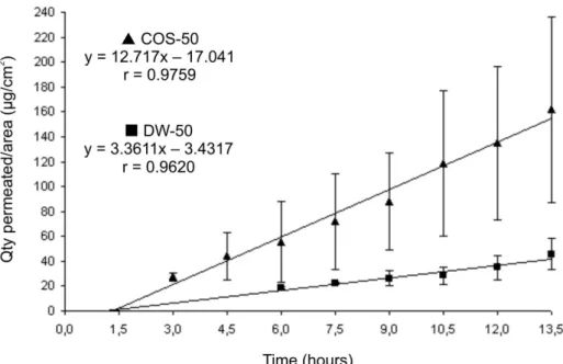
Compared with the controls, membranes pretreated with oil solutions of 25 and 50% allowed better diffusion of kojic acid from the seven and one half hour time point, and after 12 hours the amounts of kojic acid liberated were 80.2 µg/cm2 and 135.2µg/cm2, respectively (Table I).
The lines associated to these oil solutions present greater inclination indicating a larger angular coefficient, and thus a higher lux (Figures 1 and 2). Figures 1 and 2 also show variable coeficients of correlation which can be explained by the irregular thickness and tortuosity inhe-rent to the snake membrane (Ostrenga et al., 1974). The characteristics of animal membranes also inluence the values of the variation coeficients. Lopes (1999), in his study of diffusion with sodium diclophenate on treatment with 0.2% papain solution in human membranes, found a coefficient of variation of up to 73% in the liberated amounts. Sato and colleagues (2007) also observed high standard deviations in their studies assessing kojic acid formula penetration in porcine skin membrane.
Based on the regression lines in the diffusion cur-ves (Figures 1 and 2), the lux parameters, permeability parameters and latency time were extrapolated, as shown in Table II. The table demonstrates that latency times for the constant liberation of kojic acid were reduced with increased copaiba oil concentration. This represents a beneicial characteristic for depigmentation agents. The promotion factors for liberation of kojic acid obtained from treatment with 25% and 50% copaiba solutions v/v were equivalent to 4.0 and 3.7, values greater than those obtained from control solutions.
FIGURE 1- Cutaneous diffusion proile in vitro of kojic acid in shed snake skin membrane treated with 25% copaiba oil in
isopropyl-propylene glycol (95:5 v/v) alcohol solution (COS-25) or 25% distilled water in isopropyl-isopropyl-propylene glycol (95:5 v/v) alcohol solution (DW-25) and treated with kojic acid solution 3.9% p/v.
TABLE I - Kojic acid liberation in diffusion studies using shed snake skin membrane treated with 25 or 50% v/v copaiba oil solutions
(COS) or 25 or 50% distilled water (DW) in isopropyl-propylene glycol (95:5 v/v) alcohol after application of kojic acid solution 3.9% p/v
Time (hours) Pre-treatment
Quantity liberated by area (µg/cm2)
Copaíba 25% Copaíba 50%
n Mean SD N Mean SD
3:00 DW - --- --- - ---
---COS 1 20.6 --- 2 27.4 3.1
4:30 DW - --- --- - ---
---COS 1 28.3 --- 3 44.2 18.8
6:00 DW - --- --- 2 18.4 0.8
COS 1 41.3 --- 4 55.7 32.7
7:30 DW - --- --- 2 22.4 1.0
COS 3 31.2 21.2 4 71.9 38.1
9:00 DW 3 19.0 2.0 3 26.2 5.9
COS 4 39.9 24.6 4 87.8 39.1
10:30 DW 3 22.9 3.6 4 28.3 7.2
COS 4 63.8 43.7 4 118.9 58.4
12:00 DW 3 27.2 1.7 4 34.8 9.9
COS 4 80.2 53.8 4 135.2 61.6
13:30 DW - --- --- 4 45.9 12.3
COS - --- --- 4 161.9 74.5
22:30 DW 4 46.2 13.4 - ---
---COS 4 172.6 27.6 - ---
FIGURE 2- Cutaneous diffusion proile in vitro of kojic acid in shed snake skin membrane treated with 50% copaiba oil solution in
isopropyl-propylene glycol (95:5 v/v) alcohol solution (COS-50) or in 50% distilled water solution in isopropyl-propylene glycol (95:5 v/v) alcohol solution (DW-50) and treated with kojic acid solution 3.9% p/v.
TABLE II - Latency time (hours), lux (µg/cm2/h) and permeability (cm/h) of kojic acid in diffusion studies of shed snake skin
membrane treated with 25 or 50% copaiba oil solution in isopropyl-propylene glycol (95:5 v/v) alcohol (COS-25) or in 25 or 50% distilled water (DW) in isopropyl-propylene glycol (95:5 v/v) alcohol solution (DW-25) and treated with kojic acid solution 3.9% p/v
Treatment Latency time (hours) Mean Flux (µg/cm2/h) Mean Permeability (cm x 10-4/h)
DW-25 - 2.0 0.5
COS-25 1:50 8.0 2.0
DW-50 1:20 3.4 0.9
COS-50 1:00 12.7 3.3
COS - copaiba oil solution; DW – distilled water control; SD - standard deviation.
lux values from treatment studies versus those of controls revealed a decrease in these values at higher isopropyl alcohol concentrations.
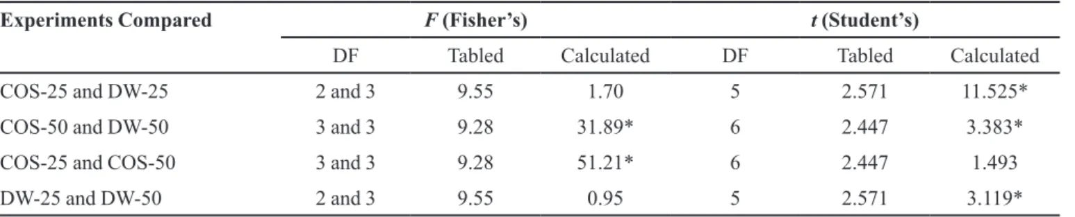
Standard deviations were calculated from the means of the lux and permeability values extrapolated on the individual graphs of each replicate experiment, and for each copaíba oil concentration (Table III). Fisher’s and Student’s t values were obtained from these values and are displayed in Table IV.
Student’s t test revealed a signiicant difference in the mean lux values of kojic acid at the different copaiba oil concentrations on diffusion testing compared to the respective distilled water solutions. However, studies using 25 and 50% v/v solutions of copaiba oil showed no signiicant differences in the mean values of these parame-ters when compared with each other. These observations agree with the analysis of promotion factors which yielded
similar values (3.7 and 4.0). Analysis using Fisher’s test in these experiments found a signiicant difference in mean lux values, probably due to the shed snake skin membrane characteristic. Fisher’stest also showed that the studies done with 25 and 50% distilled water in the treatment of snake skin membrane were homogeneous across all the experiments.
Comparing Fisher results for each experiment versus its respective control, only the 50% p/v copaíba oil solu-tion produced a statistically signiicant variasolu-tion for kojic acid lux. This shows that although test solutions and their controls were submitted to the same treatment, they did not show the same pattern of variation. This is largely due to irregularities in the snake skin membrane.
demonstrated in the study by Lin and colleagues (1992), which found permeability values 2 to 4 times higher in shed snake skin (Phyton molurus) than in isolated stra-tum corneum for sodium diclophenate, theophyline and benzoic acid (2 mg/mL or 0.2% in aqueous solution). The use of shed snake skin in a study of copaiba oil as a pro-moter of skin penetration for hydrophilic substances such as kojic acid, takes into consideration the fact that these membranes have a lower permeability coeficient (3.3 to 6.1 times) for these compounds, extending the time requi-red to carry out the experiments. With regard to lipophilic compounds, these have a permeability coeficient close those of membranes obtained from human skin (0.9 to 1.8 times) (3.3 to 6.1 times) (Ngawhirunpat et al., 2006). The percentage penetration of kojic acid for the initial amount applied was 1.63% for the 50% solution of copaiba oil after an interval of 13 and a half hours versus 1.74% for the 25 % solution of copaiba oil after a 22 and a half hour interval. These indings indicate that the 50% oil solution allowed almost the same level of kojic acid pe-netration but in a much shorter time frame. This indicated
that a 50% concentration of copaiba oil may be optimal for developing topical formulations containing kojic acid.
CONCLUSION
Copaiba oil in both the 25 and 50% v/v solutions studied proved to be a penetration promotion factor of kojic acid in 3.9%v/v solution in the shed snake skin mem-branes Crotalus durissus terriicus compared to controls. The promotion factors were 4.0 and 3.7 for 25% and 50% copaiba oil solutions, respectively. These results allowed us to conclude that this oil has the potential to be added to topical formulations as a penetration promoter for active hydrophilic substances.
ACKNOWLEDGEMENTS
We would like to thank the Department for the Con-trol and Production of Pharmaceuticals and Medications of the Medical Sciences Faculty of the University of São Paulo, where this work was conducted.
TABLE III - Mean, standard deviation, variance and coeficient of variance for mean lux and mean permeability (µg/cm2/h and
cm/h) of kojic acid from diffusion studies on shed snake skin membrane treated with 25 or 50% copaiba oil in isopropyl-propylene glycol (95:5 v/v) alcohol solution (COS-25 or COS-50) or in 25 or 50% distilled water in isopropyl-propylene glycol (95:5 v/v) alcohol solution (DW-25 or DW-50%) and treated with kojic acid solution 3.9% p/v
Experiment Pre-treatment N Flux (µg/cm2/h) Permeability (cm x 10-4/h) CV%
Mean SD S2 Mean SD S2
1 DW-25 3 2.369 0.979 0.959 0.607 0.251 0.063 41.34
COS-25 4 9.855 0.752 0.566 2.527 0.193 0.037 7.63
2 DW-50 4 4.665 0.953 0.909 1.196 0.244 0.060 20.43
COS-50 4 13.911 5.383 28.98 3.567 1.380 1.904 38.70
CV% - coeficient of variation ; SD-standard deviation ; S2 - variance; n- number of replicates
TABLE IV - Fisher’s and Student’s t test values for mean lux (µg/cm2/h) of kojic acid in diffusion membrane studies using shed
snake skin treated with 25 or 50% copaiba oil in isopropyl –propylene glycol (95:5 v/v)alcohol solution (COS-25 or COS-50) or in 25 or 50% distilled water in isopropyl –propylene glycol (95:5 v/v) alcohol solution (DW-25 or DW-50%) and treated with kojic acid solution 3.9% p/v
Experiments Compared F (Fisher’s) t (Student’s)
DF Tabled Calculated DF Tabled Calculated
COS-25 and DW-25 2 and 3 9.55 1.70 5 2.571 11.525*
COS-50 and DW-50 3 and 3 9.28 31.89* 6 2.447 3.383*
COS-25 and COS-50 3 and 3 9.28 51.21* 6 2.447 1.493
DW-25 and DW-50 2 and 3 9.55 0.95 5 2.571 3.119*
REFERENCES
AKIMOTO, T.; NAGASE, Y. Novel transdermal drug penetration enhancer: synthesis and enhancing effect of alkyldisiloxane compounds containing glucopyranosyl group. J. Control. Release, v.88, p.243-252, 2003.
ANDEGA, S.; KANIKKANNAN, N.; SINGH, M. Comparison of the effect of fatty alcohols on the permeation of melatonin between porcine and human skin. J. Control. Release, v.77, p.17-25, 2001.
ASBILL, C.S.; MICHNIAK, B.B. Percutaneous penetration enhancers: local versus transdermal activity. Pharm. Sci. Technol. Today, v.3, p.36-41, 2000
BABU, R.J.; PANDIT, J.K. Effect of cyclodextrins on the complexation and transdermal delivery of bupranolol through rat skin. Int. J. Pharm., v.271, p.155-165, 2004.
BARRY, B.W. Mode of action of penetration enhancers in human skin. In: ANDERSON, J.M., KIM, S.W., (Eds.).
Advances in drug delivery systems 3: Proceedings of the Third International Symposium on Recent Advances in Drug Delivery Systems. Salt Lake City: Elsevier, 1987. p.85-97.
BIAVATI, M.W.; DOSSIN, D.; DESCHAMPS, F.C.; LIMA, M.P. Análise de óleo-resinas de copaíba: contribuição para o seu controle de qualidade. Rev. Bras. Farmacogn., v.16, p.230-235, 2006.
BOTTER, D.A.; PAULA, G.A.; LEITE, J.G.; CORDANI, L.K.
Noções de estatística: com apoio computacional (versão preliminar). São Paulo: IME-USP, 1996. 231 p.
BRONAUGH, R.L.; COLLIER, S.W. In vitro methods for measuring skin permeation. In: ZATZ, J.L., (Ed.). Skin permeation: fundamentals and application. Wheaton: Allured, 1993. p.93-110.
BURDOCK, G.A.; SONI, M.G.; CARABIN, I.G. Evaluation of health aspects of kojic acid in food. Regul. Toxicol. Pharmacol., v.33, p.80-101, 2001.
BUSSAB, W.O.; MORETTIN, P.A. Estatística básica: métodos quantitativos. 4.ed. São Paulo: Atual, 1985. 321 p.
CABANES, J.; GARCIA-CARMONA, F. Kojic acid, a cosmetic skin whitening agent, is a slow-binding inhibitor of catecholase activity of tyrosinase. J. Pharm. Pharmacol.,
v.46, p.982-985, 1994.
CASCON, V.; GILBERT, B. Characterization of the chemical composition of oleoresins of Copaifera guianensis Desf.,
Copaifera duckei Dwyer and Copaifera multijuga Hayne.
Photochemistry, v.55, p.773-778, 2000.
CHANG, C.; ZHENG, R. Effects of ultraviolet B on epidermal morphology, shedding, lipid peroxide, and antioxidant enzymes in Cope´s rat snake (Elaphe taeniura). J. Photochem. Photobiol., B, v.72, p.79-85, 2003.
COLIPA Guidelines - Scientific Committee on Cosmetic Products and Non-Food Products. Annex 10 – Guidelines for in vitro methods to assess percutaneous absorption of cosmetic ingredients. In: Notes of Guidance for Testing of Cosmetic Ingredients for their Safety Evaluation, Monograph SCCNFP/1321/00, EC. Brussels, 2000. p.77-85.
COTTE, M.; DUMAS, P.; BESNARD, M.; TCHORELOFF, P.; WALTER, P. Synchroton FT-IR microscopic study of chemical enhancers in transdermal drug delivery: example of fatty acids. J. Control. Release, v.97, p.269-281, 2004.
EL-KATTAN, A.F.; ASBILL, C.S.; KIM, N.; MICHNIAK, B.B. The effects of terpene enhancers on the percutaneous permeation of drugs with different lipophilicities. Int. J. Pharm., v.215, p.229-240, 2001.
FRANZ, T.J.; LEHMAN, P.A.; McGUIRE, E.L. In vivo methods for the assessment of percutaneous absorption in man. In: ZATS, J.L. (Ed.). Skin Permeation: fundamentals and application. Wheaton: Allured, 1993. p.73-92.
HAIGH, J.M.; BEYSSAC, E.; CHANET, L.; AIACHE, J.M.
In vitro permeation of progesterone from a gel through the shed skin of three different snake species. Int. J. Pharm., v.170, p.151-156, 1998.
ITOH, T.; XIA, J.; MAGAVI, R.; NISHIHATA, T.; RYTTING, J.H. Use of shed snake skin as a model membrane for in vitro percutaneous penetration studies: comparison with human skin. Pharm. Res., v.7, p.1042-1047, 1990.
LARRUCEA, E.; ARELLANO, A.; SANTOYO, S.; YGARTUA, P. Combined effect of oleic acid and propylene glycol on the percutaneous penetration of tenoxicam and its retention in the skin. Eur. J. Pharm. Biopharm., v.52, p.113-119, 2001.
LOPES, P.S. Influência da papaína e do óleo de pequi na penetração percutânea do diclofenaco de sódio. São Paulo, 1999. 148 p. [Dissertação de Mestrado. Faculdade de Ciências Farmacêuticas. Universidade de São Paulo].
MAJMUDAR, G.; JACOB, G.; LABOY, Y.; FISHER, L. An
in vitro method for screening skin-whitening products. J. Cosmet. Sci., v.49, p.361-367, 1998.
NARISHETTY, S.T.K.; PANCHAGNULA, R. Transdermal delivery of zidovudine: effect of terpenes and their mechanism of action. J. Control. Release, v.95, p.367-379, 2004.
NICOLETTI, M. A.; ORSINE, E.M.A.; DUARTE, A.C.N.; BUONO, G.A. Hipercromias: aspectos gerais e uso de despigmentantes cutâneos. Cosmet. Tolet., Ed. Port., v.14 p.46-51, 2002.
NGAWHIRUNPAT, T.; PANOMSUK, S.; OPANASOPIT, P.; ROJANATA,T.; HATANAKA, T. Comparison of the percutaneous absorption of hydrophilic and lipophilic compounds in shed snake skin and human skin. Pharmazie,
v.61, p.331-335, 2006.
OLIVEIRA, R.V.M.; OHARA, M. T.; GONÇALVES, M. M.; VILA, M. M. D. C. Quantificação de ácido kójico em estudos de permeação in vitro. Lat. Am. J. Pharm., v.26, p.576-581, 2007.
OSTRENGA, J.; STEINMETZ, C.; POULSEN, B.; YETT, S. Signiicance of vehicle composition. II. Prediction of optimal vehicle composition. J. Pharm. Sci., v.67, p.1180-1183, 1974.
PINTO, A.C.; BRAGA, W.F.; REZENDE, C.M.; GARRIDO, F.M.S.; VEIGA JÚNIOR, V.F.; BERGTER, L.; PATITUCCI, M.L.; ANTUNES, O.A.C. Separation of acid diterpenes of Copaifera cearensis Huber ex Ducke by lash chromatography using potassium hydroxide impregnated silica gel. J. Braz. Chem. Soc., v.11, p.355-360, 2000.
SATO, M.E.O.; GOMARA, F.; PONTAROLO, F.G.R.; ANDREAZZA, I.F.; ZARONI, M. Penetração cutânea in vitro do ácido kójico. Rev. Bras. Cienc. Farm., v.43, p.196-203, 2007.
STUPP, T.; FREITAS, R. A.; SIERAKOWSKI, M. R.; DESCHAMPS, F. C.; WISNIEWSKI JR, A.; BIAVATTI, M. W. Characterization and potential uses of Copaifera langsdorii seeds and seed oil. Bior. Tech., v.99, p.2659-2663, 2008.
SU, E. G. Formulando com branqueadores da pele. Cosmet. Tolet., Ed. Port., v.11, p.57-63,1999
VEIGA JUNIOR, V. F.; PINTO, A. C. O gênero Copaifera L.
Quim. Nova, v.25, p.273-286, 2002.
VEIGA JUNIOR, V. F.; ROSAS, E.C.; CARVALHO, M.V.; HENRIQUES, M.G.M.O.; PINTO, A.C. Chemical composition and anti-inlammatory of copaiba oils from
Copaifera cearensis Huber ex Ducke, Copaifera reticulata
Ducke and Copaifera multijuga Hayne - A comparative study. J. Ethnopharmacol., v.112, p.248-254, 2007.
WILLIAMS, A.C.; BARRY, B.W. Penetration enhancers. Adv. Drug Delivery Rev., v.56, p.603-618, 2004.
YOURICK, J.J., BRONAUGH, R.L. (Eds.). Percutaneous penetration as it relates to the safety evaluation of cosmetic ingredients. In__: Percutaneous absorption - Drugs, cosmetics, mechanisms, methodology. 3.ed. New York: Marcel Dekker, 1999. p.659-671.