*Correspondence: G. R. Pereira. Departamento de Alimentos e Medicamentos. Faculdade de Ciências Farmacêuticas Universidade Federal de Alfenas. Rua Gabriel Monteiro da Silva, 700, 37130-001 - Alfenas- MG, Brazil. E-mail: [email protected]
A
vol. 52, n. 3, jul./sep., 2016 http://dx.doi.org/10.1590/S1984-82502016000300018
Evaluation of skin absorption of drugs from topical and
transdermal formulations
André Luís Morais Ruela
1, Aline Gravinez Perissinato
2, Mônica Esselin de Sousa Lino
3,
Paula Silva Mudrik
3, Gislaine Ribeiro Pereira
3*1Multidisciplinary Health Institute, Federal University of Bahia, Vitória da Conquista, BA, Brazil, 2School of Pharmaceutical
Sciences Universidade Estadual Paulista, Araraquara, SP, Brazil,3Departament of Pharmacy, Faculty of Pharmaceutical Sciences, Federal University of Alfenas, Alfenas, MG, Brazil
The skin barrier function has been attributed to the stratum corneum and represents a major challenge in clinical practice pertaining to cutaneous administration of drugs. Despite this, a large number of bioactive compounds have been successfully administered via cutaneous administration because of advances in the design of topical and transdermal formulations. In vitro and in vivo evaluations of these novel drug delivery systems are necessary to characterize their quality and eicacy. This review covers the most well-known methods for assessing the cutaneous absorption of drugs as an auxiliary tool for pharmaceutical formulation scientists in the design of drug delivery systems. In vitro methods as skin permeation assays using Franz-type difusion cells, cutaneous retention and tape-stripping methods to study the cutaneous penetration of drugs, and in vivo evaluations as pre-clinical pharmacokinetic studies in animal models are discussed. Alternative approaches to cutaneous microdialysis are also covered. Recent advances in research on skin absorption of drugs and the efect of skin absorption enhancers, as investigated using confocal laser scanning microscopy, Raman confocal microscopy, and attenuated total relectance Fourier-transform infrared spectroscopy, are reviewed.
Uniterms: Skin absorption/efects/study. Skin absorption/topical formulations. Skin absorption/ transdermal formulations. Skin absorptions/enhancers.
INTRODUCTION
Topical and transdermal drug delivery systems have
shown signiicant advantages in clinical practice for drug
targeting to the action site in the body; this has reduced
the systemic side efects. The administration of drugs by
through the skin is also performed to achieve controlled
or prolonged drug delivery, and this route can be explored as an alternative to the oral route. The oral route shows some limitations for drugs with irregular absorption in the gastrointestinal tract and low bioavailability and for drugs with increased irst pass metabolism and short plasma
half-life times (Barry, 2001; Wokovich et al., 2006; Prausnitz,
Langer, 2009; Alexander et al., 2012).
Many drug products applied to the skin surface
may penetrate to some extent into the skin layers, where
their effects are expected. This is the case for topical
formulations for treatment of skin disorders such as
acne and cutaneous inlammatory diseases that include
dermatitis, erythematous lupus, and psoriasis. On the other hand, transdermal formulations release drugs that permeate through the skin and enter the systemic circulation. Transdermal therapy must ensure that significant concentrations of the drug are absorbed to reach effective plasma concentrations. Permeation of drugs is targeted in some cases to body regions close to
the action site, where a regional efect is expected, e.g., in the muscles, blood vessels, and articulations. In this
way, the term “cutaneous absorption” is properly used to
characterize the sum of the amounts of drug that penetrate and permeate the skin (Barry, 2001; El Maghraby, Barry; Williams, 2008; Williams, Barry, 2012; Selzer et al., 2013).
Diferent methods have been proposed to evaluate
through the skin. These methods are important tools for assessing the efficacy and quality of topical and transdermal formulations (Lin, Ho, Chien, 1993; Manadas, Pina, Veiga, 2002; Thakker, Chern, 2003; Valenta, Auner, 2004; Cardot, Beyssac, Alric, 2007; Frum et al., 2007; Chen, Han, Lian, 2013; Schwarz, Pagitsch, Valenta, 2013). However, pharmaceutical formulation scientists must be aware of all options in the selection
of methods for intended formulations (Seki et al., 2004; Godin, Touitou, 2007; Zhang, Chan, Leong, 2013).
The aim of this study was to systematically review
the in vitro and in vivo methods applied to the assessment of skin penetration and permeation of drugs from topical
and transdermal formulations, with the goal of aiding the
pharmaceutical formulation scientists in the selection of the most appropriate formulation for a particular investigation.
Drug transport across the skin
The skin is the largest organ of the human body and has a surface area of about 2 m2 in healthy adults (Groeber
et al., 2011). It is a heterogeneous multilayer tissue, and its
primary function is to protect the body from the external
environment by functioning as an effective barrier to
absorption of exogenous molecules (Godin, Touitou,
2007; Darlenski et al., 2009; Prausnitz, Langer, 2009; Williams, Barry, 2012; Anissimov et al., 2013; Jepps et al., 2013). The epidermis and dermis layers form the main skin structure (Figure 1).
The dermis thickness ranges from 3 to 5 mm and
consists of a mixture of fibrous proteins (collagen and elastin) and an interibrillar gel of glycosaminoglycans,
salts, and water. Collagen types I and II account for approximately 75% of the dry weight of the dermis. Blood
and lymph vessels, free nerve endings, hair follicles, and
sebaceous and sweat glands are embedded in the dermis. The hair follicles and sweat gland ducts open directly to
the outside on the surface of the skin (Caspers et al., 1998; Moser et al., 2001; El Maghraby, Barry, Williams, 2008;
Cevc, Vierl, 2010; Notman, Anwar, 2013; Mathes, Rufner,
Graf-Hausner, 2014).
The epidermis, excluding the stratum corneum, which is its outermost layer, is a viable tissue. The
epidermis does not have vascularization, and nutrients
difuse from the dermoepidermal junction to maintain its viability. There are ive layers that represent the diferent
stages of cell life in the epidermis. The sequence of layers from inside to outside are the germinative (or basal) layer, stratum spinosum, stratum granulosum, stratum lucidum, and stratum corneum (Karadzovska et al., 2013; Notman,
Anwar, 2013; Mathes, Rufner, Graf-Hausner, 2014).
The stratum corneum cells are called corneocytes. These cells are dense, functionally dead, anucleated, and
illed with keratin. The stratum corneum arrangement is analogous to a wall of “bricks and mortar” in that
corneocytes represent the bricks and the intercellular lipids represent the mortar. The lipids form several bilayers surrounding the corneocytes. The intercellular
lipid consists of a mixture of ceramides, cholesterol,
cholesterol esters, fatty acids, and a small fraction of cholesterol sulfate (Barry, 2001; EL Maghraby, Barry, Williams, 2008; Anissimov et al., 2013; Jepps et al., 2013). The stratum corneum contains 15 to 20 layers of corneocytes, and, in its dry state, has a thickness of 10 to 15 µm. When hydrated, the stratum corneum
considerably swells, and its thickness may reach up to 40
µm, showing an increased permeability. Considering its
barrier characteristics and water resistance, the stratum
corneum is the main layer that limits drug absorption through the skin (El Maghraby, Barry, Williams, 2008; Cevc, Vierl, 2010; Anissimov et al., 2013; Jepps et al.,
2013). Andrews and co-workers (2013) observed that
removal of the stratum corneum drastically increased
skin permeability, whereas removal of the full epidermis
increased skin permeability by 1 to 2 orders of magnitude.
Therefore, diferent skin layers inluence permeability but at diferent magnitudes.
The skin permeation routes include transport across the epidermis and skin appendages, particularly the
hair follicles and sweat glands that form an alternative pathway to the intact epidermis. The skin appendages represent only 0.1% of the total surface of the human
skin and the contribution of this route for permeation
lux of drugs is small. Recently, it was proposed that the
route through the skin appendages contributes little to the rate of skin absorption of most drugs in the steady
state; however, this route enables permeation of charged
molecules and large polar compounds; e.g., peptide-based drugs. The major route of skin permeation is through
the intact epidermis, and two main pathways have been identiied: the intercellular route through the lipids of the
stratum corneum and the transcellular route through the
corneocytes. In both cases, the drug must difuse into the intercellular lipid matrix, which is recognized as the major determinant of drug absorption by the skin (Alexander et al., 2012; Desai et al., 2013; Frasch, Barbero, 2013;
Notman, Anwar, 2013).
According to Kalia and Guy (2001), drug transport in the skin is a process involving several steps: a) dissolution and release of drug from the formulation; b) drug partitioning into the stratum corneum; c) drug difusion
across the stratum corneum, mainly by intercellular lipids;
d) drug partitioning from the stratum corneum into viable epidermis layers; e) difusion across the viable epidermis
layers into the dermis, and f) drug absorption by capillary
vessels, which achieves systemic circulation (Kalia, Guy,
2001).
The choice of drug candidates for permeation through the skin must be based on several factors, including physicochemical properties, drug interactions
with the membrane, and pharmacokinetic considerations.
The ideal physicochemical properties of a drug selected
for cutaneous administration are low molecular weight (<600 Da) because the difusion coeicient will be high; solubility in water and oils to achieve a high concentration
gradient and increase the diffusion force across the skin; an elevated but balanced partition coefficient
because very high partition coeicients may inhibit drug
clearance from the skin and increase drug retention; and
a low melting point (<200 ºC), which is related to an appropriate solubility (Kalia, Guy, 2001; Moser et al., 2001; Farahmand, Maibach, 2009; Williams, Barry, 2012; Delgado-Charro, Guy, 2014).
In vitro evaluation of the skin permeation of
drugs
In vitro methods enable precise control of
experimental variables by using the simplest protocols. However, in vitro assays cannot fully reproduce the
complexity of biological systems, and in vivo evaluations are recommended to validate the results and, if possible, establish an in vivo–in vitro correlation. Nevertheless, in vitro evaluations are essential tools for the development
and screening of formulations, predicting in vivo cutaneous absorption (Thakker, Chern, 2003; Cardot, Beyssac, Alric, 2007; Frum et al., 2007; Godin, Touitou, 2007; Yang et al., 2015).
The diffusion cell is the most widely used experimental apparatus to assess the release and
skin permeation of drugs incorporated in topical and transdermal drug delivery systems. The main objective of these assessments lies in identifying the main variables during formulation design that may alter the in vivo bioavailability of the drug (Lewis, Paulo, Faustino, 1997; Brown et al., 2004; Godin, Touitou, 2007; Cevc,
Vierl, 2010; Hanson, 2010). The irst work in this ield was developed by Thomas J. Franz in 1970. The basic coniguration of the experimental apparatus is composed of a) the donor compartment, wherein the formulation is applied to a semipermeable membrane where the drug
released must permeate, and b) a receiver chamber,
wherein samples can be withdrawn for drug analysis
(Moser et al., 2001; Leveque et al., 2004; Hanson, 2010).
The vertical difusion cell is a modiication of the
original Franz diffusion cell. This apparatus has been used to evaluate different formulations, such as gels, creams, ointments, and patches (Thakker, Chern, 2003;
Hanson, 2010). Diferent conigurations of this difusion cell are commercially available and have diferent volume
capacities in the receiver chamber (4, 7, and 12 mL) and
efective difusion areas (1.77 and 2.54 cm2). The scheme
of the vertical difusion cell is shown in Figure 2.
The experimental conditions for assessment of skin
permeation include a receptor medium maintained under constant magnetic stirring and temperature. Important considerations must be highlighted; e.g., the composition of the medium must assure sink conditions because the
drug concentration must not reach values >10% of its
saturation (Azarmi, Roa, Löbenberg, 2007). For this, the drug must be freely soluble in the receptor medium. The sink conditions are an essential presupposition so that the
FIGURE 2 -Vertical difusion cell (Reproduced with permission
drug concentration in the receptor medium does not limit the permeation rate. The recommended receptor media are
aqueous bufers related to the physiological environment.
The use of additives in the receptor medium is necessary in some cases to increase the drug’s solubility, but these additives should not compromise the integrity of the membrane or alter the drug’s permeability. Some additives incorporated in the medium are non-ionic surfactants (e.g., Tween 80), bovine serum albumin, polyethylene glycol, and ethanol. To prevent microbiological growth
in the medium, preservatives may be added, such
as gentamicin, sodium azide (0.02%-0.05%) and formaldehyde (0.1%). Selective analytical techniques are
used for appropriate separation of contaminants from the skin, formulation, or receptor medium. High-performance liquid chromatography (HPLC) is recommended for drug
quantiication (Siewert et al., 2003; Thakker, Chern, 2003; Wang, Ma, Higgins, 2006; Hanson, 2010; Ruela et al., 2013; Selzer et al., 2013).
The Keshary-Chien cell is an experimental apparatus for evaluation of poorly water-soluble drugs, which
assures sink conditions. The apparatus has the same
coniguration as that of the vertical difusion cell; however,
it can increase the homogeneity of larger volumes (>15 mL) of the receptor medium (Tojo, Keshary, Chien, 1986).
The low-through cell is another experimental apparatus indicated for evaluation of poorly water-soluble drugs. In this apparatus, the medium is perfused in a constant low rate by using a peristaltic pump. The low-through cell is
usually applied to assure sink conditions, but drug dilution in the receptor medium may require a sensible analytical
method for its quantiication (Gupta, Jain, Varshney, 2005;
Choi et al., 2012; Selzer et al., 2013).
The diffusion cell can be modified to evaluate transdermal formulations on the basis of iontophoresis. Iontophoresis is a physical process for enhancing drug
permeation through the skin by applying an electric ield. The difusion cells are adapted with silver–silver chloride
electrodes in the donor chamber for application of a current on the skin surface. The formulations are prepared in an aqueous vehicle, such as hydrogels, and the drug is ionized. The drug diffusion is driven by the
electro-osmotic low due to the electric ield (Gratieri, Gelfuso, Lopez, 2008; Alexander et al., 2012; Patni et al., 2012; Gratieri, Kalia, 2013; Saluja et al., 2013).
The United States Pharmacopeia apparatus II (paddle) is less used for evaluating skin permeation of drugs. This assay employs a glass cylindrical tube closed
on one side of its border with excised skin, with stratum
corneum inside. The formulation is placed into the cylindrical tube over the , stratum corneum, and the dermis
side is submerged into the receptor medium. Samples of
the medium are withdrawn at different times for drug quantiication (Fouad et al., 2013).
Whole skin or dermatomed tissues have been used for in vitro permeation assays. The dermatomization technique standardizes the thickness of the skin, but it has
been associated with increased skin permeations by the
follicular route because of the opening of the bottom of the
follicles. On the other hand, when the whole skin is used, the dermis layer may take up water because of swelling of this layer. In this case, drug difusion is decreased and
drug retention in the dermis may be increased (Moser et al., 2001; Alexander et al., 2012; Selzer et al., 2013).
The most relevant model for evaluation of in vitro skin absorption of drugs is human skin excised from cadavers or obtained from plastic surgeries. However, the
availability of human skin is limited, and animal models are often employed. The skin from animal models is highly recommended for preliminary evaluations in the screening of novel formulations. The animal models used to replace human skin are domestic pigs, rats, mice, guinea pigs, and snakes (Nair et al., 1997; Zorin, Kuylenstierna, Thulin, 1999; Nair et al., 2013). According to Godin and Touitou
(2007), the porcine ear skin has shown results comparable
to those of normal human skin (Godin, Touitou, 2007).
Studies examining the thickness of various skin layers have shown that pig ear skin has a thickness similar to
that of human skin (stratum corneum and epidermis). The follicular structure, vascular anatomy, and arrangement
of collagen ibers in the dermis of the pig ear, as well as
the content of glycosphingolipids and ceramides, are also similar to those of the human skin (Nair et al., 1997; Zorin, Kuylenstierna, Thulin, 1999; Heard et al., 2006; Klang et al., 2012; Chen, Han, Lian, 2013; Delgado-Charro, Guy, 2014).
Skin permeation studies using inadequate protocols generate inaccurate data (Godin, Touitou, 2007). Thus, selection of the protocol must be performed by
using experimental conditions, such as skin model, experimental apparatus, and receptor medium,
appropriate for the formulation to be evaluated, and it must be based on the physicochemical properties of
the drug, including aqueous solubility and oil-water
partition coefficients. Several recent studies have focused on development of transdermal devices for administration of donepezil, an anti-Alzheimer drug (Choi et al., 2012; Subedi et al., 2012; Saluja et al., 2013; Liu et al., 2014). In these studies, the permeation of this
co-workers (2012) demonstrated a 1-fold reduction in the
results of donepezil permeation by using human cadaver skin relative to the results of studies using hairless mouse
skin, a skin model with increased permeability. The permeation of donepezil was also studied by Liu et al.
(2014) who used rabbit abdominal skin and by Subedi and co-workers (2012) who used isolated hairless mouse
skin. These authors also achieved satisfactory results
by using these skin models; however, the amounts of drug permeated were overestimated in relationship
to permeation across normal human skin. Different
experimental apparati were applied with satisfactory
results to evaluate the skin permeation of donepezil from
different formulations, including the Keshary–Chien
cell (Choi et al., 2012) and low-through cell (Subedi et al., 2012). In all cases, an important point is to perform
in vitro screening of drug permeation from different formulations and identify the factors that increase the
transdermal flux of the drugs to predict their in vivo behavior from diferent drug delivery systems.
Some experimental conditions for studying in vitro skin permeation were selected and are shown in the Table I. There was a huge number of experimental
conditions in the selected studies using skin permeation assays, including the skin model, composition of receptor
medium, and type of difusion cell. Comparison of skin permeation data from diferent studies is not a simple task,
and permeation data must be analyzed to determine the
efects of the formulations under the same experimental conditions. In this way, it is possible to identify the variables in formulations that increase drug lux through the skin as a way to select promising formulations for
evaluation in clinical trials.
For analysis of the permeation data, the amounts of drug permeated are plotted against the time. In many
cases, an excess of drug is applied to the skin surface
(infinite dose), and its depletion is negligible. In these
cases, drug permeation follows zero-order kinetics, which characterizes the drug difusion across the skin according to Fick’s law. In these cases, a linear equation is calculated in the steady state, and its inclination represents the lux of
drug across the skin, also denominated as the permeation
rate. In contrast, when a inite dose of drug is applied to the skin, the relationship between amounts of drug permeated
and time is not linear. When the drug amounts applied to the skin decrease along the time, the permeation rate is also
decreased. Drug lux through the skin under non-saturated conditions follows pseudo-zero order kinetics (or Higuchi kinetics), in which the drug concentration is proportional
to the square root of time (Moser et al., 2001; Selzer et al., 2013).
Evaluating the penetration of drugs into the skin
The evaluation of penetration of drugs into the skin is important in development of topical formulations
because the expected efect is targeted to the supericial
layers of the skin. The concentrations of drug in the skin layers can be determined by in vitro and in vivo assays, such as by conducting cutaneous retention studies and by using the tape-stripping technique (Moser et al., 2001; Yamashita, Hashida, 2003; Naegel, Heisig, Wittum, 2013; Nair et al., 2013; Selzer et al., 2013).
Tape-stripping technique
The tape-stripping technique is applied in vitro for analyses of drug penetration into the stratum corneum. The procedure may be applied in vivo using animal models or human volunteers, once that it is minimally invasive and painful. This technique has also been applied to assess the
potential toxicity of bioactive compounds absorbed by the
stratum corneum (Klang et al., 2012; Selzer et al., 2013; Paleco et al., 2014).
The tape-stripping technique involves sequential removal of cell layers of the stratum corneum by application of pieces of adhesive tape. The residues of the formulation are previously removed from the skin surface, and the tape is applied by using pressure to ensure its adhesion to the skin. When the tape is removed, a
portion of the stratum corneum is also removed. The irst
piece of tape is discarded to eliminate the residues of the formulation. The other pieces of tape are sequentially applied (mean of 16 applications) to strip the skin on the
site where the formulation was administered. The pressure
and speed of application should be the same during the entire process to assure the homogeneity of the relative amount of stratum corneum removed. The pieces of the
tape are collected, and drug extraction is performed by
using solvents. The solvents are selected on the basis of the drug solubility. The samples are stirred and centrifuged, and the drug is recovered in the supernatant. Validation
studies are performed by using spiked tapes with known
drug concentrations for determination of the percentage of recovery. The dermatopharmacokinetic studies of the drug may be performed in vivo by tape-stripping to evaluate the bioavailability of drugs from topical formulations (hydrogels, creams, and ointments) (Nair et al., 1997; Moser et al., 2001; Yamashita, Hashida, 2003; Anissimov
et al., 2013; Selzer et al., 2013).
Matos et al. (2015) performed an interesting tape-stripping procedure of the porcine ear skin after the
TABLE I - Some experimental conditions used for evaluation of in vitro skin permeation of drugs
Drug (Log Pa) Skin model Receptor medium/
Temperature/Stirring Difusion cell type Studied time Reference
Testosterone
(Log P = 3.47) Rat skin
Saline + 40% Polyethylene glycol
400/37 °C/NIc
Keshary-Chien 26 h Kim et al., 2001
Oxybutynin
(Log P = 5.19) Rabbit ear skin 0.9% Saline/37°C/NI
c Franz 8 h Nicoli et al., 2006
Valsartan (Log P = 4.75)
Rat and hairless mice skin, skin of Yucatan micropig (dermatomated)
PBSb pH 7.4/37°C/
600 rpm Vertical
8 h for drug suspensions,
and 48 h for transdermal
patches
Nishida et al., 2010
Ondansetron
(Log P = 2.07) Hairless mice skin PBS
b pH 7.4/37°C/NIc Franz NIc Obata et al., 2010
Nicotine (Log P = 0.72)
Human skin (200 µm, dermatomated)
Saline 0.9%/37°C/NIc
Difusion cell modiied with silver–silver chloride electrode
NIb Wu et al., 2010
Theophylline (Log P = –0.17) and hydrocortisone (Log P = 1.43)
Porcine ear skin
PBSb pH 7.4 + 0.03%
of sodium azide (preservative)/32°C/NIc
Franz
48 h (theophylline)
and 52 h (hydrocortisone)
Novotný et al., 2011
Diclofenac sodium (Log P = 4.06)
Human skin (only epidermis)
PBSb pH 7.4 + 2.5%
hydroxypropyl-β-cyclodextrin/NIb/400 rpm
Franz NIb Snorradóttir et al.,
2011
Donepezil (Log P = 4.71)
Hairless mice and human skin
PBSb pH 7.4/32°C/
600 rpm Keshary-Chien 12 h Choi et al., 2012
Timolol (Log P = 0.68)
Guinea pig skin and human skin
Phosphate bufer pH 7.4/37 °C/600 rpm
Keshary-Chien modiied with
silver–silver chloride electrode
7h Patni et al., 2012
Donepezil
(Log P = 4.71) Hairless mice skin Bufer pH 6.0/37°C/NI
c Flow-through cell 24 h Subedi et al., 2012
Theophylline (Log P = –0.17) and hydrocortisone (Log P = 1.43)
Porcine ear skin
PBSb pH 7.4 + 0.03%
of sodium azide (preservative)/32°C/NIc
Franz
48 h (theophylline)
and 52 h (hydrocortisone)
Janůšová et al., 2013
Fluoxetine (Log P = 4.09)
Hairless mice, rat and human skin
PBSb pH 7.4 + 6% of
Brij98d/37 °C/600 rpm Keshary-Chien 12 h Jung et al., 2013
Nicotine
(Log P = 0.72) Porcine ear skin
PBSb pH 7.4/32°C/
600 rpm Vertical 48 h
Ruela et al., 2013; Ruela, Figueiredo,
Pereira, 2014
Isosorbide dinitrate
(Log P = –0.90) Hairless rat skin
Saline + Polyethyleneglycol 400/37 °C/200 rpm
Franz 24 h Zhan et al., 2015
aACD/Log P values from www.chemspider.com (Royal Society of Chemistry); bPBS, phosphate-bufered saline; cNI, not
analyzing the retention of this drug in the stratum corneum and follicular casts. Analysis of the drug retained in the
stratum corneum was performed by using the conventional
tape-stripping technique, and analysis of the drug retained
in the follicular casts was performed by application of cyanoacrylate glue to the stripped skin, which was covered with a tape strip. After the glue was polymerized by exposure to ultraviolet light, the tape strip was removed, and the drug in the follicular casts was recovered from the glue and analyzed by HPLC. Similar results of diferential stripping combined with other techniques were previously
obtained by Desai et al. (2013), which demonstrated that percutaneous permeation pathways (follicular or
non-follicular) may be studied after topical administration of drugs.
In general, some considerations must be taken before the tape-stripping procedure. The application site
should be uniform and without skin damage. The adhesive tapes can be purchased from diferent manufacturers, but they must be compatible with the solvent used for drug extraction. The tapes with acrylic or silicone adhesives are widely used for this purpose. Moreover, the tape must
be innocuous and does not cause allergic reactions or
inlammation in the skin (Haag et al., 2011; Klang et al., 2012).
Recent advances in the tape-stripping technique
were achieved by its combination with other techniques,
such as attenuated total reflectance Fourier-transform infrared (ATR-FTIR) and confocal laser scanning
microscopy. These approaches will be discussed in the following sections.
Studies of cutaneous retention
Cutaneous retention of drugs and other bioactive compounds can be assessed by direct measurements of
drug concentration in the whole skin and skin layers
previously isolated or by using reconstructed skin models. The skin layers can be isolated by using several techniques. The dermis and epidermis are separated by
immersing the skin in heated water (60 °C) for 1 minute.
Next, the epidermis is detached from the dermis. The
stratum corneum also can be isolated by immersing the
whole skin in a trypsin solution at 37 °C for 24 h. After digestion of the skin by an enzymatic solution, only the stratum corneum is recovered (Snorradóttir et al., 2011; Mcauley et al., 2013; Wang et al., 2014).
Experimental protocols are based on in vitro and in vivo evaluations of skin penetration. These evaluations may be performed in vitro after the skin permeation assays
using difusion cells (Reid et al., 2013, Ruela et al., 2013;
Paleco et al., 2014; Shah et al., 2015). Animal models (mice or rats) have been employed for in vivo evaluations. These evaluations must be based on protocols previously approved by an ethics committee. In these cases, the formulations are administered to the skin of the animal, and after a predetermined time interval, the animals are euthanized and the site of application is surgically removed for analysis of drug retained in the skin (Lopes
et al., 2006; Vicentini et al., 2008). For drug analysis,
the skin is cut into small pieces that are vortexed in a solvent for drug extraction. The skin tissue may also be homogenized by using an Ultra-Turrax type homogenizer. Drug quantiication is performed by using selective and
validated analytical methods. In general, HPLC is the analytical technique usually employed (Ruela et al., 2013).
Presently, there is a trend in replacement methods
for animal experimentation, and 3D in vitro reconstructed human skin models are alternatives to animal testing (Gotz
et al., 2012; Dos Santos et al., 2015). Three-dimensional (3D) in vitro skin models have been employed in both academic and research laboratories of skin-related
industries to evaluate the toxicity and eicacy of drugs,
drug products, and cosmetics and to study interactions
between skin and its microbiome (Bojar, 2015; Dos
Santos et al., 2015). Commercial skin substitutes based on
keratinocytes and human ibroblasts are available and are
recommended for testing skin irritation and skin corrosion (Groeber et al., 2011). Despite the signiicant value of
these skin models for screening drugs and drug products, there are some limitations of 3D in vitro skin models
because they cannot provide a more complex environment
of the full thickness of the normal adult skin. 3D in vitro
skin models are usually reconstructed from primary adult
human ibroblast and keratinocytes, without incorporation of melanocytes and Langerhans’s cells, which require complex nutritional and physical requirements for each
cell type (Groeber et al., 2011; Gotz et al., 2012; Bojar, 2015; Dos Santos et al., 2015). In vitro skin substitutes have not yet achieved appreciable application in
evaluations of drug skin permeation. However, tissue engineering has shown potential for the development of more complex 3D in vitro skin models to substitute excised
skin from animals, including disease skin models, such as psoriasis, and in vitro infection models, such as herpes models, papilloma viruses, and Candida albicans (Groeber
et al., 2011; Bojar, 2015).
In vivo evaluation of the skin permeation of drugs
by using cutaneous microdialysis. In both cases, these
evaluations have been performed with human volunteers
or animal models and protocols previously approved by an ethics committee.
Preclinical pharmacokinetic evaluations
Preclinical pharmacokinetic evaluations with animal models precede clinical trials with human volunteers.
The aim of these investigations is to determine if the target plasma concentrations of the drug are achieved or sustained for prolonged periods after administration of the transdermal formulation. In many cases, in vivo–vitro correlations are determined from the in vitro
skin permeation data (Lin, Ho, Chien, 1993; Wilding et al., 1996; Costa, Sousa Lobo, 2001; Zhao et al., 2006; Farahmand, Maibach, 2009; Ren et al., 2009).
In general, the pharmacokinetic evaluations use plasma concentrations of the drug. For this, blood samples are collected at different times after administration of the transdermal delivery system. Plasma is separated immediately by centrifugation, and
the drug is extracted and analyzed by using a selective
analytical method, such as HPLC or gas chromatography. In many cases, the use of chromatographic techniques
coupled with mass spectrometry is necessary. The
pharmacokinetic parameters are determined from a plot of drug plasma concentrations versus time. The recommended parameters to be calculated are the area under the curve, the peak plasma concentration of the
drug, and the time needed to reach the maximum. Drug
concentration in the steady state and the lag time may be also determined. Rat models are generally used in these studies (hairless, Wistar, or Sprague–Dawley). The transdermal formulation is administered in the dorsal or abdominal regions of the animals (Kim et al., 2001; Nishida et al., 2010; Yang et al., 2012; Jung et al., 2013). Beagle dogs and guinea pigs are selected in some studies (Ye et al., 2008; Chen et al., 2013). Although these preclinical evaluations are important, the results cannot
be extrapolated to humans if the selected animal models show signiicant diferences in skin permeability, drug
metabolism, and elimination (Choi et al., 2012; Jung et al., 2013).
Hair removal at the site of application may be necessary before the administration of transdermal formulations in animal models and can be done by using a clipper or depilatory creams (Gupta et al., 1993; Nishida et al., 2010; Choi et al., 2012; Saluja et al., 2013). The integrity of the skin permeability must be assured.
Transepidermal water loss from the skin or electrical
conductivity measurements across the skin are useful for detection of skin damage, physical injury, or pathological
conditions. The toxicity of the drug or formulation in
the skin also may be investigated (Ashtikar et al., 2013;
Pažoureková et al., 2013; Oliveira et al., 2014; Guth et al., 2015).
Pharmacokinetic evaluations of a transdermal delivery system are essential to determine the in vivo
behavior of the drug administered by the skin route because in vitro studies cannot reproduce the complexity
of biological systems, such as metabolism, distribution, and elimination. The contribution of skin absorption
by different pathways, such as the epidermis or skin
appendages, is also important and may be characterized during the preclinical pharmacokinetic studies (Godin, Touitou, 2007; Farahmand, Maibach, 2009).
Cutaneous microdialysis
In vivo evaluations using cutaneous microdialysis h a v e b e e n u s e d f o r q u a n t i f i c a t i o n o f c e r e b r a l neurotransmitters (Shearman et al., 2006; Cerbai et al., 2007; Shannon et al., 2013), glucose monitoring in diabetes mellitus(Mader et al., 2012), measurement of antibiotics levels (Roberts et al., 2008; K. Hurtado
et al., 2014), and measurement of antineoplastic drug levels in the target tissue (Höcht, Opezzo, Taira, 2007). Among these applications, the assessment of in vivo
skin absorption of drugs from topical and transdermal formulations has also been described (Schnetz, Fartasch, 2001; Kreilgaard, 2002; Brunner, Derendorf, 2006). Although the traditional pharmacokinetic evaluations
assess the total drug in the sample (drug–protein binding
and free drug fraction), this technique measures only the free drug fraction directly in the target tissue (Nakashima
et al., 2006; Shearman et al., 2006; Zhang et al., 2007; Shinkai et al., 2011).
Cutaneous microdialysis is classified as a
semi-invasive technique in which a semipermeable microdialysis probe is inserted into deined skin layers (epidermis or
dermis), directly under the formulation (Figure 3). The physiological solution (saline or Ringer’s solution) is
slowly perfused by using a pump (1–10 µL/min). The compounds in the tissue interstitial luid difuse into the
dialysate in the probe. The samples are free of proteins and
other macromolecules because of the molecular weight cutof of the semipermeable membranes (20-100 kDa). The dialysate is collected at diferent times, and the drug is directly analyzed by for example, HPLC. However, in
some cases, because of the small amounts of sample (a
analytical techniques, such as HPLC coupled with mass
spectrometry, are required (Kreilgaard, 2002; Tang et al., 2013).
The recovery of drug from the dialysate is an important parameter to be determined. The relative recovery of the drug is evaluated by using the retrodialysis
technique. For these evaluations, a drug solution with known concentration is perfused for a deined time. Next,
the drug concentration in the dialysate is determined (Kreilgaard, 2002; Brunner, Derendorf, 2006; Shinkai et al., 2011). The initial (Cinitial) and inal (Cinal) concentrations
are used in the determination of the relative recovery according to the Equation (1):
(1)
Cutaneous microdialysis shows better results for
recovery of hydrophilic compounds, and some limitations have been reported for evaluation of lipophilic drugs. Once the dialysate is an aqueous solution, the recovery
of lipophilic and poorly water-soluble drugs is very low. Compounds with high molecular weights, such as
peptides and proteins, cannot be determined by cutaneous
microdialysis because of the molecular weight cutof of the
membranes. Overall, the technique is attractive because
of its relatively low cost relative to the costs of traditional
pharmacokinetic studies (Kreilgaard, 2002; Brunner, Derendorf, 2006).
It also has been reported that cutaneous microdialysis
ofer advantages in relationship to in vivo tape-stripping for assessment of the bioavailability of drugs from topical formulations (Seki et al., 2004). Although tape-stripping only evaluates drug absorption in the stratum corneum, cutaneous microdialysis evaluates drug absorption in the depth layers of the skin. In these cases, cutaneous microdialysis enables characterization of skin absorption
and clearance of drugs from topical formulations (Shinkai
et al., 2011; Tang et al., 2013).
Leveque et al. (2004) studied the skin absorption of salicylic acid by using cutaneous microdialysis
and compared the results with those from in vitro skin
permeation studies using a Franz difusion cell (Leveque et al., 2004). Even though the major salicylic acid levels
were determined by using the microdialysis technique, the results showed good correlation.
Bioequivalence studies of topical formulations may be performed by using cutaneous microdialysis. Tettey-Amlalo et al. (2009) evaluated the bioequivalence of ketoprofen gels by using this technique, and the results
were compared with those of three diferent commercial products with satisfactory results (Tettey-Amlalo et al., 2009).
Microscopic and spectroscopic methods applied to the percutaneous absorption of drugs
Recent advances in the evaluation of percutaneous
absorption of drugs were achieved by using microscopic and spectroscopic methods. These new trends in skin absorption studies will be discussed in the following
sections.
Attenuated total reflectance fourier-transform infrared spectroscopy
Attenuated Total Reflectance Fourier-Transform Infrared Spectroscopy (ATR-FTIR) has been applied to studies of the stratum corneum. These studies evaluated modifications in the permeability of the
horny layer by using physical methods, treatment with chemical enhancers, examining the array of lipids and
the conformation of proteins in the outermost layer of the skin. The displacement of the infrared bands of the
treated skin relative to those of the untreated skin helped to
elucidate the modiications in the stratum corneum caused
by chemical or physical treatments (Gupta, Jain, Varshney, 2005; Gannu et al., 2007; Mcauley et al., 2013).
Skin samples are directly placed in a crystal of
zinc selenite (ZnSe) without previous treatments (Moser et al., 2001). The bands in the range of 2930–2920 cm-1
and 2855–2850 cm-1 are related to asymmetric and
symmetric CH2 stretching that represents the lipid array
in the stratum corneum. The lipid matrix of the horny layer has a liquid-crystalline array, and its hexagonal or
orthorhombic conformation is related to the frequency of CH2 stretching. The hexagonal array is more luid than
is the orthorhombic one, which indicates increased skin
permeability. Complementary information is obtained from angular deformation, such as scissoring of the CH
bond (1454–1466 cm-1). When the hexagonal array is
prevalent, just one band of CH scissoring is visualized;
however, when the orthorhombic conformation is prevalent, two bands related to CH are observed in the
same spectral region. The proteins of the stratum corneum also can be characterized by ATR-FTIR. The bands in the
1640–1540 cm-1 range are related to the amine I and II
of keratin, and the protein conformation is characterized
as a random coil, α-helices, or a β-sheet. Rupture of the
secondary structure of proteins (α or β) leads to the random coil conformation, which increases the skin permeability. The α-helices conformation (bands in higher frequencies)
forms an array more densely packed than the β-sheet
conformation. In this way, the prevalence of the α-helices
conformation is indicative of a state of decreased skin permeability (Bernard et al., 2007; Schwarz, Pagitsch, Valenta, 2013; Balázs et al., 2014).
McAuley et al. (2013) studied the effect of fatty acids as chemical enhancers by ATR-FTIR (Mcauley
et al., 2013). Cyanophenol was employed as a model molecule because this compound is easily identiied in the
infrared spectrum by its CN stretching at 2227 cm-1. The
authors studied isolated skin layers (epidermis and stratum corneum) and used ATR-FTIR to characterize the Fickian
difusion of the molecule through the skin.
Wang et al. (2014) studied the efect of polymers
based on β-cyclodextrins as chemical enhancers for transdermal therapy (Wang et al., 2014). ATR-FTIR studies using stratum corneum from mice skin tissues
were performed. The efect of the polymers as chemical enhancers was related to modiication of the secondary conformation of keratin in the horny layer. Schwarz et al. (2013) performed comparative studies of mucosal membranes (buccal and vaginal) and ear porcine skin
by using ATR-FTIR (Schwarz, Pagitsch, Valenta, 2013).
The array of lipids and proteins in the stratum corneum
of different tissues was characterized along with the
effect of the vehicle (lecithin-based microemulsion) on the skin permeability of flufenamic acid. In this
case, the vehicle was prepared by using a deuterated
component because the asymmetric and symmetric CD2
stretching were observed in the 2195-2089 cm-1 range,
without spectral interference of the skin. The authors
employed deuterated phospholipids, and the ATR-FTIR
results were compared with those obtained by using the
tape-stripping technique. The determination of drug concentrations in the skin layers by using ATR-FTIR did
not show satisfactory results because of the attenuation
of the infrared signal that depended on the depth in the skin. Under these conditions, the authors observed
that the Lambert–Beer law did not show its typical
proportionality to drug concentration.
Confocal laser scanning microscopy
Confocal laser scanning microscopy (CLSM) is a non-invasive technique derived from fluorescence
microscopy. Presently, CLSM is a well-established technique for obtaining high-resolution images with
depth selectivity. CLSM has been applied in vivo and in vitro to study the skin structure without physically cutting tissue as well as to assess the effects of physical and
chemical enhancers on skin permeability. CLSM is also used to diagnose common benign skin lesions and to characterizemalignant lesions. Applications of CLSM include the characterization of keratinization and pigmentation disorders (Caspers et al., 2001; Ashtikar et al., 2013; Franzen, Anderski, Windbergs, 2015).
Fluorescent markers, such as luorescein, Nile red, and 5-bromodeoxyuridine, penetrate the skin, and their
skin localization can be characterized by CLSM. These compounds may be incorporated in nanostructured drug
delivery systems in which they are encapsulated. The efect
of these drug delivery systems can be studied by CLSM to
characterize the permeation proiles of these luorescent
markers across the skin or skin appendages. CLSM is based on a laser source that emits a monochromatic beam
to excite the luorescent markers. The luorescence emitted
by the specimen in a single plane is filtered through a
dichroic mirror to reach the objective lens, which gives
a high-resolution image. The main advantage of CLSM is that it enables characterization of the skin in depth
at diferent focal planes. Thus, the images generated at diferent planes are combined to obtain a 3D image of the
sample (Caspers et al., 1998; Pereira et al., 2002; Das,
Some limitations of CLSM have been reported
and include the limited number of luorescent markers
available for these studies and that only semi-quantitative assessments based on fluorescence signals can be performed. The images are also limited to determined points of the skin at determined times, and the images do not represent the dynamic process of skin permeation for prolonged times (Darlenski et al., 2009; Franzen et al., 2013; Franzen, Anderski, Windbergs, 2015).
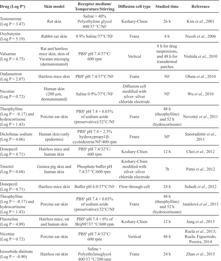
Raman confocal microscopy
Raman spectroscopy is a technique analogous to ATR-FTIR, but it does not suffer from interference
by water. Infrared spectroscopy is based on absorption
of light, but Raman spectroscopy is based on inelastic scattering of monochromatic light from a laser beam. From
the inelastic scattering, the alterations in the wavelength of the photons, which provide a fingerprint by which molecules can be identiied in the sample, are analyzed. The excitation or deactivation of molecular vibrations is
related to the energetic variations in the photons. Thus, it is possible to obtain detailed information about the chemical
structure, electronic coniguration, and molecular bonds
of a compound. Raman spectroscopy has been applied in
diferent ields as pathology, cosmetics, and pharmaceutical
sciences (Caspers et al., 2001; Darlenski et al., 2009). The association of CLSM and Raman spectroscopy
in the last two decades has contributed to the recent
advances in the study of skin barrier properties (Caspers
et al., 1998; Caspers et al., 2001). This association has led to the development of Raman confocal microscopy (RCM), a non-invasive technique that has the combined advantages of Raman spectroscopy and CLSM (Figure 4) (Baena, Lendl, 2004; Darlenski et al., 2009).
R C M h a s e n a b l e d s t u d y o f t h e q u a l i t a t i v e biochemical composition of the skin regarding the lipids in the stratum corneum and of natural moisturizing factor
components. The thickness of the horny layer and its water
concentration also has been determined by RCM. Recent studies using RCM have been conducted to determine the skin absorption of drugs. These studies are limited by signal attenuation in the depth layers of the skin. In the
same way as ATR-FTIR signals, refraction index values
are altered in the depth layers, and the light scattering
becomes more difuse with increasing skin depth (Franzen et al., 2013).
Mateus et al. (2013) studied the dermato pharma-cokinetics of ibuprofen in human volunteers by using
RCM and compared the results with those obtained
by using the tape-stripping technique (MATES et al.,
2013). In both cases, drug diffusion was characterized according to Fick’s law. When RCM was employed,
relative measurements of drug concentrations in the skin
layers were made and compared with the initial amount of drug on the skin surface. This procedure did not allow
the precise determination of the drug concentrations in each skin layer.
Franzen et al. (2013) studied mathematical algorithms for correction of the signal attenuation at different skin layers for quantitative analysis of the
skin penetration of cafeine (Franzen et al., 2013). For
this, the authors created a simulated matrix of keratin, water, and various lipids to obtain a sample with optical
properties similar to those of the stratum corneum. The
drug was incorporated in the simulated matrix at known
concentrations, and the concentrations at different
depths were quantitatively determined. The results were promising for quantitative determination of cafeine by
using RCM. Recently, the validation of an in vitro method
by RCM for quantitative analysis of cafeine in skin was
reported as an interesting non-invasive strategy for drug
quantiication directly in the skin by using a non-invasive
technique (Franzen, Anderski, Windbergs, 2015).
CONCLUSIONS
Although there are different in vitro and in vivo
methods for evaluation of the cutaneous absorption of drugs and bioactive molecules, pharmaceutical formulation scientists must select those best suited to their investigations. Thus, the success of topical and transdermal administration of drugs is directly related to the methods used for evaluation of the formulations,
which enable optimization of the skin absorption of the drug so that it can reach efective drug concentrations at
the therapeutic site.
ACKNOWLEDGMENTS
The authors are grateful for CAPES (Brasília, Brazil), CNPq (Brasília, Brazil), and Unifal-MG (Alfenas,
Brazil) for research fellowships.
REFERENCES
ANDREWS, S.N.; JEONG, E.; PRAUSNITZ, M.R. Transdermal delivery of molecules is limited by full epidermis, not just stratum corneum. Pharm. Res., v.30, n.4, p.1099-109, 2013.
ALEXANDER, A.; DWIVEDI, S.; AJAZUDDIN; GIRI, T.K.; SARAF, S.; SARAF, S.; TRIPATHI, D.K. Approaches for breaking the barriers of drug permeation through transdermal drug delivery. J. Control. Rel., v.164, n.1, p.26-40, 2012.
ANISSIMOV, Y.G.; JEPPS, O.G.; DANCIK, Y.; ROBERTS, M.S. Mathematical and pharmacokinetic modelling of epidermal and dermal transport processes. Adv. Drug Deliv. Rev., v.65, n.2, p.169-190, 2013.
ASHTIKAR, M.; MATTHÄUS, C.; SCHMITT, M.; KRAFFT, C.; FAHR, A.; POPP, J. Non-invasive depth proile imaging of the stratum corneum using confocal Raman microscopy: First insights into the method. Eur. J. Pharm. Sci., v.50, n.5, p.601-608, 2013.
AZARMI, S.; ROA, W.; LÖBENBERG, R. Current perspectives in dissolution testing of conventional and novel dosage forms. Int. J. Pharm., v.328, n.1, p.12-21, 2007.
BAENA, J.R.; LENDL, B. Raman spectroscopy in chemical bioanalysis. Curr. Opin. Chem. Biol., v.8, n.5, p.534-539, 2004.
BALÁZS, B.; FARKAS, G.; BERKESI, O.; GYULAI, R.; BERKÓ, S.; BUDAI-SZŰCS, M.; SZABÓ-RÉVÉSZ, P.; KEMÉNY, L.; CSÁNYI, E. Protein structure is changed in psoriatic skin on the unafected region - Imaging possibility
with ATR-FTIR spectroscopy. Microchem. J., v.117,
p.183-186, 2014.
BARRY, B.W. Novel mechanisms and devices to enable successful transdermal drug delivery. Eur. J. Pharm. Sci., v.14, n.2, p.101-114, 2001.
BERNARD, G.; AUGER, M.; SOUCY, J.; POULIOT, R. Physical characterization of the stratum corneum of an in vitro psoriatic skin model by ATR-FTIR and Raman spectroscopies. Biochim. Biophys. Acta, v.1770, n.9, p.1317-1323, 2007.
BOJAR, R.A. Studying the human skin microbiome using 3D in vitro skin models. Applied In Vitro Toxicol., v.1, n.2, p.165-71, 2015.
BROWN, C.K.; CHOKSHI, H.P.; NICKERSON, B.; REED, R.A; ROHRS, B.R.; SHAH, P.A. Acceptable analytical practices for dissolution testing of poorly soluble compounds. Pharm. Technol., v.28, n.12, p.56, 2004.
BRUNNER, M.; DERENDORF, H. Clinical microdialysis: Current applications and potential use in drug development. TrAC Trends in Anal. Chem., v.25, n.7, p.674-680, 2006.
CARDOT, J.; BEYSSAC, E.; ALRIC, M. In vitro-in vivo correlation: Importance of dissolution in IVIVC. Dissol. Technol., p.15-19, 2007.
CASPERS, P.J.; LUCASSEN, G.W.; CARTER, E.A; BRUINING, H.A; PUPPELS, G.J. In vivo confocal Raman microspectroscopy of the skin: Noninvasive determination
of molecular concentration proiles. J. Invest. Dermatol.,
v.116, n.3, p.434-442, 2001.
CERBAI, F.; GIOVANNINI, M.G.; MELANI, C.; ENZ, A.; PEPEU, G. N1phenethyl-norcymserine, a selective butyrylcholinesterase inhibitor, increases acetylcholine Release in rat cerebral cortex: A comparison with donepezil and rivastigmine. Eur. J. Pharmacol., v.572, n.2-3, p.142-50, 2007.
CEVC, G.; VIERL, U. Nanotechnology and the transdermal
route. A state of the art review and critical appraisal. J.
Control. Release, v.141, n.3, p.277-299, 2010.
CHEN, J.; HU, W.; QU, Y.Q.; DONG, J.; GU, W.; GAO, Y.; FANG, Y.; FANG, F.; CHEN, Z.P.; CAI, B.C. Evaluation of the pharmacodynamics and pharmacokinetics of brucine
following transdermal administration. Fitoterapia, v.86,
n.1, p.193-201, 2013.
CHEN, L.; HAN, L.; LIAN, G. Recent advances in predicting skin permeability of hydrophilic solutes. Adv. Drug Deliv. Rev., v.65, n.2, p.295-305, 2013.
CHOI, J.; CHOI, M.-K.; CHONG, S.; CHUNG, S.-J.; SHIM, C.-K.; KIM, D.-D. Efect of fatty acids on the transdermal delivery of donepezil: In vitro and in vivo evaluation. Int. J. Pharm., v.422, n.1-2, p.83-90, 2012.
COSTA, P.; SOUSA LOBO, J.M. Modeling and comparison of
dissolution proiles. Eur. J. Pharm. Sci., v.13, n.2,
p.123-133, 2001.
DARLENSKI, R.; SASSNING, S.; TSANKOV, N.; FLUHR, J. W. Non-invasive in vivo methods for investigation of the skin barrier physical properties. Eur. J. Pharm. Biopharm., v.72, n.2, p.295-303, 2009.
DAS, R.S.; AGRAWAL, Y.K. Raman spectroscopy: Recent advancements, techniques and applications. Vib. Spectrosc., v.57, n.2, p.163-176, 2011.
DELGADO-CHARRO, M.B.; GUY, R.H. Effective use of transdermal drug delivery in children. Adv. Drug Deliv. Rev., v.73, p.63-82, 2014.
DESAI, P.R.; SHAH, P.P.; HAYDEN, P.; SINGH, M. Investigation of follicular and non-follicular pathways for
polyarginine and oleic acid modiied nanoparticles. Pharm.
Res., v.30, n.4, p.1037-49, 2013.
DOS SANTOS, M.; METRAL, E.; BOHER, A.; ROUSSELLE, P.; THEPOT, A.; DAMOUR, O. In vitro 3-Dmodel based on extending time of culture for studying chronological epidermis aging. Matrix Biol., v.47, p.85-97, 2015.
EL MAGHRABY, G.M.; BARRY, B.W.; WILLIAMS, A.C. Liposomes and skin: From drug delivery to model membranes. Eur. J. Pharm. Sci., v.34, n.4-5, p.203-222, 2008.
FARAHMAND, S.; MAIBACH, H.I. Transdermal drug pharmacokinetics in man: Interindividual variability and partial prediction. Int. J. Pharm., v.367, n.1-2, p.1-15, 2009.
FOUAD, S.A.; BASALIOUS, E.B.; EL-NABARAWI, M.A.; TAYEL, S.A. Microemulsion and poloxamer microemulsion-based gel for sustained transdermal delivery of diclofenac epolamine using in-skin drug depot: In vitro/in vivo evaluation. Int. J. Pharm., v.453, n.2, p.569-578, 2013.
FRANZEN, L.; ANDERSKI, J.; WINDBERGS, M. Quantitative detection of caffeine in human skin by confocal Raman spectroscopy - A systematic in vitro validation study. Eur. J. Pharm. Biopharm., v.95, pt.A, p. 110-116, 2015.
FRANZEN, L.; SELZER, D.; FLUHR, J.W.; SCHAEFER, U.F.; WINDBERGS, M. Towards drug quantiication in human
skin with confocal Raman microscopy. Eur. J. Pharm.
Biopharm., v.84, n.2, p.437-444, 2013.
FRASCH, H.F.; BARBERO, A.M. Application of numerical methods for difusion-based modeling of skin permeation. Adv. Drug Deliv. Rev.,v.65, n.2, p.208-220, 2013.
F R U M , Y. ; K H A N , G . M . ; S E F C I K , J . ; R O U S E , J . ; ECCLESTON, G.M.; MEIDAN, V.M. Towards a correlation between drug properties and in vitro transdermal
lux variability. Int. J. Pharm., v.336, n.1, p.140-147, 2007.
GANNU, R.; VISHNU, Y.V.; KISHAN, V.; RAO, Y.M. Development of nitrendipine transdermal patches: In vitro
and ex vivo characterization. Curr. Drug Deliv., v.4, n.1,
p.69-76, 2007.
GOTZ, C.; PFEIFFER, R.; TIGGES, J.; BLATZ, V.; JACKH, C.; FREYTAG, E.; FABIAN, E.; LANDSIEDEL, R.; MERK, H.F.; KRUTMANN, J.; EDWARDS, R.J.; PEASE, C.; GOEBEL, C.; HEWITT, N.; FRITSCHE, E. Xenobiotic metabolism capacities of human skin in comparison with a 3D epidermis model and keratinocyte-based cell culture as in vitro alternatives for chemical testing: activating enzymes (Phase I). Exp. Dermatol., v.21, n.5, p.364-9, 2012.
GRATIERI, T.; KALIA, Y.N. Mathematical models to describe iontophoretic transport in vitro and in vivo and the efect of current application on the skin barrier. Adv. Drug Deliv. Rev., v.65, n.2, p.315-329, 2013.
GRATIERI, T.; GELFUSO, G.M.; LOPEZ., R.V. F. Basic principles and aplications of iontophoresis for cutaneous penetration of drugs. Quim. Nova, v.31, n.6, p.1490-1498, 2008.
GROEBER, F.; HOLEITER, M.; HAMPEL, M.; HINDERER, S.; SCHENKE-LAYLAND, K. Skin tissue engineering - In vivo and in vitro applications. Adv. Drug Deliv. Rev., v.63, n.4, p.352-366, 2011.
GUPTA, R.R.; JAIN, S.K.; VARSHNEY, M. AOT Water-in-oil microemulsions as a penetration enhancer in transdermal
drug delivery of 5-luorouracil. Colloids Surf. B Biointerf.,
v.41, n.1, p.25-32, 2005.
GUPTA, S.K.; BENOWITZ, N.L.; JACOB, P.; ROLF, C.N.; GORSLINE, J. Bioavailability and absorption kinetics of
nicotine following application of a transdermal system. Br.
J. Clin. Pharm., v.36, n.3, p.221-227, 1993.
GUTH, K.; SCHÄFER-KORTING, M.; FABIAN, E.; LANDSIEDEL, R.; VAN RAVENZWAAY, B. Suitability of skin integrity tests for dermal absorption studies in vitro. Toxicol. In Vitro, v.29, n.1, p.113-123, 2015.
HAAG, S.F.; FLEIGE, E.; CHEN, M.; FAHR, A.; TEUTLOFF, C.; BITTL, R.; LADEMANN, J.; SCHÄFER-KORTING, M.; HAAG, R.; MEINKE, M.C. Skin penetration enhancement of core-multishell nanotransporters and invasomes measured by electron paramagnetic resonance spectroscopy. Int. J. Pharm., v.416, n.1, p.223-228, 2011.
HANSON, R. A primer on release-rate testing of semisolids. Dissol. Technol., v.17, n.4, p.33-35, 2010.
HEARD, C.M.; JOHNSON, S.; MOSS, G.; THOMAS, C.P. In vitro transdermal delivery of caffeine, theobromine, theophylline and catechin from extract of guarana, Paullinia Cupana. Int. J. Pharm., v.317, n.1, p.26-31, 2006.
HÖCHT, C.; OPEZZO, J.A.W.; TAIRA, C.A. Applicability of reverse microdialysis in pharmacological and toxicological studies. J. Pharmacol. Toxicol. Meth., v.55, n.1, p.3-15, 2007.
JANŮŠOVÁ, B.; ŠKOLOVÁ, B.; TÜKÖROVÁ, K.; WOJNAROVÁ, L.; ŠIMŮNEK, T.; MLADĚNKA, P.; FILIPSKÝ, T.; ŘÍHA, M.; ROH, J.; PALÁT, K.; HRABÁLEK, A.; VÁVROVÁ, K. Amino acid derivatives as transdermal permeation enhancers. J. Control. Release, v.165, n.2, p.91-100, 2013.
JEPPS, O.G.; DANCIK, Y.; ANISSIMOV, Y.G.; ROBERTS, M.S. Modeling the human skin barrier - Towards a better understanding of dermal absorption. Adv. Drug Deliv. Rev., v.65, n.2, p.152-158, 2013.
JUNG, E.; KANG, Y.P.; YOON, I.S.; KIM, J.S.; KWON, S.W.; CHUNG, S.J.; SHIM, C.K.; KIM, D.D. Efect of permeation enhancers on transdermal delivery of luoxetine: In vitro and in vivo evaluation. Int. J. Pharm., v.456, n.2, p.362-369, 2013.
K. HURTADO, F.; LAUREANO, J.V.; DE A. LOCK, G.; DERENDORF, H.; DALLA COSTA, T. Enhanced penetration of moxifloxacin into rat prostate tissue evidenced by microdialysis. Int. J. Antimicrob. Agents, v.44, n.4, p.327-333, 2014.
KALIA, Y.N.; GUY, R.H. Modeling transdermal drug release. Adv. Drug Deliv. Rev., v.48, n.2-3, p.159-172, 2001.
KARADZOVSKA, D.; BROOKS, J.D.; MONTEIRO-RIVIERE, N.A.; MONTEIRO-RIVIERE, J.E. Predicting skin permeability
from complex vehicles. Adv. Drug Deliv. Rev., v.65, n.2,
p.265-277, 2013.
KIM, M.K.; ZHAO, H.; LEE, C.H.; KIM, D.D. Formulation of a reservoir-type testosterone transdermal delivery system. Int. J. Pharm., v.219, n.1-2, p.51-59, 2001.
KREILGAARD, M. Assessment of cutaneous drug delivery using microdialysis. Adv. Drug Deliv. Rev., v.54 Suppl 1, p.S99-121, 2002.
LEVEQUE, N.; MAKKI, S.; HADGRAFT, J.; HUMBERT, P. Comparison of Franz cells and microdialysis for assessing salicylic acid penetration through human skin. Int. J. Pharm., v.269, n.2, p.323-328, 2004.
LEWIS, D.; PAULO, M.; FAUSTINO, E. In vitro comparative studies of transdermal nicotine delivery systems. Int. J. Pharm., v.148, n.2, p.177-189, 1997.
LIN, S.; HO, H.; CHIEN, Y.W. Development of a new nicotine transdermal delivery system: In vitro kinetics studies and clinical pharmacokinetic evaluations in two ethnic groups. J. Control. Release, v.26, n.3, p.175-193, 1993.
LIU, N.; ZHANG, Y.; CUN, D.; QUAN, P.; FANG, L. Efect of Backing Films on the Transdermal Delivery of Conepezil from Patches. AAPS Pharm. Sci. Tech., v.15, p.1569-1573, 2014.
LOPES, L.B.; FERREIRA, D.A.; DE PAULA, D.; GARCIA, M.T.J.; THOMAZINI, J.A.; FANTINI, M.C.A; BENTLEY, M.V.L.B. Reverse hexagonal phase nanodispersion of monoolein and oleic acid for topical delivery of peptides: In vitro and in vivo skin penetration of cyclosporin A. Pharm. Res., v.23, n.6, p.1332-1342, 2006.
MADER, J.K.; FEICHTNER, F.; BOCK, G.; KÖHLER, G.; SCHALLER, R.; PLANK, J.; PIEBER, T.R.; ELLMERER, M. Microdialysis-A versatile technology to perform metabolic monitoring in diabetes and critically ill patients. Diabetes Res. Clin. Pract., v.97, n.1, p.112-118, 2012.
MANADAS, R.; PINA, M. E.; VEIGA, F. A dissolução in vitro na previsão da absorção oral de fármacos em formas
farmacêuticas de liberação modiicada. Rev. Bras. Ciênc.
Farm., v.38, n.4, 2002.
MATEUS, R.; ABDALGHAFOR, H.; OLIVEIRA, G.; HADGRAFT, J.; LANE, M.E. A New Paradigm in dermatopharmacokinetics - Confocal Raman spectroscopy. Int. J. Pharm., v.444, n.1-2, p.106-8, 2013.
MATHES, S.H.; RUFFNER, H.; GRAF-HAUSNER, U. The use of skin models in drug development. Adv. Drug Deliv. Rev., v.69-70, p.81-102, 2014.
MATOS, B.N.; REIS, T.A.; GRATIERI, T.; GELFUSO, G.M. Chitosan nanoparticles for targeting and sustaining
minoxidil sulphate delivery to hair follicles. Int. J. Biol.
Macromol., v.75, p.225-9, 2015.
MCAULEY, W.J.; CHAVDA-SITARAM, S.; MADER, K.T.; TETTEH, J.; LANE, M.E.; HADGRAFT, J. The efects of esteriied solvents on the difusion of a model compound across human skin: An ATR-FTIR spectroscopic study. Int. J. Pharm., v.447, n.1-2, p.1-6, 2013.
MOSER, K.; KRIWET, K.; NAIK, A.; KALIA, Y.N.; GUY, R.H. Passive skin penetration enhancement and its quantiication in vitro. Eur. J. Pharm. Biopharm., v.52, n.2, p.103-112, 2001.
NAEGEL, A.; HEISIG, M.; WITTUM, G. Detailed modeling of
skin penetration- An overview. Adv. Drug Deliv. Rev., v.65,
n.2, p.191-207, 2013.
NAIR, A.; JACOB, S.; AL-DHUBIAB, B.; ATTIMARAD, M.; HARSHA, S. Basic considerations in the dermatokinetics of topical formulations. Braz. J. Pharm. Sci., v.49, n.3, p.423-434, 2013.
NAIR, M.K.; CHETTY, D.J.; HO, H.; CHIEN, Y.W. Biomembrane permeation of nicotine: Mechanistic studies
with porcine mucosae and skin. J. Pharm. Sci., v.86, n.2,
p.257-262, 1997.
NAKASHIMA, K.; ITOH, K.; KONO, M.; NAKASHIMA, M.N.; WADA, M. Determination of donepezil hydrochloride in human and rat plasma, blood and brain microdialysates
by HPLC with a short C30 column. J. Pharm. Biom. Anal.,
v.41, n.1, p.201-206, 2006.
NICOLI, S.; PENNA, E.; PADULA, C.; COLOMBO, P.; SANTI, P. New transdermal bioadhesive ilm containing
oxybutynin: In vitro permeation across rabbit ear skin. Int.
J. Pharm., v.325, n.1-2, p.2-7, 2006.
NISHIDA, N.; TANIYAMA, K.; SAWABE, T.; MANOME, Y. Development and evaluation of a monolithic drug-in-adhesive patch for valsartan. Int. J. Pharm., v.402, n.1-2, p.103-109, 2010.
NOVOTNÝ, M.; KLIMENTOVÁ, J.; JANŮŠOVÁ, B.; PALÁT, K.; HRABÁLEK, A.; VÁVROVÁ, K. Ammonium carbamates as highly active transdermal permeation
enhancers with a dual mechanism of action. J. Control.
Release, v.150, n.2, p.164-170, 2011.
OBATA, Y.; ASHITAKA, Y.; KIKUCHI, S.; ISOWA, K.; TAKAYAMA, K.A Statistical approach to the development of a transdermal delivery system for ondansetron. Int. J. Pharm., v.399, n.1-2, p.87-93, 2010.
OLIVEIRA, G.; LEVERETT, J.C.; EMAMZADEH, M.; LANE, M.E. The efects of heat on skin barrier function and in vivo dermal absorption. Int. J. Pharm., v.464, n.1-2, p.145-151, 2014.
PALECO, R.; VUČEN, S.R.; CREAN, A.M.; MOORE, A.; SCALIA, S. Enhancement of the in vitro penetration of quercetin through pig skin by combined microneedles and lipid microparticles. Int. J. Pharm., v.472, n.1-2, p.206-213, 2014.
PATNI, M.; PURANIK, P.; SONAWANE, A.; PANZADE, P. Transdermal iontophoretic delivery of timolol maleate. Braz. J. Pharm. Sci., v.48, n.4, p.819-827, 2012.
PAŽOUREKOVÁ, S.; HOJEROVÁ, J.; KLIMOVÁ, Z.; LUCOVÁ, M. Dermal absorption and hydrolysis of methylparaben in different vehicles through intact and damaged skin: Using a pig-ear model in vitro. Food Chem. Toxicol., v.59, p.754-765, 2013.
P E R E I R A , G . R . ; C O L L E T T, J . H . ; G A R C I A , S . B . ; THOMAZINI, J.A.; VITÓRIA, M.; BADRA, L. Glycerol monooleate/solvents systems for progesterone transdermal delivery: Invitro permeation and microscopic studies. Rev. Bras. Ciênc. Farm.., v.38, n.1, p.55-62, 2002.
PRAUSNITZ, M.R.; LANGER, R. Transdermal drug delivery. Nat. Biotechnol., v.26, n.11, p.1261-1268, 2009.
REID, M.L.; BENAOUDA, F.; KHENGAR, R.; JONES, S.A.; BROWN, M.B. Topical corticosteroid delivery into human skin using hydroluoroalkane metered dose aerosol sprays. Int. J. Pharm., v.452, n.1-2, p.157-165, 2013.
REN, C.; FANG, L.; LING, L.; WANG, Q.; LIU, S.; ZHAO, L.; HE, Z. Design and in vivo evaluation of an indapamide transdermal patch. Int. J. Pharm., v.370, n.1-2, p.129-135, 2009.
ROBERTS, J.A.; ROBERTS, M.S.; ROBERTSON, T.A.; CROSS, S.E.; LIPMAN, J. A novel way to investigate the effects of plasma exchange on antibiotic levels: Use of microdialysis. Int. J. Antimicrob. Ag., v.31, n 3, p.240-244, 2008.
RUELA, A.L.M.; PEREIRA, G.R. Design and evaluation of molecularly imprinted polymers as drug delivery systems. In: TIWARI, A.; UZUN, L. (Eds.) Advanced molecularly imprinting materials. Beverly: Wiley-Scrivener, 2017. p.413-454.
RUELA, A.L.M.; FIGUEIREDO, E.C.; PEREIRA, G.R. Molecularly imprinted polymers as nicotine transdermal delivery systems. Chem. Eng. J., v.248, p.1-8, 2014.
RUELA, A.L.M.; FIGUEIREDO, E.C.; PERISSINATO, A.G.; LIMA, A.C.Z.; PEREIRA, G.R. In vitro evaluation of transdermal nicotine delivery systems commercially available in Brazil. Braz. J. Pharm. Sci., v.49, n.3, p.579-588, 2013.
SALUJA, S.; KASHA, P.C.; PATURI, J.; ANDERSON, C.; MORRIS, R.; BANGA, A.K. A novel electronic skin patch for delivery and pharmacokinetic evaluation of donepezil
following transdermal iontophoresis. Int. J. Pharm., v.453,
n.2, p.395-399, 2013.
SCHNETZ, E.; FARTASCH, M. Microdialysis for the evaluation of penetration through the human skin barrier - a promising tool for future research? Eur. J. Pharm. Sci., v.12, n.3, p.165-174, 2001.
SCHWARZ, J.C.; PAGITSCH, E.; VALENTA, C. Comparison of ATR-FTIR spectra of porcine vaginal and buccal mucosa with ear skin and penetration analysis of drug and vehicle components into pig ear. Eur. J. Pharm. Sci., v.50, n.5, p.595-600, 2013.