*Correspondence to: G. M. Gelfuso. Laboratório de Tecnologia de Medica-mentos, Alimentos e Cosméticos (LTMAC). Faculdade de Ciências da Saúde. Universidade de Brasília. Campus Universitário Darcy Ribeiro, s/n, 70910-900 - Brasília - DF, Brazil. E-mail: [email protected]
A
vol. 51, n. 4, oct./dec., 2015 http://dx.doi.org/10.1590/S1984-82502015000400018
Influence of monoolein on progesterone transdermal delivery
Wanessa de Souza Cardoso Quintão
1,2, Breno Noronha Matos
1, Thaiene Avila Reis
1, Lívia Cristina
de Sá Barreto
2, Taís Gratieri
1, Guilherme Msrtins Gelfuso
1,*1Laboratory of Food, Drugs and Cosmetics, LTMAC, School of Health Sciences, University of Brasília, Brasília, DF, Brazil, 2School of Ceilândia, University of Brasília, Brasília, DF, Brazil
This work aimed to investigate in vitro the inluence of monoolein (MO) on progesterone (PG) transdermal
delivery and skin retention. Information about the role of MO as an absorption enhancer for lipophilic molecules can help on innovative product development capable of delivering the hormone through the skin in a consistent manner, improving transdermal therapy of hormonal replacement. MO was dispersed in propylene glycol under heat at concentrations of 0% (control), 5% w/w, 10% w/w and 20% w/w. Then, 0.6% of PG (w/w) was added to each formulation. Permeation proile of the hormone was determined in vitro for 48 h using porcine skin in Franz difusion cells. PG permeation doubled when 5% (w/w) of MO
was present in formulation in comparison to both the control and higher MO concentrations (10% and 20% w/w). An equal trend was observed for PG retention in stratum corneum (SC) and reminiscent skin (E+D). PG release rates from the MO formulations, investigated using cellulose membranes, revealed that concentrations of MO higher than 5% (w/w) hindered PG release, which indeed negatively relected on the hormone permeation through the skin. In conclusion, this work demonstrated the feasibility of MO addition (at 5% w/w) in formulations as a simple method to increase transdermal PG delivery for therapies of hormonal replacement. In contrast, higher MO concentrations (from 10% to 20% w/w) can control active release, and this approach could be extrapolated to other lipophilic, low-molecular-weight molecules.
Uniterms: Monoolein/inluence/transdermal therapy. Progesterone/transdermal delivery. Progesterone/
skin retention. Hormonal replacement/transdermal therapy. Skin permeation.
Este trabalho teve como objetivo investigar in vitro a inluência de monooleína (MO) na permeação
transdérmica de progesterona (PG), bem como sobre a retenção cutânea desse hormônio a im de (i) liberar de maneira mais consistente hormônio através da pele para melhorar a terapia transdérmica de reposição hormonal e (ii) trazer mais informações sobre o papel da MO como promotor da absorção cutânea de moléculas lipofílicas, tema ainda pouco explorado na literatura. MO foi dispersa em propilenoglicol, a concentrações de 0% (controle), 5%, 10% e 20% (p/p). Adicionou-se, em seguida, 0,6% (p/p) de PG a cada uma das formulações. O peril de permeação do hormônio foi então determinado in vitro durante 48 h, utilizando pele de porco em células de difusão do tipo Franz. MO a 5% (p/p) foi capaz de duplicar a permeação de PG em comparação ao controle e às concentrações mais elevadas de MO, assim como a retenção de PG no estrato córneo (SC) e epiderme e derme remanescentes (E+D). A velocidade de liberação de PG a partir das formulações foi investigada usando membranas de celulose e este estudo revelou que concentrações de MO superiores a 5% (p/p) impediram a liberacão de PG, o que de fato reletiu de forma negativa na permeação cutânea do hormônio. Concluindo, este trabalho demonstrou a viabilidade da adição de MO a uma formulação como um método simples para aumentar a permeação transdérmica de PG para uso em terapias de reposição hormonal. Por outro lado, altas concentrações de MO (de 10% a 20% p/p) controlam a liberação de PG e este efeito pode ser extrapolado para outras moléculas lipofílicas de baixa massa molecular.
INTRODUCTION
The use of oral progesterone (PG, Figure 1) is
approved by the FDA and by other international regulatory agencies and is the most conventional strategy for hormone replacement therapy in menopausal women (Studd, 2014).
It is undeniable, however, that transdermal delivery
of PG would be more advantageous than oral therapy,
if considered the possibility to obtain a controlled and prolonged hormone delivery to the blood and the avoidance of first-pass metabolism in the liver, which guarantee a more consistent bioavailability of the hormonal substance (Silva et al., 2010). In addition, the painless, non-invasive and easy administration of transdermal patches and formulations can improve patient acceptance (Martins, Veiga, 2002).
Many papers evaluating transdermal creams and gels
containing PG for treating menopausal related symptoms
have been published over the years (Chang et al., 1995; Du etal., 2013; Leonetti et al., 2005). However, PG potential in preventing estrogen-stimulated cell proliferation has not yet been fully explored because current formulations and devices are unable to deliver therapeutically relevant progesterone concentrations (Zava et al., 2014).
In this context, some research groups have devoted
their eforts in using many diferent strategies to overcome
the stratum corneum (SC) barrier of skin. Swarnakar et al. (2007), for instance, prepared and characterized
lipid-based hexosomes for oral administration of PG, but, to
our knowledge, no further study from the group evaluated cutaneous permeation. A more recent study determined
the eicacy of a transdermal nanostructured formulation of PG combined with estriol, and preliminary clinical studies demonstrated the system was safe and efective
(Botelho et al., 2014).
A simpler approach for PG delivery would consist on the use of absorption enhancers incorporated in PG
transdermal formulations. Absorption enhancers reversibly decrease skin barrier resistance, allowing the drug to penetrating viable tissues to act locally and/or reach the
systemic circulation (Alexander et al., 2012). Monoolein
(MO, Figure 2) is one of the most studied absorption
enhancer (Simonetti et al., 2009; Herai et al., 2007; Steluti et al., 2001; Lopes, Collett, Bentley, 2005; Pereira et al., 2002; Puglia et al., 2013). It is pharmacologically inert, non-toxic, immediate and reversible in action, non-irritating, non-allergenic, odorless, colorless and chemically and physically compatible with many drugs and pharmaceutical excipients (Hadgraft, 1999; Qiu,
Cafrey, 2000). MO is able to interact with phospholipid
bilayers of SC lipid matrix and destabilize its structure
momentarily (Pereira et al., 2002). However, due to
its high lipophilicity, MO is rarely incorporated in
formulations containing lipophilic active agents, such as
PG, since high interaction with the absorption enhancer
could hinder drug release, hindering drug transdermal permeation.
In this way, this work aimed to investigate in vitro
MO influence on PG transdermal delivery and skin
retention, which could bring more information about
the role of MO as absorption enhancer for lipophilic
molecules, a still little explored matter in scientific literature.
MATERIAL AND METHODS
Material
PG was kindly provided by “Farmacotécnica Farmácia de Manipulação” (Brasília, Brazil). MO (gliceril oleate, ≥ 99%) was purchased from Sigma-Aldrich (Steinheim, Germany). Propylene glycol used to prepared
formulations, as well as to be placed in receptor solution,
was purchased from Dinâmica Química Ltda. (São Paulo,
Brazil). Monobasic and dibasic sodium phosphate (Vetec, Rio de Janeiro, Brazil), and sodium chloride (Serva, Rio
de Janeiro, Brazil) were all used in bufer preparation, and sodium hydroxide (Dinâmica Química Ltda., São Paulo, Brazil) was used to adjust the pHs of the bufer
and formulations. Regenerated Cellulose Dialysis Tubing
(Dialysis Tubing MWCO 12000-14000, Fisherbrand)
used in release studies was purchased from Fisher
Scientiic (Leicestershire, United Kingdom). Methanol
FIGURE 1 - Chemical structure of progesterone (PG) (MW =
314.46 g/mol; water solubility = 16.8 mg/L at 25 oC).
FIGURE 2 - Chemical structure of MO (MW = 356.53 g/mol;
and acetonitrile, used for extraction and chromatographic
analyses, were of HPLC grade and purchased from Tedia
Brazil Ltda. (Rio de Janeiro, Brazil). The water used in all preparations was of Milli Q grade (Millipore, France).
Skin
Porcine skin was kindly provided by the Frigoriic Bonasa (Brasília, Brazil). Porcine ears were obtained soon after animal sacriice, before the scalding process.
The whole skin was removed from the outer region of the ear, separated from its underlying layer with scissors and stored frozen at -4 °C for a maximum of 1 month before use.
Formulations
Formulations were prepared by adding MO at 5%,
10% or 20% (w/w) to propylene glycol and heated to 40 °C for complete homogenization. After cooling, 0.6%
(w/w) of PG was incorporated to each formulation and
the pH adjusted to 5 with an aqueous solution of sodium hydroxide at 1 M. A control formulation was prepared
without MO, i.e., simply dissolving 0.6% (w/w) of PG in
propylene glycol and adjusting the pH to 5.
In vitro skin permeation
Permeation studies were carried out in modified “Franz” diffusion cells, mounted with small pieces
(2 cm × 2 cm) of porcine ear skin separating the donor and
receptor compartments. The receptor chamber was illed with a solution prepared with phosphate bufer (pH 7.4)
and propylene glycol in the ratio of 60:40 (v/v). The skin
was hydrated for 30 minutes by adding 1 mL of bufer to
the donor chamber prior the beginning of the experiments.
After draw of the bufer, it was added the same volume
for each of the formulations to be tested or the control formulation to the donor compartment, which was closed
with Parailm®to minimize formulation evaporation. The
receptor solution was continuously stirred at 500 rpm for
48 h. Samples were collected in deined time intervals of
1 h, 3 h, 6 h, 12 h, 24 h, 30 h, 36 h and 48 h. A minimum of four replicates was performed for each formulation.
PG recovery from SC and reminiscent epidermis and dermis (E+D)
At the end of each permeation experiment, tape-stripping technique was performed to determine and
diferentiate PG penetration in SC from that in E+D. The
skin was removed from the difusion cell and placed onto
a flat surface with the SC facing up. That part of skin,
which had been in contact with the PG donor formulation,
was cleaned with deionized water (water used to clean the skin was discarded). A plastic template with 1.7 cm2
of exhibition area was placed on the skin in order to leave exposed only the drug transport area. Then, the skin was tape-stripped 15 times, using Scotch Book Tape (3 M, St
Paul, Minnesota). PG was determined after extraction of
the drug from the tapes with methanol over a 12 h period. The remaining skin was cut into small pieces and placed in plastic tube along with methanol also over a period of 12 h for drug extraction. The resulting suspensions were
iltered on 0.22 μm ilters and quantiied by HPLC. The PG recovery from SC and E+D was previously
validated and showed error percentages within the limits accepted for validation of methods involving extractive processes (± 15%) (FDA, 2000).
In vitro release
These studies were performed using the same modified Franz-type diffusion cells, but mounted with synthetic hydrophilic membranes of cellulose acetate separating the donor and receptor compartments instead
of skin. The receptor compartment was illed with 15 mL of a receptor solution prepared with phosphate bufer (pH
7.4) and propylene glycol in the ratio of 60:40 (v/v). The receptor solution was continuously stirred by means of a spinning bar magnet, at 500 rpm. 1 mL of each tested formulation was placed on donor compartment and, from this time, samples from receptor compartment were collected every hour for 12 h. At the end of the experiment, the amount of the drug released across the membrane, i.e.,
the amount of PG released in the receptor solution, was
determined analytically as described below, and release
kinetics of PG from each MO formulation was determined.
Analytical analysis
PG was quantiied by an analytical method using a high performance liquid chromatograph (HPLC) Shimadzu
LC-20 AD, composed by two pumps (model LC 20-AT),
automatic gun (model 9SIL-20AD) and oven (model CTO
- 20th century), coupled to a spectrophotometric detector
(model SPD-M20A) and a computer equipped with the
chromatographic analysis program Shimadzu LC. A
reverse-phase column (Dionex 4.0 × 125 mm, 5 µm) was
used as stationary phase, and mobile phase consisted of a mixture of water: acetonitrile (70: 30) (v/v). The mobile
was 50 μL, the oven was used at 40 ºC and detection was made at 244 nm. The method was validated in terms of
linearity, precision, accuracy, speciicity/selectivity and limit of quantiication, according to current legislation
(FDA, 2000).
Data analyses
Three to ive replicates of each transport experiment
were performed. Results are presented as mean ± standard deviation (SD) and expressed in terms of the quantity of
PG per unit area of skin (μg/cm2). Linear regressions were
obtained with Microsoft Excel 2007. Statistical analyses
were performed with the program GraphPad Prism. Statistical signiicance was ixed at p < 0.05.
RESULTS AND DISCUSSION
PG is a lipophilic, relatively small and non-charged molecule. It presents log P = 3.5, features which are
generally required for proper SC permeation. However, its potency is not high enough to assure high blood concentrations for long time periods (Zava et al., 2014).
This study aimed to investigate the feasibility of MO
to be used as permeation enhancer in formulations
incorporating PG for enhancing its transdermal delivery. For that, MO was dispersed in propylene glycol in three diferent concentrations and PG permeation from these
formulations were evaluated relatively to a control
(without MO addition).
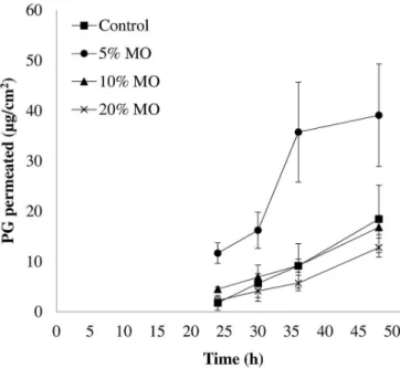
Figure 3 shows PG in vitro permeation proiles for
48 h as a function of MO concentration in the formulations. Although PG does permeate the skin passively, permeated levels up to the irst 24 h of experiment were under method’s limit of quantiication (LOQ = 500 ng/ mL). Even until 36 h, PG levels were near the LOQ for
control formulation and formulations with 10% and 20%
of MO.
It is clear from Figure 3 that 5% (w/w) of MO in formulation was suicient to increase in approximately 2-fold PG permeation when compared to control. Pereira et al. (2002) studied transdermal delivery of PG from MO
dispersed in mineral oil (at 20%) and observed a 3-fold
enhance in PG delivery over control (240 and 80 µg/cm2,
respectively, after 48 h of experiment). However, authors used mice skin as a model, whose SC is much thinner than porcine and human skin, compromising the comparison with the data presented in this paper.
Despite of the increase in PG permeation provided by 5% (w/w) MO in formulation, 20% (w/w) of MO dramatically decreased (in about 50%) PG permeation
through porcine skin (P > 0.05). Steluti et al. (2001) observed an increase in permeation of aminolevulinic acid (ALA) through mouse skin from all formulations
containing the same range of MO concentrations (5%,
10%, and 20%), while Simonetti et al. (2009), evaluating
the effect MO on cisplatin (CIS) permeation, showed an increase in drug lux with the increase, up to 10%, of
the enhancer. It is interesting to notice that, as molecule
hydrosolubility decreases, (ALA>CIS>PG), a progressive annulment of MO enhancement effect is observed. A possible reason for this is that high MO concentrations
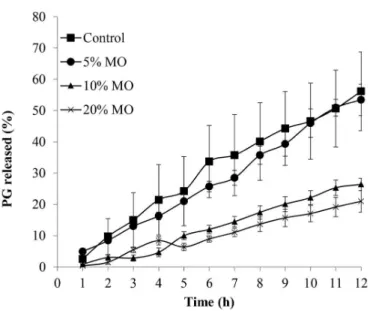
increase formulation lipophilicity, hindering drug release. To test the above-mentioned hypothesis, release
proile of PG from MO formulations was assessed using
cellulose membrane, and the results are presented in Figure 4.
As expected, only 5% (w/w) of MO in the formulation did not increase lipophilicity highly enough to inluence PG release in comparison with control (P > 0.05). Instead, MO at 10% or 20% (w/w) hindered the hormone release (P < 0.05). As it can be seen in Figure 3, this efect had a signiicant inluence on the permeation of PG through
the skin.
The data presented in Table I shows clearer that MO at 5% (w/w) was not able to inluence PG release kinetics,
while at 10% and 20% (w/w) decreased by 2 and 2.5-fold
the hormone release lux.
FIGURE 3 - PG permeation proile through porcine skin from
TABLE I - PG release flux (µg/cm2/h) from the formulations
containing 0% (control), 5% MO, 10% MO and 20% of MO
Formulation Release lux
(μg/cm2/h)
Correlation
coeicient (r)*
Control 28.2 ± 5.0 0.992
5% MO 27.4 ± 2.7 0.997
10% MO 15.0 ± 1.2 0.993
20% MO 11.1 ± 2.0 0.989
*all formulations release PG in a zero order kinetics.
FIGURE 4 - PG release proiles of the PG in propylene glycol
formulations (0.6% m/m) containing diferent concentrations of MO: Control (■); 5% (●), 10% (▲) and 20% (×). Each point represents the average of 4 experiments and the vertical bars represent the standard deviation of the mean (± SD).
Similar to our results, Herai et al. (2007) observed
that the addition of 5% (w/w) of MO in formulation did not alter signiicantly the release rate of the chemotherapeutic agent, doxorubicin, while higher MO concentrations
did. Herai et al. attributed this effect to an increase in
lipophilicity or viscosity provided by MO in the vehicle
(Herai et al., 2007). Taken the data together, the efect
of lipophilicity provided by MO must be much more
pronounced than any viscosity increment it could provide to the formulation, since Simonetti et al. (2009)
demonstrated that the addition of MO did not alter release
rate of the more hydrophilic CIS, in any concentration it was tested.
Figure 5 summarizes the data of PG recovered from
SC, E+D and receptor solution after 48 h of skin treatment with formulations containing 0%, 5%, 10% or 20% (w/w)
of MO.
FIGURE 5 - PG recovered from the SC, reminiscent E+D and receptor solution after 48 h of treatment with formulations
containing MO at diferent concentrations. Bars represent the
average of 4-5 experiments and vertical bars represent the standard deviation of the mean (± SD). *Signiicant diference
from control in SC samples (P < 0.05); #Signiicant diference
from control in E + D samples (P < 0.05); $Signiicant diference
from control in receptor samples (P < 0.05).
The formulation containing 5% MO (w/w) not only doubled the PG amount permeated, as it also
increased hormone retention in reminiscent skin (E +
D). Interestingly, 10% of MO (w/w), even restricting PG
release, was still capable of retaining a great amount of
PG in E + D, even though this high skin retention was not able to guarantee higher transdermal lux of the hormone.
CONCLUSION
In conclusion, the data presented in this work
demonstrated the feasibility of MO addition as a simple method to increase transdermal delivery of PG for the
therapy of hormonal replacement. It was shown that, depending on the concentration added to the topical and
transdermal formulation, MO could cause opposite efects, i.e., while small concentration of MO (5% w/w) can
enhance about 2-fold transdermal delivery of the lipophilic molecule, higher concentrations (from 10% to 20% w/w)
of MO signiicantly reduces PG skin permeation as a result of a more pronounced controlling efect in drug release.
Such conclusion is corroborated by the 2-fold reduction in
PG release observed at higher MO concentrations.
ACKNOWLEDGEMENTS
Funding Agencies CAPES, CNPq and FAP-DF, and the
University of Brasília for supporting this research.
REFERENCES
ALEXANDER, A.; DWIVEDI S.; AJAZUDDIN; GIRI, T.K.; SARAF, S.; SARAF, S.; TRIPATHI, D.K. Approaches
for breaking the barriers of drug permeation through transdermal drug delivery. J. Control. Release, v.164, n.1, p.26-40, 2012.
BOTELHO, M.A.; QUEIROZ, D.B.; BARROS, G.; GUERREIRO, S.; FECHINE, P.; UMBELINO, S.; LYRA, A.; BORGES, B.; FREITAS, A.; QUEIROZ, D.C.; RUELA, R.; ALMEIDA, J.G.; QUINTANS, L. JR. Nanostructured
transdermal hormone replacement therapy for relieving menopausal symptoms: a confocal Raman spectroscopy study. Clinics, v.69, n.2, p.75-82, 2014.
CHANG, K.J.; LEE, T.T.; LINARES-CRUZ, G.; FOURNIER, S.; DE LIGNIERES, B. Influences of percutaneous
administration of estradiol and progesterone on human breast epithelial cell cycle in vivo. Fertil. Steril., v.63, n.4, p.785-791, 1995.
DU, J.Y.; SANCHEZ, P.; KIM, L.; AZEN, C.G.; ZAVA, D.T.; STANCZYK, F.Z. Percutaneous progesterone delivery
via cream or gel application in postmenopausal women: a randomized cross-over study of progesterone levels in serum whole blood saliva capillary blood. Menopause, v.20, p.1107-1226, 2013.
FOOD AND DRUG ADMINISTRATION. FDA. Guidance for Industry: analytical procedures and methods validation: chemistry, manufacturing, and controls documentation. Rockville: US Food and Drug Administration, 2000. 33 p.
HADGRAFT, J. Passive enhancement strategies in topical and
transdermal drug delivery. Int. J. Pharm., v.184, n.1, p.1-6, 1999.
HERAI, H.; GRATIERI, T.; THOMAZINE, J.A.; BENTLEY, M.V.L.B.; LOPEZ, R.F.V. Doxorubicin skin penetration
from monoolein-containing propylene glycol formulations. Int. J. Pharm., v.329, n.1-2, p.88-93, 2007.
LEONETTI, H.B.; LANDES, J.; STEINBERG, D.; ANASTI, J.N. Transdermal progesterone cream as an alternative
progestin in hormone therapy. Altern. Ther. Health Med., v.11, n.6, p.36-38, 2005.
LOPES, L.B.; COLLETT, J.H.; BENTLEY, M.V.L.B. Topical
delivery of cyclosporin A: an in vitro study using monoolein as a penetration enhancer. Eur. J. Pharm. Biopharm., v.60, n.1, p.25-30, 2005.
MARTINS, M.R.; VEIGA, F. Promotores de permeação para a liberação transdérmica de fármacos: uma nova aplicação
para as ciclodextrinas. Braz. J. Pharm. Sci., v.38, n.1, p.33-54, 2002.
PEREIRA, G.R.; COLETT, J.H.; GARCIA, S.B., THOMAZINI, J.A.; BENTLEY, M.V. Glycerol monooleate/solvents
systems for progesterone transdermal delivery: in vitro permeation and microscopic studies. Braz. J. Pharm. Sci., v.38, n.1, p.55-62, 2002.
PUGLIA, C.; CARDILE, V.; PANICO, A.M.; CRASCÌ, L.; OFFERTA, A.; CAGGIA, S.; DRECHSLER, M.; MARIANI, P.; CORTESI, R.; ESPOSITO, E. Evaluation
of monooleine aqueous dispersions as tools for topical administration of curcumin: characterization, in vitro and ex-vivo studies. J. Pharm. Sci., v.102, n.1, p.2349-2361, 2013.
QIU, H.; CAFFREY, M. The phase diagram of the monoolein/
water system: metastability and equilibrium aspects. Biomaterials, v.21, n.3, p.223-234, 2000.
STUDD J. Hormone therapy for reproductive depression in women. Post Reprod. Health, v.20, n.4, p.132-137, 2014.
SILVA, J.A.; APOLINÁRIO, A.C.; SOUZA, M.S.R.; DAMASCENO, B.P.G.L.; MEDEIROS, A.C.D. Administração cutânea de fármacos: desaios e estratégias para o desenvolvimento de formulações transdérmicas.
Rev. Ciênc. Farm. Básica Apl., v.31, n.3, p.125-131, 2010.
SIMONETTI, L.D.; GELFUSO, G.M.; BARBOSA, J.C.; LOPEZ, R.F. Assessment of the percutaneous penetration
of cisplatin: the effect of monoolein and the drug skin penetration pathway. Eur. J. Pharm. Biopharm., v.73, n.1, p.90-94, 2009.
STELUTI, R.; DE ROSA, F.S.; COLLET, J.H.; BENTLEY,
M.V.L.B. Influence of monoolein on 384 in vivo protoporphyrin IX accumulation in hairless mouse skin induced by 5- 385 aminolevulinic acid. In: VI
PHARMATECH: ANUAL MEETING OF THE SBTF, 6.,
SWARNAKAR, N.K.; JAIN, V.; DUBEY, V.; MISHRA, D.; JAIN, N.K. Enhanced oromucosal delivery of progesterone
via hexosomes. Pharm. Res., v.24, n.12, p.2223-2230, 2007.
ZAVA, D.T.; GROVES, M.N.; STANCZYK, F.Z. Percutaneous
absorption of progesterone. Maturitas, v.77, n.2, p.91-92, 2014.