RBCCV 44205-1628 DOI 10.5935/1678-9741.20140086
Atrioventricular block in coronary artery
bypass surgery: perioperative predictors
and impact on mortality
Bloqueio atrioventricular pós-cirurgia de revascularização do miocárdio: fatores preditores perioperatórios
e impacto na mortalidade
Ricardo Medeiros Piantá
1, MD; Andres Di Leoni Ferrari
2, MD; Aline Almeida Heck
3, MD; Débora
Klein Ferreira
3, MD; Jacqueline da Costa Escobar Piccoli
4, PhD; Luciano Cabral Albuquerque
5, PhD;
João Carlos Vieira da Costa Guaragna
6, PhD; João Batista Petracco
7, MD, MSc
1Specialist in Cardiovascular Surgery by the Brazilian Society of Cardio-vascular Surgery, CardioCardio-vascular Surgeon at the Sao Lucas Hospital of the
Pontiical Catholic University of Rio Grande do Sul (PUCRS), Porto Alegre,
RS, Brazil.
2Specialist in Internal Medicine by the Brazilian Medical Association and in Cardiology through the Brazilian Society of Cardiology, Assistant
Cardiolo-gist of the Cardiac Pacing Unit at the Sao Lucas Hospital - PUCRS, Porto
Alegre, RS, Brazil.
3Resident Physician for Cardiovascular Surgery of the Medical Residency
Program for Cardiovascular Surgery of Sao Lucas Hospital - PUCRS, Porto
Alegre, RS, Brazil.
4Adjunct Professor at the Federal University of Pampa - Uruguaiana Cam -pus, RS, Brazil.
5PhD in Health Sciences with Area of Concentration in Cardiology at the
Federal University of Rio Grande do Sul, Cardiovascular Surgeon at the Sao Lucas Hospital - PUCRS, Porto Alegre, RS, Brazil.
6Chief of Cardiology at the Sao Lucas Hospital - PUCRS, Head of the Post
-operative Cardiac Surgery at the Sao Lucas Hospital - PUCRS, Porto Alegre,
RS, Brazil.
7Master of Internal Medicine - Cardiology at the Federal University of Rio Grande do Sul, Chief of the Department of Cardiovascular Surgery at the
São Lucas Hospital - PUCRS, Porto Alegre, RS, Brazil.
This study was carried out at the Department of Cardiology, Electrophysiol-ogy Laboratory, Department of Cardiovascular Surgery, SSao Lucas
Hos-pital of the Pontiical Catholic University of Rio Grande do Sul (PUCRS),
Porto Alegre, RS, Brazil.
No inancial support.
Correspondence address: Ricardo Medeiros Piantá
Av. Ipiranga, 6690 – Conj. 615 – Jardim Botânico – Porto Alegre, RS, Brazil Zip Code: 90610-000
E-mail: [email protected]
Article received on December 16th, 2013 Article accepted on Juny 29th, 2014 Abstract
Introduction: Disturbances of the cardiac conduction sys-tem are frequent in the postoperative period of coronary artery bypass surgery. They are mostly reversible and associated with some injury of the conduction tissue, caused by the ischemic heart disease itself or by perioperative factors.
Objective: Primary: investigate the association between perioperative factors and the emergence of atrioventricular block in the postoperative period of coronary artery bypass surgery. Secondary: determine the need for temporary pacing and of a permanent pacemaker in the postoperative period of coronary artery bypass surgery and the impact on hospital stay and hospital mortality.
Methods: Analysis of a retrospective cohort of patients sub-mitted to coronary artery bypass surgery from the database of the Postoperative Heart Surgery Unit of the Sao Lucas Hospital
of the Pontiical Catholic University of Rio Grande do Sul, using the logistic regression method.
INTRODUCTION
Disturbances of the cardiac conduction system are
relative-ly frequent in the postoperative period (PO) of coronary artery bypass grafting (CABG), with an incidence ranging from 18 to
55% of cases[1-6]. Atrioventricular block (AVB) is one of these
conduction disturbances and its incidence ranges from 0.5 to 16%[3,5-9]. Most patients have transitory and reversible
conduc-perioperative acute myocardial infarction (OR=1.70; CI 95% 1.26-2.29; P<0.0001) and with the use of the intra-aortic balloon in the postoperative period of coronary artery bypass surgery (OR=1.92; CI 95% 1.21-3.05; P=0.006). The presence of atrio-ventricular block resulted in a signiicant increase in mortality (17.9% vs. 7.3% in those who did not develop atrioventricular block) (OR=2.09; CI 95% 1.46-2.99; P<0.0001) and a longer hos-pital stay (12.75 days x 10.53 days for those who didn’t develop atrioventricular block) (OR=1.01; CI 95% 1.00-1.02; P=0.01).
Conclusions: In most cases, atrioventricular block in the postoperative period of coronary artery bypass surgery is transient and associated with several perioperative factors: age above 60 years, female sex, chronic kidney disease, atrial ibrillation, New York Heart Association functional class III or IV, perioperative acute myocardial infarction and use of an in -tra-aortic balloon. Its occurrence prolongs hospitalization and, above all, doubles the risk of mortality.
Descriptors: Atrioventricular block. Artiicial Pacemaker. Coronary Artery Bypass. Postoperative Complications.
Resumo
Introdução: Os distúrbios do sistema de condução cardíaca são frequentes no pós-operatório de cirurgia de revasculariza-ção do miocárdio. Majoritariamente reversíveis, estão associa-dos com alguma injúria do tecido de condução, causada pela própria cardiopatia isquêmica ou por fatores perioperatórios.
Objetivo: Primário: investigar a associação entre fatores pe-rioperatórios com o surgimento de bloqueio atrioventricular no pós-operatório de cirurgia de revascularização do miocárdio. Secundários: determinar a necessidade de estimulação cardíaca
artiicial temporária e de marca-passo deinitivo no pós-opera-tório de cirurgia de revascularização do miocárdio e seu impac-to na permanência e na mortalidade hospitalar.
Métodos: Análise de Coorte retrospectiva de pacientes sub -metidos à cirurgia de revascularização do miocárdio, do banco de dados da unidade de Pós-Operatório de Cirurgia Cardíaca do Hospital São Lucas da Pontifícia Universidade Católica do Rio Grande do Sul, pelo método de regressão logística.
Resultados: No período de janeiro de 1996 a dezembro de 2012, foram realizadas 3532 cirurgias de revascularização do miocárdio. Duzentos e oitenta e oito (8,15%) pacientes apresen-taram bloqueio atrioventricular durante o pós-operatório de ci-rurgia de revascularização do miocárdio, necessitando de estimu-lação cardíaca artiicial temporária. Oito dos que apresentaram bloqueio atrioventricular evoluíram para implante de marcapas-so deinitivo (0,23% do total da amostra). A análise multivaria-da evidenciou associação signiicativa de bloqueio atrioventricu-lar com idade acima de 60 anos (OR=2,34; IC 95% 1,75-3,12; P<0,0001), sexo feminino (OR=1,37; IC 95% 1,06-1,77; P=0,015), doença renal crônica (OR=2,05; IC 95% 1,49-2,81; P<0,0001), i -brilação atrial (OR=2,06; IC 95% 1,16-3,66; P=0,014), classe fun-cional III e IV da New York Heart Association (OR=1,43; IC 95% 1,03-1,98; P=0,031), infarto agudo do miocárdio perioperatório (OR=1,70; IC 95% 1,26-2,29; P<0,0001) e com o uso do balão intra-aórtico no pós-operatório de cirurgia de revascularização do miocárdio (OR=1,92; IC 95% 1,21-3,05; P=0,006). A presença de bloqueio atrioventricular acarretou um aumento signiicativo da mortalidade (17,9% vs. 7,3% nos que não desenvolveram blo -queio atrioventricular) (OR=2,09; IC 95% 1,46-2,99; P<0,0001) e um tempo mais prolongado de permanência hospitalar (12,75 dias vs. 10,53 dias nos que não desenvolveram bloqueio atrioven-tricular) (OR=1,01; IC 95% 1,00-1,02; P=0,01).
Conclusão: O bloqueio atrioventricular, no pós-operatório de cirurgia de revascularização do miocárdio, é, na maioria dos casos, transitório, sendo associado a diversos fatores periope-ratórios: idade acima de 60 anos, sexo feminino, doença renal crônica, ibrilação atrial, classe funcional III e IV da New York Heart Association, infarto agudo do miocárdio perioperatório e uso do balão intra-aórtico. Sua ocorrência prolonga a interna-ção hospitalar e, sobretudo, duplica o risco de mortalidade.
Descritores: Bloqueio Atrioventricular. Marcapasso Artiicial. Revascularização do Miocárdio. Complicações Pós-Operatórias.
Abbreviations, acronyms & symbols
AVB Atrioventricular block CABG Coronary artery bypass surgery NYHA New York Heart Association PO Postoperative period
POHS Postoperative Heart Surgery Unit PPM Permanent pacemaker
PUCRS Pontiical Catholic University of Rio Grande do Sul
TP Temporary pacing
tion disorders that require temporary pacing (TP). 0.4 to 1.1%
of patients, however, when faced with the irreversibility of the
condition, will have to undergo a permanent pacemaker (PPM)
implant during their hospital stay[10]. This study, which is
un-precedented in the national literature, tries to identify the
rela-tionship between pre-, intra and postoperative (perioperative)
METHODS
Population and sample
Between January 1996 and December 2012, 3532
CABGs with cardiopulmonary bypass (CPB) were per
-formed at the Sao Lucas Hospital of the Pontiical Catholic University of Rio Grande do Sul (PUCRS).
Study Design
Historical cohort observation study. Data were collected prospectively and entered into the database of the
Postoper-ative Heart Surgery Unit (POHS) of the São Lucas Hospital of PUCRS.
Inclusion Criteria
Patients with age equal to or greater than 18 years who were submitted to isolated CABG.
Exclusion Criteria
Patients who had been undergo valvular surgery, for left ventricular aneurysmectomy or correction of the interventric-ular communication associated with CABG.
Study Variables
Age - the mean age was calculated and also divided into groups for analysis: less than 60 years and greater than or equal to 60 years, according to the reference in the litera-ture[11,12]; gender (male and female); left ventricular ejection
fraction (EF) - evaluated by echocardiography or radio -cardiography, with the values being subdivided for
analy-sis into ≤ 40% and > 40 %; chronic kidney disease (CKD) - diagnosed through serum creatinine level > 1.5 mg/dl,
according to the reference in the literature[11,12]; Diabetes
Mellitus (DM); atrial ibrillation (AF); previous use of be -ta-blockers; previous use of statins; previous use of other
antiarrhythmics (propafenone and/or amiodarone); acute myocardial infarction (AMI) prior to CABG; New York Heart Association functional (NYHA) class; presence of calciication of the aorta; time of cardiopulmonary bypass (CPB); aortic clamping time; need for the use of intra-aor
-tic balloon (IAB) in the PO of CABG; perioperative AMI;
length of hospital stay and hospital death.
Outcome
Development of AVB in the PO of CABG and the need for TP and implantation of a PPM.
Procedures
The CABGs were performed under general anesthesia. In all cases, a hyperkalemic cardiac arrest was induced using a
cold cardioplegic blood solution in the anterograde low, with
the infusion being repeated every 20 minutes. A mild
system-ic hypothermia (32ºC) was used. After surgery, all patients
were transferred to the ICU of the POHS with mechanical
ventilation.
Statistical Analysis
The data was plotted in a digital Microsoft Access®
spreadsheet and analyzed using version 17.0 of the statisti-cal software SPSS. The descriptive analysis was performed through frequency and mean ± standard deviation analysis, according to the case. For the univariate analysis the follow-ing tests were performed: chi-square and/or Fisher’s Exact Test for ordinal variables and the Student’s T-test for quan-titative data. The multivariate analysis was performed using
logistic regression (backward conditional method). The dif
-ference was considered as statistically signiicant for the val -ue of P<0.05.
Ethical Considerations
The design of this study was submitted to the Research
Ethics Committee of the Faculty of Medicine of the PUCRS,
under registration number 060/3478.
RESULTS
Of the 3532 patients undergoing CABG in the period
un-der analysis, 288 (8.15%) presented the clinical and electro -cardiographic signs of AVB during the postoperative period,
with an indication for temporary pacing (TP).
Table 1 shows the demographic proile of the patients
studied. The univariate analysis of the preoperative data revealed a greater need for TP in the PO of CABG in
pa-tients above 60 years of age (OR=2.48; CI 95% 1.90-3.24; P<0.0001), of the female gender (OR=1.03; CI 95%
1.00-1.05; P=0.012), with CKD (OR=2.03; CI 95% 1.55-2.65;
P<0.0001), the presence of AF (OR=2.38; CI 95% 1.49-3.72; P<0.0001) and in patients with NYHA functional class III or IV (OR=1.60; CI 95% 1.21-2.12; P=0.001).
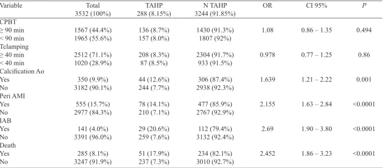
Table 2 presents the trans and postoperative data, together with their univariate analysis. Here we can observe the asso-ciation of AVB with the need of TP in patients who
present-ed calciication of the aorta, perioperative AMI and the nepresent-ed for the use of an IAB. Of statistically signiicant relevance,
the univariate analysis also revealed the association of TP
caused by AVB with increased mortality (17.9% vs. 7.3%) and with a longer hospital stay (mean hospitalization time
of 12.75 days compared to 10.53 days for those who did not
require TP).
These data were submitted to multivariate analysis
(Table 3), which revealed a higher risk of AVB in the PO of CABG in patients with: age > 60 years, female sex,
CKD, AF, NYHA functional class III or IV, perioperative
AMI and with the use of an IAB. Patients with EF≤40
times didn’t prove to be independent risk variables for the development of AVB in the PO of CABG.
In the multivariate analysis, the presence of AVB resulted
in a longer hospital stay (12.75 days vs. 10.53 days for those who didn’t develop AVB) (OR=1.01; CI 95% 1.00-1.02; P=0.01) and in a signiicant increase in the risk of mortali
-Table 1. Preoperative characteristics of the groups and univariate analysis.
AF=atrial ibrillation; AMI=acute myocardial infarction; BB=beta-blockers; CI=conidence interval; CKD=chronic kidney disease; DM=diabetes mellitus; EF=left ventricular ejection fraction; FC=functional class; NYHA=New York Heart Association; NTP=no use of temporary pacing; OR=odds ratio; P=statistical signiicance; TP=temporary pacing
Variable Age ≥ 60 <60 Gender Male Female EF ≤ 40 > 40 CKD (Creat>1.5) Yes No DM Yes No AF Yes No Antiarrhythmic Agents Yes No BB Yes No Digoxin Yes No Previous AMI Yes No NYHA FC III and IV I and II
Total 3532 (100%) 2030 (57.5%) 1502 (42.5%) 2393 (67.8%) 1139 (32.2%) 730 (20.7%) 2802 (79.3%) 398 (11.3%) 3134 (88.7%) 1129 (32.0%) 2403 (68.0%) 86 (2.4%) 3446 (97.6%) 90 (2.5%) 3442 (97.5%) 2500 (70.8%) 1032 (29.2%) 204 (5.8%) 3328 (94.2%) 1600 (45.3%) 1932 (54.7%) 453 (12.8%) 3079 (87.2%) TP 288 (8.15%) 222 (10.9%) 66 (4.4%) 176 (7.4%) 112 (9.8%) 64 (8.8%) 224 (8.0%) 59 (14.8%) 229 (7.3%) 98 (8.7%) 190 (7.9%) 16 (18.6%) 272 (7.9%) 12 (13.3%) 276 (8.0%) 208 (8.3%) 80 (7.8%) 23 (11.3%) 265 (8.0%) 117 (7.3%) 171 (8.9%) 55 (12.1%) 233 (7.6%) NTP 3244 (91.85%) 1808 (89.1%) 1436 (95.6%) 2217 (92.6%) 1027 (90.2%) 666 (91.2%) 2578 (92.0%) 339 (85.2%) 2905 (92.7%) 1031 (91.3%) 2213 (92.1%) 70 (81.4%) 3174 (92.1%) 78 (86.7%) 3166 (92.2%) 2292 (91.7%) 952 (92.2%) 181 (88.7%) 3063 (92.0%) 1483 (92.7%) 1761 (91.1%) 398 (87.9%) 2846 (92.4%) CI 95%
1.90 – 3.24
0.57 – 0.94 1.00 – 1.05
0.85 – 1.45
1.55 – 2.65
0.87 – 1.39
1.49 – 3.72
0.97 – 2.85
0.84 – 1.37
0.95 – 2.12
0.66 – 1.03
1.21 – 2.12 OR 2.48 0.75 1.03 1.11 2.03 1.09 2.38 1.66 1.07 1.42 0.826 1.604 P <0.0001 0.012 0.44 <0.0001 0.433 <0.0001 0.069 0.575 0.093 0.096 0.001 ty (17.9% vs. 7.3% for patients without AVB) (OR=2.09; CI
95% 1.46-2.99; P<0.0001).
In the subgroup of 288 patients who had AVB and who had
Table 3. Multivariate analysis of the risk factors and outcomes of AVB in the PO of CABG.
AMI=acute myocardial infarction; AVB=atrioventricular block; CABG=coronary artery bypass grafting; CKD=chronic kidney disease; FC=functional class; IAB=intra-aortic balloon; PO=postoperative
Variable Age > 60 years Female Gender Atrial Fibrillation Previous CKD FC III and IV Perioperative AMI IAB
Hospitalization Time Death OR 2.34 1.37 2.06 2.05 1.43 1.70 1.92 1.01 2.09 CI 95% 1.75 – 3.12 1.06 – 1.77 1.16 – 3.66 1.49 – 2.81 1.03 – 1.98 1.26 – 2.29 1.21 – 3.05 1.00 – 1.02 1.46 – 2.99
P < 0.0001 0.015 0.014 < 0.0001 0.031 < 0.0001 0.006 0.01 < 0.0001 Table 2. Trans and postoperative data of groups and univariate analysis.
Calciication Ao=calciication of the aorta; CPBT=cardiopulmonary bypass time; IAB=intra-aortic balloon; Peri AMI=perioperative acute myocardial infarction; Tclamping=aortic clamping time; Others: see Table 1
Variable
CPBT ≥ 90 min < 90 min Tclamping ≥ 40 min < 40 min Calciication Ao Yes No Peri AMI Yes No IAB Yes No Death Yes No Total 3532 (100%) 1567 (44.4%) 1965 (55.6%) 2512 (71.1%) 1020 (28.9%) 350 (9.9%) 3182 (90.1%) 555 (15.7%) 2977 (84.3%) 141 (4.0%) 3391 (96.0%) 285 (8.1%) 3247 (91.9%) TAHP 288 (8.15%) 136 (8.7%) 157 (8.0%) 208 (8.3%) 87 (8.5%) 44 (12.6%) 244 (7.7%) 78 (14.1%) 210 (7.1%) 29 (20.6%) 259 (7.6%) 51 (17.9%) 237 (7.3%) N TAHP 3244 (91.85%) 1430 (91.3%) 1807 (92%) 2304 (91.7%) 933 (91.5%) 306 (87.4%) 2938 (92.3%) 477 (85.9%) 2767 (92.9%) 112 (79.4%) 3132 (92.4%) 234 (82.1%) 3010 (92.7%) P 0.494 0.86 0.001 <0.0001 <0.0001 <0.0001 OR 1.08 0.978 1.639 2.155 2.69 2.452 CI 95%
0.86 – 1.35
0.77 – 1.25
1.21 – 2.22
1.63 – 2.84
1.90 – 3.80
1.86 – 3.23
DISCUSSION
CABG is a proven therapeutic strategy for the treatment
of Coronary Artery Disease (CAD). Although it is well toler -ated by most patients, perioperative complications can occur,
among which we ind disturbances in the cardiac conduction
system in varying degrees, including AVB.
Previous studies have reported an incidence of
conduc-tion disturbances (CD) after CABG that varies from 18 to
55% of cases[1-6], with the right bundle branch block being
the most common[4]. Atrioventricular block (AVB) is one of
these conduction disturbances and its incidence ranges from 0.5 to 16%[3,5-9]. Our incidence of AVB is in line with this
data, since 8.15% of our patients developed AVB in the PO of CABG.
The etiology of AVB seems to be multifactorial. The
pa-tient’s age (>60 years), hypertension, number of revascular -ized vessels, aortic clamping time, total time of CPB, use of digitalis and beta-blockers, type of cardioplegia and previ-ously existing left bundle branch block may be related to its appearance[1-4,8,9,13,14].
Myocardial ischemia seems to be the factor that is most implicated in the emergence of AVB, since there is a
cor-relation with coronary artery disease (CAD) and preopera -tive AMI[4]. Studies[3,10] have demonstrated that perioperative
AMI also increases the incidence of AVB in the PO of CABG. Caspi et al.[7] reported a higher occurrence of AVB in patients
with AMI in the PO of CABG (12% vs. 2 %, P<0.05). However, AMI before CABG was not a signiicant fac -tor for the appearance of AVB in our study, which is con-sistent with the world literature[3,7,8,15,16]. This shows there is
no difference in the incidence of AVB between patients who had preoperative AMI and those who didn’t, regardless of its electrocardiographic location.
Caspi et al.[7] have shown that the combination of left
main disease and proximal obstruction of a dominant right coronary artery was more frequent in patients who exhibited
AVB (32%) than in those who without it (12%, P<0.05). The
their high degree of obstruction, which compromises myocar-dial protection and, in some cases, because of the impossibility of bypassing the right coronary artery.
The impairment of myocardial irrigation gets worse with age, just as the frequency of degenerative diseases of the con-duction system, increasing the probability of AVB[9,11,15,17-19].
In this scenario, our patients above 60 years of age presented
a signiicant risk (OR=2.34; CI 95% 1.75-3.12; P<0.0001)
for the development of AVB in the PO of CABG,
corroborat-ing the indcorroborat-ings of other studies[7,8,13,14].
The electrical cardiac conduction tissue differs from car-diac myocytes by being less tolerant to the effects of
isch-emia, hyperkalemia and hypothermia (whether these are sys -temic or, mostly, induced by a cardioplegic solution that is
cold and rich in potassium). This may cause a transient block
of the conduction system[11]. The advent of cold
cardiople-gia as a method of myocardial protection has increased the incidence of CD from 20 to 58%[13]. The more signiicant in
-cidence of conduction disturbances occurred in patients who
received cold cardioplegia, as opposed to warm (19.6% vs. 1.7 %, respectively)[13], a inding that has also been described
by Sirlak et al.[20]. Speciically with respect to AVB, the inci
-dence was of 3.8% in the hypothermia group and zero in the normothermal group[13]. All patients in our study underwent
surgery with the myocardial protection performed by infu-sion of a cold cardioplegic blood solution at the root of the aorta every 20 minutes, which contributed to the genesis of AVB cases.
As such, the perfusion injury determined by the myocardial ischemia and the hypothermic injury caused by the cardiople-gic solution are the mechanisms that are most involved in the genesis of AVB, acting on the proximal portions of the bundle of His, which are more sensitive to this type of aggression than the more distal conduction tissue, determining the emergence of bundle branch blocks and increasing the risk of AVB[4].
In this scenario, the extent of the CAD, the duration of CPB and the aortic clamping time could compromise myo-cardial protection during surgery, increasing the risk of an ischemic injury and of metabolic damage to the conduction tissue[11]. However, our CPB time of ≥ 90 min and aortic
clamping time of ≥ 40 min showed no inluence on the devel -opment of AVB, which is supported by the literature[5-7,16,19].
Baerman et al.[1], however, demonstrated that patients with
lower CPB (101±32min x 121±34min; P<0,01) and aortic clamping (44±19min x 53±17min; P<0.05) times didn’t
show evidence of AVB in the PO of CABG.
Our study has shown that the female gender is a risk
fac-tor for the occurrence of AVB (OR=1.37, CI 95% 1.06-1.77; P=0.015), which contrasts with the results of Gordon et
al.[19] who observed a higher need for PPM implants in men
(P=0.041). Other studies[3,8,15,20], however, didn’t point to
any of the genders as risk factor. Cadore et al.[12] had already
pointed to the female gender as a risk predictor for mortality
in CABG, which can be an expression of the greater severity of the ischemic impairment in this gender and explain their greater tendency for developing the block, as seen in our study.
The presence of CKD was also veriied to be a risk factor for the development of AVB (OR = 2.05; CI 95% 1.49-2.81; P<0.0001). A previous study[19] indicated the presence of
CKD as more signiicant among those patients who required
PPM implantation in the PO of CABG. Like the female gen-der variable, CKD was also found to be a predictor of mor-tality in patients underwent CABG according to the score by Cadore et al.[12], expressing its potential for increasing the
risk of complications in the PO of CABG.
Another risk predictor for the occurrence of AVB was
the more advanced functional class of the NYHA (III and IV) (OR = 1.43; CI 95% 1.03-1.98; P=0.031). Studies[18,19]
have corroborated this inding, indicating that patients who
underwent heart surgery and needing a PPM implant were
in the more severe functional class of the NYHA (III and IV) when compared to patients who did not require such an implant (57% vs. 35%, respectively, P<0.0001)[18]. Bateman
et al.[6] showed that of those patients who passed away within
the irst 30 days of the PO of CABG and who had devel -oped some degree of blockage, 90% were into class IV of the NYHA in the preoperative period.
The patients in this study who had EF≤40% did not pres
-ent a signiicant risk for the appearance of AVB in the PO of CABG (OR=1.11; CI 95% 0.85-1.45; P=0.44), a inding sup -ported by Gordon et al.[19] who didn’t observe any signiicant
impact of EF on the need for PPM implantation in the PO of isolated CABG. Caspi et al.[7], however, found a greater
susceptibility to the development of atrioventricular block in patients submitted to CABG with a lower EF.
Although Merin et al.[18] mention that the use of
antiar-rhythmic agents is more frequent in the group of patients that
develops blockage after heart surgery (CABG, valve or com
-bined), our data does not relect this inluence. Regarding the use of beta-blockers, we also didn’t ind any association with
the development of AVB, which has already been described by other authors[2,3,16].
The need for the use of an IAB in the PO of CABG
oc-curred in 141 patients (4%) of the total sample of 3532 pa -tients, of which 20.6% developed AVB, leading to the need for
TP (OR=1.92; CI 95% 1.21-3.05; P=0.006). The need for the
use of the IAB has been associated with a greater probability of developing blocking and has been indicated as a predictor of its occurrence and of the need for a PPM implant[6,17,19].
Proba-bly because its use is an expression of a more signiicant isch -emic cardiopathy, i.e., of patients with more severe
compro-mising. This inding is important because the patients did not have AVB in the preoperative period, presumably relecting a
greater perioperative myocardial injury[6].
Perioperative AMI was a risk factor for the emergence of
corroborated by the study of Caspi et al.[7] who identiied the oc
-currence of low cardiac output (34% vs. 3%) and perioperative AMI (12% x 2%) as risk factors for AVB in the PO of CABG.
Perioperative AMI also increases the need for a PPM implant[10],
relecting acute ischemic damage of the conduction tissue. The need for TP showed a signiicant association with mortality (OR=2.09; CI 95% 1.46-2.99; P<0.0001), which
was 17.7% for patients with AVB and 7.2% for those who didn’t develop it. Zeldis et al.[3] had already reported a
mor-tality of 19.2% in the group of patients who developed block
of the left conduction system (left bundle branch block or left anterior hemiblock or both), compared with a 7% mortality rate in the group of patients without such block. Speciically
with respect to AVB, Caspi et al.[7] observed a signiicantly
higher mortality in the group of patients who developed AVB
(7% vs. 0.6%). On the other hand, patients who develop right
bundle branch block or fascicular block have a more favor-able prognosis, because these are more transient disorders and because they do not increase mortality[6,21].
The patients who developed AVB had a signiicantly lon
-ger hospital stay (mean hospitalization time of 12.75 days
compared to 10.53 days for those who did not need TP for
AVB (OR=1.01 CI 95% 1.00-1.02; P=0.01). Gordon et al.[19]
have shown that the need for a PPM implant signiicantly increased hospital stay (23.3±18.7 days vs. 9.6±9.0 days for patients without need of implant, P=0.0001) and ICU stay
(5.6±10.5 days vs. 2.2±3.3 days, P=0.0258). Other stud -ies[11,18] also corroborate this inding of a longer hospital stay
in the presence of AVB and the need of TP.
In our study, the need for a PPM implant occurred in 08
of the 3532 patients studied (0.23%), which is lower than the
rate found in the literature, which points to the need for PPM implants in 0.49% of AVB cases[8]. Gordon et al.[19]
implant-ed PPMs in 50 of their 6859 patients submittimplant-ed to CABG
(0.73%). When other types of post-CABG conduction blocks
are considered, the incidence of implants rises and ranges from 0.4 to 1.1%[10]. The calculated risk for need of a PPM
implant in the PO of non-complicated CABG is 0.9%[19].
Nascimento et al.[22] couldn’t identify any prognosis
cri-terion for the reversibility of AVB in the PO of heart surgery. The ideal moment for the implantation of a PPM in the PO of CABG hasn’t yet been properly established. According to the Brazilian Guidelines for Implantable Electronic Heart Devices[23], patients with asymptomatic AVB with wide QRS
after cardiac surgery that persists after 15 days, are indicated
for a PPM implantation (Class I, level of evidence C). In the
cases of asymptomatic AVB persisting after 15 days, result-ing from cardiac surgery, with narrow QRS or nodal escape rhythm and good chronotropic response, and in those cases
without the prospect of reversal (< 15 days) PPM implanta
-tion is also indicated (Class IIa, level C).
According to the criteria of the American College of Car-diology and the American Heart Association, a PPM implant
is indicated in 3rd and advanced 2nd degree AVB in the
post-operative period of heart surgery, in addition to cases without expectation of resolution. The decision regarding the time of the implant should be taken by the physician[24].
The European Society of Cardiology recommends a wait-ing period of 5 to 7 days for the resolution of transient brady-arrhythmias after cardiac surgery, before the decision for the implant is made[25].
According to Pires et al.[13] and Merin et al.[18], the
deci-sion to perform the implant should be taken between the 4th
and 5th day of the PO, because if the AVB or dysfunction of
the sinus node are still present up to this moment, then they tend to be permanent. This would facilitate the early mobili-zation of patients and shorten their hospitalimobili-zation time.
Of the 288 patients in our study who had AVB, 08 re-ceived a PPM implant after an average of 12.25 days into
the PO, which is in line with the Brazilian (Class IIa, level of evidence C), American and European (Class I, level C)
guidelines. Emlein et al.[8] described a series of 8 patients
who underwent a PPM implant after developing AVB with an average of 10.5±6.5 days into the PO.
Limitations of the Study
The limitations of this study are those inherent to a
ret-rospective database analysis, but they relect the signiicant years of experience of an academic institution. Within these limitations we can cite the relative dificulty of accessing
the full data, which causes a potential risk of not measuring some random variables. The fact that the results come from the sample of a single center can also represent some degree of bias in the treatment. Another limitation of this study is the absence of more precise information regarding the height of the atrioventricular conduction disorder and the existence or not of any escape rhythm.
Regarding the PPM implants performed in our study, they followed the recommendations of Brazilian, American and European guidelines almost strictly. In this small group of patients a more thorough analysis was compromised, but this could be the target of a more detailed study to be developed in the future.
CONCLUSION
This work sheds light on the risk factors associated with the development of AVB in the PO of CABG and the
conse-quent need for TP and a deinitive pacemaker. Based on this
REFERENCES
1. Baerman JM, Kirsh MM, Buitleir M, Hyatt L, Juni JE, Pitt B, et al. Natural history and determinants of conduction defects following coronary artery bypass surgery. Ann Thorac Surg.
1987;44(2):150-3.
2. Wexelman W, Lichstein E, Cunningham JN, Hollander G, Greengart A, Shani J. Etiology and clinical signiicance of new
fascicular conduction defects following coronary bypass surgery.
Am Heart J. 1986;111(5):923-7.
3. Zeldis SM, Morganroth J, Horowitz LN, Michelson EL, Josephson ME, Lozner EC, et al. Fascicular conduction disturbances after
coronary bypass surgery. Am J Cardiol. 1978;41(5):860-4.
4. Kumbhani DJ, Sharma GV, Khuri SF, Kirdar JA. Fascicular conduction disturbances after coronary artery bypass surgery:
a review with a meta-analysis of their long-term signiicance. J Card Surg. 2006;21(4):428-34.
5. Kirdar JA, Sharma GV, Khuri SF, Josa M, Parisi AF. Pathogenesis
and prognostic signiicance of conduction abnormalities after coronary bypass surgery. Cardiovasc Surg. 1996;4(6):832-6.
6. Bateman TM, Weiss MH, Czer LS, Conklin CM, Kass RM, Stewart
ME, et al. Fascicular conduction disturbances and ischemic heart disease: adverse prognosis despite coronary revascularization. J
Am Coll Cardiol. 1985;5(3):632-9.
7. Caspi J, Amar R, Elami A, Safadi T, Merin G. Frequency and
signiicance of complete atrioventricular block after coronary artery bypass grafting. Am J Cardiol. 1989;63(9):526-9. Authors’ roles & responsibilities
RMP Analysis and/or interpretation of data; inal approval of the manuscript; study design; conduct of operations and/or ex-periments; writing of the manuscript or critical review of its content
ADLF Analysis and/or interpretation of data; study design; conduct of operations and/or experiments; writing of the manuscript or critical review of its content
AAH Analysis and/or interpretation of data; actual operations and/ or experiments; writing of the manuscript or critical review of its content
DKF Analysis and/or interpretation of the data
JCEP Analysis and/or interpretation of the data; statistical analysis LCA Final approval of the manuscript; conduct of operations and/ or experiments; writing of the manuscript or critical review of its content
JCVCG Final approval of the manuscript; writing of the manuscript or critical review of its content
JBP Conduct of operations and/or experiments; writing of the manuscript or critical review of its content
8. Emlein G, Huang SK, Pires LA, Roino K, Okike ON, Vander
Salm TJ. Prolonged bradyarrhythmias after isolated coronary
artery bypass graft surgery. Am Heart J. 1993;126(5):1084-90.
9. Rocha AS, Pitella FJ, Lorenzo AR, Barzan V, Colafranceschi AS,
Brito JO, et al. Age inluences outcomes in 70-year or older
patients undergoing isolated coronary artery bypass graft surgery.
Rev Bras Cir Cardiovasc. 2012;27(1):45-51.
10. Emkanjoo Z, Mirza-Ali M, Alizadeh A, Hosseini S, Jorat MV, Nikoo MH, et al. Predictors and frequency of conduction disturbances after
open-heart surgery. Indian Pacing Electrophysiol J. 2008;8(1):14-21.
11. Ferrari AD, Süssenbach CP, Guaragna JC, Piccoli JC, Gazzoni GF, Ferreira DK, et al. Atrioventricular block in the postoperative period of heart valve surgery: incidence, risk factors and hospital
evolution. Rev Bras Cir Cardiovasc. 2011;26(3):364-72.
12. Cadore MP, Guaragna JC, Anacker JF, Albuquerque LC, Bodanese LC, Piccoli Jda C, et al. A score proposal to evaluate surgical risk in patients submitted to myocardial revascularization surgery.
Rev Bras Cir Cardiovasc. 2010;25(4):447-56.
13. Pires LA, Wagshal AB, Lancey R, Huang SK. Arrhythmias
and conduction disturbances after coronary artery bypass graft surgery: epidemiology, management, and prognosis. Am Heart
J. 1995;129(4):799-808.
14. Baraka AS, Taha SK, Yazbeck VK, Rizkallah PA, Zughbi JP, Aouad MJ, et al. Transient atrioventricular block after release of
aortic cross-clamp. Anesth Analg. 1995;80(1):54-7.
15. Mosseri M, Meir G, Lotan C, Hasin Y, Applebaum A, Rosenheck S, et al. Coronary pathology predicts conduction disturbances after
coronary artery bypass grafting. Ann Thorac Surg. 1991;51(2):248-52.
16. Hippeläinen M, Mustonen P, Manninen H, Rehnberg S. Predictors of conduction disturbances after coronary bypass grafting. Ann
Thorac Surg. 1994;57(5):1284-7.
17. Cook DJ, Bailon JM, Douglas TT, Henke KD, Westberg JR,
Shirk-Marienau ME, et al. Changing incidence, type, and natural history of conduction defects after coronary artery bypass grafting. Ann
Thorac Surg. 2005;80(5):1732-7.
18. Merin O, Ilan M, Oren A, Fink D, Deeb M, Bitran D, et al. Permanent pacemaker implantation following cardiac surgery: indications and long-term follow-up. Pacing Clin Electrophysiol.
2009;32(1):7-12.
19. Gordon RS, Ivanov J, Cohen G, Ralph-Edwards AL. Permanent cardiac pacing after a cardiac pperation: predicting the use of permanent pacemakers. Ann Thorac
Surg. 1998;66(5):1698-704.
R, et al. Conduction disturbances in coronary artery bypass
surgery. Int J Cardiol. 2003;92(1):43-8.
21. Vogler J, Breithardt G, Eckardt L. Bradiarritmias y bloqueos de
laconducción. Rev Esp Cardiol. 2012;65(7):656-67.
22. Nascimento CS, Viotti Junior LA, Silva LHF, Araújo AM, Bragalha AMLA, Gubolino LA. Bloqueio atrioventricular de alto grau induzido pela cirurgia cardíaca: estudo de critérios de
reversibilidade. Rev Bras Cir Cardiovasc. 1997;12(1):56-61.
23. Martinelli Filho M, Zimerman LI, Lorga AM, Vasconcelos JTM, Lorga Filho A, Fagundes AA, et al. Guidelines for Implantable Electronic Cardiac Devices of the Brazilian Society of Cardiology.
Arq Bras Cardiol. 2007;89(6):e210-38.
24. Epstein AE, DiMarco JP, Ellenbogen KA, Estes NA 3rd, Freedman RA, Gettes LS, et al; American College of Cardiology
Foundation; American Heart Association Task Force on Practice Guidelines; Heart Rhythm Society. 2012 ACCF/AHA/ HRS focused update incorporated into the ACCF/AHA/HRS 2008 guidelines for device-based therapy of cardiac rhythm abnormalities: a report of the American College of Cardiology Foundation/American Heart Association Task Force on Practice Guidelines and the Heart Rhythm Society. J Am Coll Cardiol.
2013;61(3):e6-75.
25. European Society of Cardiology (ESC); European Heart Rhythm Association (EHRA), Brignole M, Auricchio A,
Baron-Esquivias G, Bordachar P, Boriani G, Breithardt OA, et al. 2013 ESC guidelines on cardiac pacing and cardiac resynchronization therapy: the task force on cardiac pacing and resynchronization therapy of the European Society of