Incidence and risk factors for central nervous system relapse in children and adolescents
with acute lymphoblastic leukemia
Camila Silva Peres Cancela Mitiko Murao
Marcos Borato Viana Benigna Maria de Oliveira
Universidade Federal de Minas Gerais – UFMG, Belo Horizonte, MG, Brazil
Conlict-of-interest disclosure:
The authors declare no competing inancial interest
Submitted: 4/15/2012 Accepted: 9/3/2012
Corresponding author: Camila Silva Peres Cancela
Departamento de Pediatria da Faculdade de Medicina - Universidade Federal de Minas Gerais – UFMG
Av. Alfredo Balena, 190, sala 267 30130-100 Belo Horizonte, MG, Brazil [email protected]
www.rbhh.org or www.scielo.br/rbhh
DOI: 10.5581/1516-8484.20120109
Introduction
The survival of children with acute lymphoblastic leukemia (ALL) has greatly improved in the last 40 years with rates of up to 80% among the main referral centers(1).
Relapse of ALL is the main cause of treatment failure(2). It may occur at a single extramedullary site or concomitantly with marrow relapse(1,3).
The central nervous system (CNS) is the most frequently affected extramedullary site at diagnosis (< 5%) and at relapse (up to 30% to 40%)(4). Risk factors at diagnosis related to subsequent CNS relapse are: T-cell immunophenotype, presence of high-risk cytogenetic abnormalities [t(9;22) and t(4;11)], hyperleukocytosis (above 100 x 109/L) and overt CNS leukemia at diagnosis(4). Some authors have observed that the identification of blasts in the cerebrospinal fluid (CSF) at diagnosis, regardless of the number of cells, is associated with a higher accumulated incidence of CNS relapse(3,5,6). Other authors have shown a lower probability of event-free survival (EFS) in patients with traumatic lumbar puncture at diagnosis (≥ 10 red blood cells per µL) associated with the detection of blasts(5,7,8).
Treatment directed at the CNS was one of the major advances for improving survival of ALL during the last decades. It consists in the administration of intrathecal chemotherapy, high dose systemic chemotherapy and/or cranial radiotherapy(1). Nowadays, the majority of treatment protocols restricts radiotherapy to a small group of patients, based on already established risk factors(4,9,10).
Despite all the advances in the treatment of ALL, CNS relapse remains an important obstacle to curing these patients(11). Patients who relapse have a better prognosis if it occurs as a irst isolated CNS relapse, particularly if there has been a period of remission of 18 months or more and no previous cranial irradiation had been given(12,13).
The study presented herein analyzes the incidence of CNS relapse and the risk factors for its occurrence in children treated according to the Grupo Brasileiro para Tratamento de Leucemias na Infância LLA-99 (GBTLI-LLA99) protocol at the Hematology Service of Hospital das Clínicas, Universidade Federal de Minas Gerais (HC-UFMG).
Background: Despite all the advances in the treatment of childhood acute lymphoblastic leukemia, central nervous system relapse remains an important obstacle to curing these patients. This study analyzed the incidence of central nervous system relapse and the risk factors for its occurrence in children and adolescents with acute lymphoblastic leukemia.
Methods: This study has a retrospective cohort design. The studied population comprised 199 children and adolescents with a diagnosis of acute lymphoblastic leukemia followed up at Hospital das Clinicas, Universidade Federal de Minas Gerais (HC-UFMG) between March 2001 and August 2009 and submitted to the Grupo Brasileiro de Tratamento de Leucemia da Infância - acute lymphoblastic leukemia (GBTLI-LLA-99) treatment protocol.
Results: The estimated probabilities of overall survival and event free survival at 5 years were 69.5% (± 3.6%) and 58.8% (± 4.0%), respectively. The cumulative incidence of central nervous system (isolated or combined) relapse was 11.0% at 8 years. The estimated rate of isolated central nervous system relapse at 8 years was 6.8%.
In patients with a blood leukocyte count at diagnosis ≥ 50 x 109/L, the estimated rate of isolated or combined
central nervous system relapse was higher than in the group with a count < 50 x 109/L (p-value = 0.0008). There
was no difference in cumulative central nervous system relapse (isolated or combined) for the other analyzed
variables: immunophenotype, traumatic lumbar puncture, interval between diagnosis and irst lumbar puncture
and place where the procedure was performed.
Conclusions: These results suggest that a leukocyte count > 50 x 109/L at diagnosis seems to be a
signiicant prognostic factor for a higher incidence of central nervous system relapse in childhood acute
lymphoblastic leukemia.
Methods
This study has a retrospective cohort design. The studied population comprised children and adolescents with a diagnosis of ALL between March 2001 and August 2009 followed up at HC-UFMG and submitted to the GBTLI-LLA-99 treatment protocol(14). The study included all under 18-year-old ALL patients who had not been previously treated.
According to the treatment protocol, the irst lumbar puncture (LP) – with collection of CSF for blast cell analysis and intrathecal chemotherapy (MADIT: methotrexate, cytarabine and dexametasone) – should be performed on the irst day of treatment. The procedure was preferably performed in the institution´s surgical center using general anesthesia. Intrathecal chemotherapy was intensiied for cases with overt CNS leukemia at diagnosis. Cranial irradiation was limited to patients who were classiied as having a ‘high relapse risk’(14) and had overt CNS leukemia at diagnosis.
According to the GBTLI-LLA-99 protocol, overt CNS leukemia is deined as “leukocyte count in CSF ≥ 5/µL and the undoubtful detection of leukemic blasts after centrifugation, or the occurrence of clinical signs, such as cranial nerve palsy, hypothalamic syndrome or evidence of spinal compression, even if the leukocyte count in CSF is normal”.
Clinical and laboratory data were collected by a revisit of patient records and institutional laboratory registers. The following data were analyzed: age at diagnosis, gender, risk classiication at initial diagnosis [low risk of relapse (LR), high risk of relapse (HR)], leukocyte count at diagnosis, immunophenotype, cytogenetics, date and site of irst relapse, date and cause of death, present situation of the patient (alive and in irst remission, alive but having relapsed at least once, or dead), place of irst LP for CSF collection (hospital ward without anesthesia or surgical center with anesthesia), platelet count at irst LP and platelet transfusion at time of irst LP.
Data on irst CSF cytological examination were retrieved for all patients. Patients were retrospectively classiied according to the CNS status and the occurrence, or not, of traumatic bleeding during LP(6,7):
• CNS 1 – puncture not traumatic (< 10 red blood cells per µL) and no identiiable leukemic blast cells after cytocentrifugation;
• CNS 2 – puncture not traumatic (< 10 red blood cells per µL), < 5 white blood cells (WBCs) per µL with leukemic blast cells after cytocentrifugation;
• CNS 3 – puncture not traumatic (< 10 red blood cells per µl), ≥ 5 WBCs per µL with leukemic blast cells after cytocentrifugation;
• Negative traumatic lumbar puncture (TLP) – puncture traumatic (≥ 10 red blood cells per µL) with no leukemic blast cells after cytocentrifugation;
• Positive TLP – puncture traumatic (≥ 10 red blood cells per µL) with leukemic blast cells after cytocentrifugation. Relapses were conirmed by bone marrow aspirate analysis, CSF analysis or tissue biopsy, according to the site of relapse.
Possible associations between “traumatic LP” and categorical variables were analyzed by the chi-square or Fisher´s exact test. The association with continuous non-Gaussian variables was analyzed using the Mann-Whitney test. The Kaplan-Meier method was used to estimate overall survival (OS) and EFS. Outcome events for EFS were death or relapse. Patients who did not suffer any of these events were censored at the time of data analysis. The log-rank test was used to compare survival curves.
The cumulative incidence of CNS relapse was estimated according to the method reported by Gray(15) considering the presence of competing events at the time of CNS relapse(16). For this analysis, treatment failure was classiied as: a) isolated CNS relapse; b) combined CNS and bone marrow relapse; c) other isolated or combined relapses; d) death while in remission. Categories ‘a’ and ‘b’ were the events of interest in this analysis and the remainder (‘c’ and ‘d’) were considered competitive events. Patients who did not achieve a complete clinical remission were excluded from the analyses. The Cmprsk statistics package as part of the R program was used to compare between the cumulative incidence curves of CNS relapse (isolated or combined) according to the analyzed subgroups. This program also tests regression models when competing risks are involved(17). A p-value ≤ 0.05 in relation to the alpha error was considered statistically signiicant. In order to analyze the risk factors for CNS relapse, a competing risks regression analysis was used(17).
The study was approved by the institution´s Research Ethics Committee. Informed consent was obtained from the parents or guardians of all the patients who were still being followed at our institution.
Results
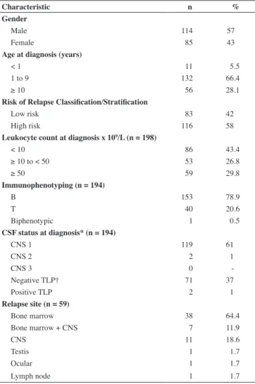
From March 2001 to August 2009, 199 children were diagnosed with ALL. All of these patients were treated according to the GBTLI-LLA-99 protocol and were included in this study. The follow-up period varied between two days and 8.7 years (median: 3.3 years). There were no losses to follow up in this study. The characteristics of the study population are summarized in Table 1.
Of the 199 patients, 191 achieved remission (96%). Up to the time of data analysis, 59 relapses and 55 deaths had occurred (5 deaths while in remission and 50 related to the disease). The estimated OS at 5 years for the whole group was 69.5% (± 3.6%). The estimated EFS at 5 years for the whole group was 58.8% (± 4.0% - Figure 1) with estimated probabilities of 76.2% (± 5.4%) and 46.6% (± 5.3%) for the LR and HR groups, respectively (p-value = 0.00002).
Probability of event free survival
Time (years)
0.0 0.2 0.4 0.6 0.8 1.0
0 2 4 6 8 10
The interval between diagnosis and the irst LP varied from two to 33 days (median: seven days). In 77 patients, the irst LP was performed after the irst week of treatment. The main reasons for the delay in the irst LP were: hyperleukocytosis (29 patients), unsuccessful previous attempt at irst LP (seven patients), tumor lysis syndrome (three patients), coagulopathy (two patients) and chickenpox (one patient). In 35 patients, it was not possible to determine the reason for this delay.
In 155 patients, data on the place where the procedure was performed were retrieved. LP was performed without anesthesia in the hospital ward in 43% of patients (67/155) and with general anesthesia in the surgical center in 57% (88/155).
The platelet count at irst LP was determined in 158 patients and varied between 1 x 10⁹/L and 482 x 109/L (median: 49 x 109/L). In 53% of patients (84/158), LP was performed after platelet transfusion. Of the 194 patients for whom data on irst LP was obtained, traumatic LP was observed in 38% (73/194) of the cases. These 73 children comprised the traumatic LP group, which was compared to the 121 children of the non-traumatic LP group. A signiicant association between age and traumatic LP was observed; the median age for those who suffered a traumatic LP was 3.3 years and for those who did not it was 6.1 years (p-value = 0.0004).
There was no statistically signiicant difference between the platelet count in patients who had a traumatic LP and those who did not (p-value = 0.56). Moreover no association was found between the leukocyte count at diagnosis and traumatic LP (p-value = 0.30). Likewise, no association was found when the leukocyte count at diagnosis was stratiied as less than 10 x 109/L, between 10 and 50 x 109/L or ≥ 50 x 109/L (p-value = 0.31).
There was no association between traumatic LP and the place where the procedure was performed (p-value = 0.24).
CNS relapse occurred in 18 (9.4%) out of 191 patients who achieved remission; in 11 it was an isolated relapse. At the time of data analysis, 12 deaths had been recorded among the group of patients who had had a CNS relapse. One of these patients was in remission and died of infectious complications (septic shock).
The cumulative incidence of CNS relapse (isolated or combined) for the whole group of 191 children was 11.0% at 8 years (Figure 2). The estimated rate of isolated CNS relapse at 8 years was 6.8% (95% conidence interval – 95% CI: 3.6%-11.5%). In patients with a blood leukocyte count at diagnosis ≥ 50 x 109/L, the estimated rate of isolated or combined CNS relapse was 23.9%; in the group with a count < 50 x 109/L, the estimated rate was 6.2% (p-value = 0.0008; Figure 3). The same estimates for isolated CNS relapse were 15.7% and 3.4%, respectively (p-value = 0.006).
There was no statistically signiicant difference in cumulative CNS relapse (isolated or combined) for the other analyzed variables: immunophenotype (T versus B; p-value =0.71), traumatic or non-traumatic LP (p-value = 0.52), interval between diagnosis and irst LP (up to seven days versus eight or more days; p-value = 0.88) and place where LP was performed (hospital ward or surgical center; p-value = 0.45). A multivariate regression model containing all the variables analyzed through the univariate model conirmed that leukocyte count at diagnosis ≥ 50 x 109/L was the only risk factor for a higher cumulative incidence of CNS relapse. The risk of such event in this group increased seven-fold (CI 95%: 2.5-19.8) when compared to the group with a leukocyte count < 50 x 109/L.
*CNS1: puncture not traumatic (< 10 red blood cells per µL) and no identiiable
leukemic blast cells after cytocentrifugation; CNS2: puncture not traumatic (< 10
red blood cells per µL), < 5 white blood cells per µL with leukemic blast cells after cytocentrifugation; CNS3: puncture not traumatic (< 10 red blood cells per µL), ≥ 5 white blood cells per µL with leukemic blast cells after cytocentrifugation
† TLP = traumatic lumbar puncture
Table 1 - Characteristics of the 199 patients diagnosed with acute lymphoblastic leukemia
Characteristic n %
Gender
Male 114 57
Female 85 43
Age at diagnosis (years)
< 1 11 5.5
1 to 9 132 66.4
≥ 10 56 28.1
Risk of Relapse Classiication/Stratiication
Low risk 83 42
High risk 116 58
Leukocyte count at diagnosis x 109/L (n = 198)
< 10 86 43.4
≥ 10 to < 50 53 26.8
≥ 50 59 29.8
Immunophenotyping (n = 194)
B 153 78.9
T 40 20.6
Biphenotypic 1 0.5
CSF status at diagnosis* (n = 194)
CNS 1 119 61
CNS 2 2 1
CNS 3 0
-Negative TLP† 71 37
Positive TLP 2 1
Relapse site (n = 59)
Bone marrow 38 64.4
Bone marrow + CNS 7 11.9
CNS 11 18.6
Testis 1 1.7
Ocular 1 1.7
Lymph node 1 1.7
Discussion
This retrospective study demonstrated the characteristics of CNS leukemia in children and adolescents with ALL who were treated at a Brazilian institution according to a multicentric study protocol, the GBTLI-LLA99 protocol(14).
In the present study, none of the patients were classiied as CNS3(6). The main explanation for this inding may be the relatively small sample size of patients. Another possible contributing factor may be the delay in performing LP in 40% of the patients. In a Japanese study in which the irst LP was set to be performed on the eighth day of the chemotherapy induction phase, only 0.5% of patients were classiied as CNS3(18). The results of both of these studies differ from most of the literature in childhood ALL, as demonstrated by Table 2.
Evaluation of the incidence of traumatic LP and the risk factors for its occurrence were two of the aims of this study, since traumatic LP is known to interfere in the prognosis of children with ALL(7). Traumatic LP at diagnosis occurred in 38% of patients, which is in accordance with the results of different Brazilian ALL referral centers(19,20). As to international referral centers, a lower incidence of traumatic LP is observed with rates varying from 3.4% to 21% at diagnosis(5,7,8,18). Of the studied factors, low “age” was the only risk factor shown to have an association with traumatic LP as anesthesia did not reduce the incidence of traumatic LP.
It should be pointed out that in the initial period of the study most LPs were performed without anesthesia. Because the study was carried out in a university teaching hospital many LPs were actually performed by medical residents. Since, during the study, new evidence in the literature showed that traumatic LPs can potentially interfere with the outcome of ALL(5,7,8), most of the procedures were then performed by an experienced member of the staff, under general anesthesia.
Despite the large number of traumatic LP at diagnosis, this was not associated with a worse EFS or increased CNS relapse. It was unable to determine the effect of positive TLP associated with the presence of blasts on EFS due to the fact that only two patients had these characteristics and received intensiied intrathecal chemotherapy so as to minimize the risk of relapse. This was also the case of the two patients who were classiied as CNS2.
As mentioned before, there was a delay in the irst LP for a signiicant number (40%) of patients; LP was performed more
*CNS1: puncture not traumatic (< 10 red blood cells per µL) and no identiiable leukemic blast cells after cytocentrifugation; CNS2: puncture not traumatic (< 10 red blood cells per µL), < 5 white blood cells per µL with leukemic blast cells after cytocentrifugation; CNS3: puncture not traumatic (< 10 red blood cells per µL), ≥ 5 white blood cells per µL with leukemic blast cells
after cytocentrifugation
† TLP = traumatic lumbar puncture
# CNS1 - 669 patients (88.7%) and CNSs (CNS1 with symptoms due to intracranial iniltrates) - four patients (0.5%)
Table 2 - Comparison between CNS statuses at diagnosis of acute lymphoblastic leukemia among different studies
Authors Number
of Patients CNS1*n (%) CNS2*n (%) CNS3*n (%)
Negative TLP† n (%)
Positive TLP† n (%)
Gajjar et al., (2000)(7) 546 336 (61.5) 80 (14.6) 16 (2.9) 54 (10.0) 60 (11.0)
Te Loo et al., (2006)(8) 526 304 (57.8) 111 (21.1) 10 (1.9) 39 (7.4) 62 (11.8)
Bürger et al., (2003)(5) 2021 1605 (79.5) 103 (5) 58 (2.9) 111 (5.5) 135 (6.7)
Hasegawa et al., (2012)(18) 754 673 (89.2)# 8 (1.1) 4 (0.5) 6 (0.8) 54 (7.2)
Current study 194 119 (61.0) 2 (1.0) 0 71 (37.0) 2 (1.0)
Figure 2 - Cumulative probability of isolated or combined central nervous system relapse in children with ALL. At a 4-year follow-up, a plateau is observed around 11% (continuous line), with a 95% conidence interval (hatched lines) between 6.8% and 16.5%.
than seven days after the diagnosis of ALL. The main reason for this delay was the presence of hyperleukocytosis followed by tumor lysis syndrome and coagulopathy, which shows that these patients were included in the high relapse risk group or had a worse clinical status at diagnosis. Nonetheless, the cumulative incidence of CNS relapse was not inluenced by this delay.
The cumulative incidences of CNS relapse (isolated or combined) and of isolated CNS relapse for the patients in the present study were higher than the overall results obtained by the GBTLI-LLA99 multicentric study in which these single-institution patients are included: 11% versus 6.4% at ive years and 6.8% versus 4.7% at ive years (preliminary results – personal communication), respectively. These results are also unfavorable when compared to those of international referral centers, in which the cumulative incidence of CNS relapse (isolated or combined) now varies from 2.0% to 6.8% at ive years and of isolated CNS relapse from 0.4 to 4.5% at ive years (21-23).
As to the risk factors for ALL CNS relapse referred to in the literature (T-cell immunophenotype, hyperleukocytosis, high risk cytogenetic abnormalities and presence of overt CNS leukemia at diagnosis) only the immunophenotype and the leukocyte count at diagnosis were evaluated in the present study. Although the studied population had a greater proportion of T-cell immunophenotype ALL (20.6%) when compared to that reported in the literature (15%)(1), a greater cumulative incidence of CNS relapse was not shown for these patients when compared to those with a B-lineage ALL phenotype. Furthermore, the GBTLI-LLA99 protocol did not determine any different approach to B- or T-cell phenotype ALL with regard to prophylaxis for CNS leukemia. Hyperleukocytosis was the only risk factor shown to be associated with a higher CNS relapse incidence in univariate and multivariate analyses. According to the treatment protocol used in this study (GBTLI-LLA99), radiotherapy was limited to patients with overt CNS leukemia at diagnosis. Withdrawal of prophylactic radiotherapy from the treatment protocol may have contributed to this result.
The exclusion of prophylactic radiotherapy from the treatment protocol in ALL, for which there is nowadays ample supporting evidence in the literature, may not only protect patients from its well-known deleterious effects but offer the possibility of salvage therapy for patients who have an isolated CNS relapse and have not been previously treated with radiotherapy. Withdrawal of prophylactic radiotherapy may also allow a better understanding of the risk factors directly related to CNS relapse in ALL. Moreover, since CNS relapse acts as a competitive event in relation to bone marrow relapse, preventing it through the use of radiotherapy could increase the likelihood of marrow relapse, which, in turn, has been shown to have a worse prognosis(9). However, the results of the present study emphasize the need for adjustments in the treatment protocol so as to intensify therapy directed to the CNS, especially for patients with an initial leukocyte count above 50 x 109/L.
The worse results observed in this study may be related to several factors, so it is dificult to draw deinite conclusions. The major drawbacks of this study are that it is based on a retrospective analysis and was undertaken in a single institution. Its main limitations concern dificulties in reviewing patient records and the relatively small sample size of the study. It is noteworthy to recall the high incidence of leukocyte counts (equal to or above 50 x 109/L) at diagnosis in
this study (30% of the studied cases) which has been shown to be associated with a higher CNS relapse rate and lower EFS. Therefore, these results suggest that a leukocyte count of more than 50 x 109/L at diagnosis seems, indeed, to be a signiicant prognostic factor for a higher incidence of CNS relapse in childhood ALL.
Despite the study population presenting with others characteristics associated to risk of CNS relapse, hyperleukocytosis was the only risk factor shown to be associated with a higher CNS relapse incidence in univariate and multivariate analyses. These results point to the need of better identifying the population that would beneit from more intensiied prophylaxis of the CNS such as patients with leukocyte counts above 50 x 109/L at diagnosis.
Acknowledgements
We gratefully acknowledge the help of Antonio Vaz de Macedo in preparing the English version of the manuscript. This study was partially supported by grants from Fundação de Amparo à Pesquisa do Estado de Minas Gerais (FAPEMIG) and Conselho Nacional de Desenvolvimento Cientíico e Tecnológico (CNPq). Marcos Borato Viana has a research grant from CNPq, Brazil.
References
1. Pieters R, Carroll WL. Biology and treatment of acute lymphoblastic leukemia. Hematol Oncol Clin North Am. 2010;24(1):1–18.
2. Gaynon PS. Childhood acute lymphoblastic leukemia and relapse. Br J Haematol. 2005;131(5):579-87.
3. Pui CH. Central nervous system disease in acute lymphoblastic leukemia: prophylaxis and treatment. Hematology Am Soc Hematol Educ Program. 2006:142-6.
4. Pui CH, Howard SC. Current management and challenges of malignant disease in the CNS in paediatric leukaemia. Lancet Oncol. 2008;9(3):257–68.
5. Bürger B, Zimmermann M, Mann G, Kühl J, Löning L, Riehm H, et al. Diagnostic cerebrospinal luid examination in children with acute lymphoblastic leukemia: signiicance of low leukocyte counts with blasts or traumatic lumbar puncture. J Clin Oncol. 2003; 21(2):184-188. Comment in: J Clin Oncol. 2003;21(2):179-81.
6. Mahmoud HH, Rivera GK, Hancock ML, Krance RA, Kun LE, Behm FG, et al. Low leukocyte counts with blast cells in cerebrospinal luid of children with newly diagnosed acute lymphoblastic leukemia. N Engl J Med. 1993;329(5):314-9.
7. Gajjar A, Harrison PL, Sandlund JT, Rivera GK, Ribeiro RC, Rubnitz JE, et al. Traumatic lumbar puncture at diagnosis adversely affects outcome in childhood acute lymphoblastic leukemia. Blood. 2000; 96(10):3381-4. Comment in: Blood. 2001;98(12):3496-7.
8. Te Loo DM, Kamps WA, van der Does-van den Berg A, van Wering ER, de Graaf SS; Dutch Childhood Oncology Group. Prognostic signiicance of blasts in the cerebrospinal luid without pleiocytosis or a traumatic lumbar puncture in children with acute lymphoblastic leukemia: experience of the Dutch Childhood Oncology Group. J Clin Oncol. 2006;24(15):2332-6.
9. Pui CH, Campana D, Pei D, Bowman WP, Sandlund JT, Kaste SC, et al. Treating childhood acute lymphoblastic leukemia without prophylactic cranial irradiation. N Engl J Med. 2009;360(26): 2730–41. Comment in: N Engl J Med. 2009;361(13):1310-1; author reply 1311-2; N Eng J Med. 2009;361(13):1310; author reply 1311-2.
xxx
van der Pal K, Stijnen T, van Weel Sipman MH, van Weerden JF, van Wering ER, van der Does-van den Berg A; Dutch Childhood Oncology Group. Dexamethasone-based therapy for childhood acute lymphoblastic leukaemia: results of the retrospective Dutch Childhood Oncology Group (DCOG) protocol ALL-9 (1997–2004). Lancet Oncol. 2009;10(10):957–66. Comment in: Lancet Oncol. 2009;10(10):932-3.
11. Krishnan S, Wade R, Moorman AV, Mitchell C, Kinsey SE, Eden TO, et al. Temporal changes in the incidence and pattern of central nervous system relapses in children with acute lymphoblastic leukaemia treated on four consecutive Medical Research Council trials, 1985–2001. Leukemia. 2010;24(2):450-9.
12. Barredo JC, Devidas M, Lauer SJ, Billett A, Marymont M, Pullen J, et al. Isolated CNS relapse of acute lymphoblastic leukemia treated with intensive systemic chemotherapy and delayed CNS radiation: a pediatric oncology group study. J Clin Oncol. 2006;24(19):3142-9.
13. Ribeiro RC, Rivera GK, Hudson M, Mulhern RK, Hancock ML, Kun L, et al. An intensive re-treatment protocol for children with an isolated CNS relapse of acute lymphoblastic leukemia. J Clin Oncol. 1995;13(2):333-8. 14. Brandalise SR, Pinheiro VR, Aguiar SS, Matsuda EI, Otubo R, Yunes JA,
et al. Beneits of the intermittent use of 6-mercaptopurine and methotrexate in maintenance treatment for low-risk acute lymphoblastic leukemia in children: randomized trial from the Brazilian Childhood Cooperative Group—Protocol ALL-99. J Clin Oncol. 2010;28(11):1911- 8.
15. Gray RJ. A class of K-sample tests for comparing the cumulative incidence of a competing risk. Ann Stat. 1988;16(3):1141-54.
16. Viana MB, Oliveira BM, Cancela CS. Kaplan–Meier method is inappropriate for estimating cumulative incidence of graft-versus-host disease in the presence of competing events. Am J Hematol. 2010;85(6):458-9. Comment in: Am J Hematol. 2008;83(7):528-30.
17. Fine JP, Gray RJ. A proportional hazards model for the subdistribution of a competing risk. JASA. 1999;94:496-509.
18. Hasegawa D, Manabe A, Ohara A, Kikuchi A, Koh K, Kiyokawa N, Fukushima T, Ishida Y, Saito T, Hanada R, Tsuchida M; Tokyo Children’s Cancer Study Group. The utility of performing the initial lumbar puncture on day 8 in remission induction therapy for childhood acute lymphoblastic leukemia: TCCSG L99-15 Study. Pediatr Blood Cancer. 2012;58(1):23-30.
19. Rech A, de Carvalho GP, Meneses CF, Hankins J, Howard S, Brunetto AL. The inluence of traumatic lumbar puncture and timing of intrathecal therapy on outcome of pediatric acute lymphoblastic leukemia. Pediatr Hematol Oncol. 2005;22(6):483-8.
20. Biojone E, Queiróz Rde P, Valera ET, Odashima NS, Takayanagui OM, Viana MB, et al. Minimal residual disease in cerebrospinal luid at diagnosis: a more intensive treatment protocol was able to eliminate the adverse prognosis in children with acute lymphoblastic leukemia. Leuk Lymphoma. 2012;53(1):89-95.
21. Escherich G, Horstmann MA, Zimmermann M, Janka-Schaub GE; COALL study group. Cooperative study group for childhood acute lymphoblastic leukaemia (COALL): long-term results of trials 82,85,89,92 and 97. Leukemia. 2010;24(2):298-308.
22. Gaynon PS, Angiolillo AL, Carroll WL, Nachman JB, Trigg ME, Sather HN, Hunger SP, Devidas M; Children’s Oncology Group. Long-term results of the children’s cancer group studies for childhood acute lymphoblastic leukemia 1983–2002: a Children’s Oncology Group Report. Leukemia. 2010;24(2):285-97.