Enhanced levels of the apoptotic BAX/BCL-2 ratio in children
with acute lymphoblastic leukemia and high-risk features
Maria Kaparou, Despoina Choumerianou, Chrysoula Perdikogianni, Georgia Martimianaki,
Maria Kalmanti and Eftichia Stiakaki
Department of Pediatric Hematology-Oncology, University Hospital of Heraklion, Heraklion, Crete, Greece.
Abstract
It has been suggested that leukemia is characterized by an impaired balance between the proliferation of blood cells and their capacity to undergo apoptosis. The aim of this study was to examine the expression of key molecules re-lated to apoptosis (BCL-2, BAX, FAS, FAS-L) in children with acute lymphoblastic leukemia (ALL). Measurement of BCL-2 and BAX mRNA was performed by quantitative real-time PCR, and membrane expression of FAS and FAS-L was assessed by flow cytometry in bone marrow mononuclear cells, both at diagnosis and at remission following in-duction chemotherapy. At diagnosis, increased levels of the apoptoticBAX/BCL-2 ratio were observed in children older than 10 years and with higher white blood cell counts. A DNA index < 1.16 was associated with increased BAX/BCL-2, both at diagnosis and at remission, and the del(9p) chromosome abnormality with increased BAX/BCL-2 at remission. The expression of the apoptotic receptor FAS was significantly higher at remission com-pared to diagnosis, which might reflect enhanced sensitivity of the leukemic clone to apoptosis and response to treat-ment. Altogether, our results highlight the association of apoptosis-related genes with clinical and cytogenetic prognostic parameters in pediatric ALL. A better understanding of the mechanisms and regulation of apoptosis should enable the design of novel targeted therapies for these patients.
Keywords: leukemia, real-time PCR analysis, protein.
Received: June 19, 2012; Accepted: August 30, 2012.
Introduction
Acute lymphoblastic leukemia (ALL) accounts for nearly 1/3 of all pediatric malignancies and 75% of all childhood leukemias. The annual incidence of ALL has been estimated at 30 cases per million, with a peak inci-dence in children aged two to five years (Pui and Evans, 2006). Progress in diagnosis provided by novel molecular techniques, risk classification, and treatment strategy in ALL has led to cure rates that now exceed 80% (Pui and Ev-ans, 2006). However, a significant proportion (20%) of pa-tients fails to respond to therapy, and treatment failure can occur even in patients with favorable prognostic features (Winicket al., 2004; Carrollet al., 2006).
It has been suggested that leukemia results from an im-balance between the proliferation of blood cells and their ca-pacity to undergo apoptotic death (Peterset al., 1998). At the cellular level, apoptosis is regulated by two major signaling pathways: a) the receptor-mediated (extrinsic), and b) the mitochondria-mediated (intrinsic) pathway (Hacker, 2000). The susceptibility of cells to apoptosis depends on the rela-tive expression of intracellular molecules that enhance
apoptosis (pro-apoptotic), with BAX being the major repre-sentative (Farrow and Brown, 1996), and of molecules that inhibit apoptosis (anti-apoptotic), such as BCL-2 (Hocken-beryet al., 1993; Yang and Korsmeyer, 1996), BCL-XL, and
MCL-1 (Hacker, 2000). Activation of the above-mentioned pathways may be mediated by membrane receptors, such as FAS (APO-1/CD95), that is significantly expressed in neo-plastic cells and interacts with FAS ligand (FAS-L) ex-pressed in activated T-lymphocytes (Woodet al., 2003).
From a clinical standpoint, expression of apopto-sis-related genes has been associated with outcome in vari-ous hematological malignancies. To this end, high BAX
levels correlate with favorable prognosis in acute myelo-blastic leukemia (AML) (Ong et al., 2000), whereas en-hanced expression ofBCL-2is a poor prognostic factor in lymphomas and in chronic lymphocytic leukemia (Hogarth and Hall, 1999). In the case of ALL, studies existing so far have yielded conflicting results. Thus, Arefet al.(2004) re-ported thatBCL-2expression at the time of diagnosis is cor-related with responsiveness to induction chemotherapy, but not patient outcome. In contrast, another study (Coustan-Smithet al., 1996) found no association betweenBCL-2 levels and disease aggressiveness or resistance to therapy. Similarly, although high expression ofBAXhas been asso-ciated with increased risk for relapse in one study (Hogarth Send correspondence to Eftichia Stiakaki. Department of Pediatric
Hematology-Oncology, University Hospital of Heraklion, 71110 Heraklion Crete, Greece. E-mail: [email protected].
and Hall, 1999), other researchers have suggested that the
BAX/BCL-2ratio - rather than individualBAX or BCL-2
levels-may be a more reliable prognostic indicator in ALL (Prokopet al., 2000).
The aim of this study was to assess the expression of the apoptosis-related genesBCL-2andBAX in childhood ALL, both at the time of diagnosis and at remission achieved post induction treatment. In addition, we mea-sured the levels of the apoptotic receptors FAS, FAS ligand, and their co-expression in patients’ leukemic cells. To explore the prognostic significance of apoptosis-related genes in childhood ALL, we examined associations be-tween expression levels and established clinical and cyto-genetic disease parameters.
Materials and Methods
Patients
The study included 26 children (18 boys, eight girls) with newly diagnosed ALL (23 B-ALL, three T-ALL). All patients were diagnosed, treated and followed-up at the De-partment of Pediatric Hematology-Oncology, University of Crete, and received chemotherapy according to the ALL BFM 2000 protocol. Bone marrow specimens were ob-tained from all children, under informed consent signed by the parents/legal guardians. Cytogenetic abnormalities were screened by conventional karyotype analysis and FISH. Disease remission following induction therapy was assessed by bone marrow microscopic evaluation and flow cytometry. The study was approved by the Institute’s Eth-ics Committee.
Bone marrow mononuclear cell isolation
Bone marrow mononuclear cells (MNCs) were iso-lated by Ficoll Hypaque separation (Lymphoprep-Ny-comed d = 1077 g/mL) at the time of ALL diagnosis, upon remission on day 33, and at the end of therapy. The MNCs isolated consisted almost exclusively of leukemic blasts (> 90% bone marrow infiltration at the time of diagnosis). Cells were stored at -80 ºC immediately after collection.
RNA isolation and cDNA synthesis
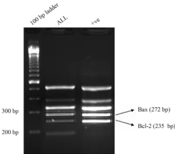
Total RNA was isolated from frozen cells using the SV Total RNA Isolation kit (Promega, Madison, WI, USA). RNA was quantified by spectrophotometry at 260 nm, and the quality was evaluated by 1% agarose elec-trophoresis (Figure 1). An amount of 500 ng of RNA was used for cDNA synthesis with the IM-PROM Reverse Transcription System (Promega).
Quantitative real-time PCR
BAXandBCL-2mRNA levels were assessed by quan-titative real-time PCR, usingGAPDHas housekeeping gene for data normalization. A plot of Ct vs. log was used for quantification, using a standard curve obtained from serial
dilutions of a reference cell line cDNA (HL-60). A total reac-tion volume of 20mL was set up, containing 1x Quantitect SYBR Green mix (QIAGEN, Hilden, Germany), 1x Quantitect Primer mix (Bax QT00997381, bcl-2
QT00025011,GapdhQT01192646 (all from QIAGEN), and 1.2 mL cDNA. All reactions were performed in an ABI PRISM 7700 Sequence Detection System (Applied Bio-systems, Foster City, CA, USA). The thermal cycling condi-tions included a hot start step at 95 ºC for 15 min followed by 40 cycles at 95 ºC for 15 s, 55 ºC for 30 s, and 72 ºC for 30 s.
Flow cytometry
Anticoagulated whole bone marrow samples were transferred into polystyrene round-bottom tubes and incu-bated with 10mL of PC5-anti-CD45 (A07785, Beckman Coulter, Brea, CA, USA), 20mL FITC-anti-Fas (sc-52524, Santa Cruz Biotechnology Inc,, Santa Cruz, CA, USA), and 20 mL PE-anti-Fas-L (sc-19681, Santa Cruz Biotechnol-ogy). Control samples were stained with anti-CD45, PE-mouse IgG1 (A07796, Beckman Coulter) and FITC-PE-mouse IgG1 (A07795, Beckman Coulter) isotype controls. The samples were incubated for 15 min at room temperature in the dark, washed with PBS supplemented with 2% FBSSV (Hyclone, Thermo Scientific, Logan, UT, USA) and 5% so-dium azide (106688, Merck, Darmstadt, Germany) for 5 min at 290 x g. Supernatants were discarded and red blood cells were lysed by Immunoprep Reagent System (Beckman Coulter). Cells were analyzed on an Epics Elite Coulter cell sorter. The CD45+ population was gated and lymphocytes were identified based on forward scatter and side scatter properties. The expression of surface FAS, FAS-L and their co-expression were determined.
Statistical analysis
The differences in gene expression among groups were evaluated with the nonparametric Mann-WhitneyU test. Correlations were calculated with the Spearman test. A p-value < 0.05 was considered as statistically significant. Data are presented as mean±standard error of the mean (SE), unless stated otherwise.
Results
Expression of the apoptosis-related genesBAXand
BCL-2in children with ALL and association with clinical characteristics
We measured the levels of the BAX, BCL-2, and
BAX/BCL-2ratio in bone marrow MNCs from 26 children (18 boys, eight girls) with ALL. The patients’ demographic and clinical characteristics are presented in Table 1. At di-agnosis,BAXlevels were correlated withBCL-2(r= 0.46, p = 0.001) andBAX/BCL-2(r= 0.47, p < 0.001). A signifi-cant negative correlation between white blood cell count andBCL-2mRNA (r= -0.57, p = 0.007) was observed, whereas patients’ age and hemoglobin showed no associa-tion withBAXorBCL-2. According to the BFM protocol criteria, 12 ALL patients were classified in the high-risk group and 14 in the median-risk group. We found no differ-ence inBAX,BCL-2, orBAX/BCL-2levels between high-risk and median-high-risk patients. Older children (> 10 years) in the high-risk group presented higher values ofBAX/BCL-2
ratio compared to younger counterparts (0.71 ± 0.20 vs.
0.15±0.24, p = 0.031).
Correlation ofBAXandBCL-2levels at diagnosis with cytogenetic abnormalities in children with ALL
Hyperdiploidy (defined as DNA index³1.16) was correlated with higher levels of the anti-apoptoticBCL-2at diagnosis (r= 0.71, p = 0.05). In line with this finding, the DNA index showed negative correlation with the
BAX/BCL-2ratio (r= -0.88, p = 0.004) (Figure 2).
Con-versely, the presence of a t(12;21)(p13;q22) translocation, observed in four children, was associated with significantly higher BAX/BCL-2 ratios (1.45 ± 0.23 vs. 0.46 ± 0.30, p = 0.030). A deletion in chromosome 9p was also observed in four children, but did not correlate with a differential
BAX,BCL-2, orBAX/BCL-2ratio.
Expression ofBAXandBCL-2in ALL children at remission post induction therapy
Measurement of BAX and BCL-2 in bone marrow MNCs was repeated in the ALL children at disease remis-sion on day 33 after treatment initiation. The apoptotic
BAX/BCL-2ratio was significantly lower at remission than at the time of diagnosis in the high-risk (0.05±0.02vs.0.32 ±0.12, p = 0.019), but not in the median-risk group. In line with this finding, patients in the high-risk group had lower
BAX/BCL-2 at remission compared to their median-risk counterparts (0.05±0.02vs.0.58±0.13, p = 0.009).
Expression of the apoptosis-related genes once remis-sion has been achieved is correlated with certain baseline Table 1- Demographic and clinical characteristics of the ALL patients.
No. of patients 261
Age (years) 7.1±1.2
Gender (female/male) 8/18
ALL type (B/T) 23/3
White blood cell count (K/mL) 27.5±10.6
Hemoglobin (g/dL) 9.1±0.6
Cytogenetics
Hyperdiploidy (no. of patients) 7
t(12;21) (no. of patients) 4
del(9p21) (no. of patients) 4
Other abnormalities 5
ALL risk (median/high) 17/9
1
Data presented asNor mean±SE.
cytogenetic findings. First, a negative correlation was ob-served between the pro-apoptotic BAX and DNA index (r= -0.85, p = 0.015). Accordingly, significantly higher levels ofBAX/BCL-2were observed in patients with DNA index < 1.16 compared to those with DNA index³1.16 (hyperdiploidy) (0.80± 0.78vs. 0.08±0.03, p = 0.025). Moreover, patients with deletion in chromosome 9p had significantly higherBAX/BCL-2levels than those without the deletion (0.51vs.20, p = 0.007).
Expression of FAS and FAS-L in children with ALL
The membrane expression of the pro-apoptotic recep-tors FAS and FAS-L in bone marrow CD45+ MNCs was determined in 13 children with B-ALL both at the time of diagnosis and at disease remission (day 33 after treatment initiation). The proportion of FAS+ MNCs was signifi-cantly increased at remission compared to the time of diag-nosis (24.0±6.1%vs.8.0±1.9%, p = 0.035) (Figure 3). In contrast, a non-significant reduction in FAS-L+cells was observed at remission (2.4±0.8%vs.4.6±1.4% at diagno-sis). We found no association between FAS/FAS-L expres-sion and clinical or cytogenetic parameters.
Discussion
Deregulation of apoptosis disrupts the balance be-tween cell survival and death and may be a key patho-genetic aspect in hematologic malignancies. Herein, we re-port increased levels of the apoptoticBAX/BCL-2ratio in ALL children with high-risk features, such as higher white blood cell count, DNA index < 1.16, and the del(9p) chro-mosomal abnormality. Previous studies have failed to dem-onstrate a significant association between clinical prognos-tic parameters, such as age at diagnosis, gender, or white blood cell count, andBAXorBCL-2in pediatric ALL (Gala
et al., 1994; Hogarth and Hall, 1999; Narayanet al., 2007). In our study, although the expression of the apoptosis-related genes did not vary significantly according to the risk group classification, a higher white blood cell count was
correlated with reduced expression of the anti-apoptotic
BCL-2. Another high-risk feature, namely age above 10 years, showed inverse association with theBAX/BCL-2
ratio, albeit only in the high-risk group of patients. Alto-gether, in children with ALL, clinical parameters of ad-verse outcome are associated with increased levels of pro-apoptoticvs.anti-apoptotic genes in bone marrow MNCs.
Ploidy has been recently recognized as a prognostic factor in ALL, with hyperdiploidy (DNA index³1.16) be-ing associated with a favorable prognosis (Aricò et al., 2008). In our analysis,BCL-2 mRNA showed a positive correlation with the DNA index, both at diagnosis and at the time of remission following induction therapy, whereas
BAXexpression was inversely correlated with the DNA in-dex at remission. TheBAX/BCL-2ratio was inversely cor-related with the DNA index at diagnosis and remission, suggesting that the persistence of a highBAX/BCL-2ratio in bone marrow MNCs may be related to a less favorable prognosis.
Cytogenetic abnormalities have significant prognos-tic value in ALL. Abnormalities in chromosome 9 are com-mon in children with ALL (11% according to large series) (Heeremaet al., 1999). Although in adult B-precursor ALL with abnormal chromosome 9p the prognosis has been re-ported to be poor (Nahiet al., 2008), data in children are scarce and controversial (Heerema et al., 1999). Since del(9p) was found in 15% of our patients, we investigated its association with apoptosis-related genes. Children with del(9p) had higher levels ofBAXat diagnosis than children without this chromosome abnormality. Moreover, the
BAX/BCL-2ratio at the time of remission was significantly higher in the del(9p) group than in patients without this ab-normality. Although the small number of patients in this study precludes definitive conclusions, these data may sug-gest that the presence of del(9p) in children with ALL indi-cates a poor prognosis.
In our study, the ALL patients in the high-risk group had a higherBAX/BCL-2ratio at diagnosis than at remis-sion, achieved after induction chemotherapy, whereas no significant change was observed in the median-risk group. This finding might be a further indication that a high
BAX/BCL-2ratio at diagnosis points to a poor prognosis in children with ALL. Alternatively, longitudinal changes in the expression of apoptosis-related genes could reflect al-terations in the apoptosis burden of the disease as response to chemotherapeutic regimens. Thus, changes in apoptosis during induction therapy may be indicative of the effective-ness of treatment and of the disease outcome (Schuler and Szende, 2004). Higher BAX levels were associated with in-creased risk of relapse in one study (Hogarth and Hall, 1999), whereas another one (Narayanet al., 2007) showed that lowBAX/BCL-2ratio correlates with favorable prog-nosis.
It has been suggested thatFASexpression could con-trol the response of leukemic cells to chemotherapy and Figure 3- Expression of the apoptotic receptors FAS and FAS-L in bone
have an impact on prognosis (Aref et al., 2004). In our study, the ALL patients had low levels ofFASat diagnosis, followed by a significant increase at disease remission. In some pediatric ALL studies, increased levels ofFAShave been associated with longer survival in complete remission (Baryshnikovet al., 1999), whereas other studies found no association ofFAS levels with outcome (Wuchter et al., 2000; Arefet al., 2004; Fulda, 2009). At this time, we sug-gest that increasedFASlevels at remission post induction treatment could discriminate patients who are not resistant to chemotherapy, but additional studies with long-term fol-low-up are needed to further address this issue.
In conclusion, our study highlights the association be-tween the apoptoticBAX/BCL-2ratio with high-risk fea-tures, such as older age, higher white blood cell count, the del(9p) abnormality, and DNA index < 1.16, in children with ALL. The increase inFASexpression, once remission has been achieved after induction treatment, could repre-sent a prognostic factor of favorable response to chemo-therapy and deserves further investigation. A better under-standing of the role of apoptosis in pathogenesis and prognosis of pediatric ALL should enable the design of novel targeted therapies.
References
Aref S, Salama O, Al-Tonbary Y and Mansour A (2004) Assess-ment of Bcl-2 expression as modulator of Fas mediated apoptosis in acute leukemia. Hematology 9:113-121. Aricò M, Valsecchi MG, Rizzari C, Barisone E, Biondi A, Casale
F, Locatelli F, Lo Nigro L, Luciani M, Messina C, et al.
(2008) Long-term results of the AIEOP-ALL-95 TRIAL for childhood acute lymphoblastic leukemia: Insight on the prognostic value of DNA Index in the framework of Berlin-Frankfurt-Muenster-based chemotherapy. J Clin Oncol 26:283-289.
Baryshnikov AY, Polosukhina ER, Tupitsin NN, Gavrikova NV, Andreeva LY, Zabotina TN, Mayakova SA, Kurmashov VI, Syrkin AB, Kadagidze ZG,et al.(1999) CD95 (FAS/APO-1) antigen is a new prognostic marker of blast cells of acute lymphoblastic leukemia patients. Adv Exp Med Biol 457:251-258.
Carroll WL, Bhojwani D, Min DJ and Raetz EA (2006) Childhood acute lymphoblastic leukemia in the age of genomics. Pediatr Blood Cancer 46:570-578.
Coustan-Smith E, Kitanaka A, Pui CH, McNinch L, Evans WE, Raimondi SC, Behm FG, Aricò M and Campana D (1996) Clinical relevance of BCL-2 overexpression in childhood acute lymphoblastic leukemia. Blood 87:1140-1146. Farrow SN and Brown R (1996) New members of the Bcl-2
fam-ily and their protein partners. Curr Opin Genet Dev 8:1960-1966.
Fulda S (2009) Therapeutic opportunities for counteracting apoptosis resistance in childhood leukemia. Br J Haematol 145:441-454.
Gala JL, Vermylen C, Cornu G, Ferrant A, Michaux JL, Philippe M and Martiat P (1994) High expression of bcl-2 is the rule in acute lymphoblastic leukemia, except in Burkitt subtype
at presentation, and is not correlated with the prognosis. Ann Haematol 69:17-24.
Hacker G (2000) The morphology of apoptosis. Cell Tissue Res 301:5-17.
Heerema NA, Sather HN, Sensel MG, Liu-Mares W, Lange BJ, Bostrom BC, Nachman JB, Steinherz PG, Hutchinson R, Gaynon PS,et al.(1999) Association of chromosome arm 9p abnormalities with adverse risk in childhood acute lym-phoblastic leukemia: A report from the Children’s Cancer Group. Blood 94:1537-1544.
Hockenbery DM, Oltavi ZN, Yin XM, Milliman CL and Korsmeyer SJ (1993) Bcl-2 functions in an antioxidant path-way to prevent apoptosis. Cell 75:241-251.
Hogarth AL and Hall GA (1999) Increased BAX expression is as-sociated with an increased risk of relapse in childhood acute lymphoblastic leukemia. Blood 93:2671-2678.
Nahi H, Hägglund H, Ahlgren T, Bernell P, Hardling M, Karlsson K, Lazarevic VLj, Linderholm M, Smedmyr, B, Aström, M,
et al.(2008) An investigation into whether deletions in 9p reflect prognosis in adult precursor B-cell acute lym-phoblastic leukemia: A multi-center study of 381 patients. Haematologica 93:1734-1738.
Narayan S, Chandra J, Sharma M, Naithani R and Sharma S (2007) Expression of apoptosis regulators Bcl-2 and Bax in child-hood acute lymphoblastic leukemia. Hematology 12:39-43. Ong YL, McMullin MF, Bailie KE, Lappin TR, Jones FG and Irvine
AE (2000) High Bax expression is a good prognostic indicator in acute myeloid leukemia. Br J Haematol 111:182-189. Peters R, Leyraz S and Perey L (1998) Apoptotic regulation in
primitive hematopoietic precursors. Blood 92:2041-2052. Prokop A, Wieder T, Sturm I, Essmann F, Seeger K, Wuchter C,
Ludwig WD, Henze G, Dörken B and Daniel PT (2000) Re-lapse in childhood acute lymphoblastic leukemia is associ-ated with a decrease of the Bax/Bcl-2 ratio and loss of
spon-taneous caspase-3 processing in vivo. Leukemia
14:1606-1613.
Pui CH and Evans WE (2006) Treatment of acute lymphoblastic leukemia. N Engl J Med 354:166-178.
Schuler D and Szende B (2004) Apoptosis in acute leukemia. Leuk Res 28:661-666.
Winick NJ, Carroll WL and Hunger SP (2004) Childhood leuke-mia-new advances and challenges. N Engl J Med 351:601-603.
Wood CM, Goodman PA, Vassilev AO and Uckun FM (2003) CD95 (APO-1/FAS) deficiency in infant acute lympho-blastic leukemia: Detection of novel soluble Fas splice vari-ants. Eur J Haematol 70:156-171.
Wuchter C, Karawajew L, Ruppert V, Schrappe M, Harbott J, Ratei R, Dörken B and Ludwig WD (2000) Constitutive ex-pression levels of CD95 and Bcl-2 as well as CD95 function and spontaneous apoptosis in vitrodo not predict the re-sponse to induction chemotherapy and relapse rate in child-hood acute lymphoblastic leukemia. Br J Haematol 110:54-160.
Yang S and Korsmeyer SJ (1996) Molecular thanaptosis: A dis-course on bcl-2 family and cell death. Blood 88:386-401.
Associate Editor: Anamaria Aranha Camargo