Terazaki CRT et al. / Synovial chondromatosis of the shoulder
Radiol Bras. 2014 Jan/Fev;47(1):38–42 38
Review Article
Synovial chondromatosis of the shoulder: imaging findings
*
Osteocondromatose sinovial no ombro: achados por métodos de imagem
Terazaki CRT, Trippia CR, Trippia CH, Caboclo MFSF, Medaglia CRM. Synovial chondromatosis of the shoulder: imaging findings. Radiol Bras. 2014 Jan/ Fev;47(1):38–42.
Abstract
R e s u m o
Synovial chondromatosis is a benign condition characterized by synovial proliferation and metaplasia, with development of cartilaginous or osteocartilaginous nodules within a joint, bursa or tendon sheath. In the shoulder, synovial osteochondromatosis may occur within the glenohumeral joint and its recesses (including the tendon sheath of the biceps long head), and in the subacromial-deltoid bursa. Such condition can be identified either by radiography, ultrasonography or magnetic resonance imaging, showing typical features according to each method. Radiography commonly shows ring-shaped calcified cartilages and periarticular soft tissues swelling with erosion of joint margins. Ultrasonography demonstrates hypoechogenic cartilaginous nodules with progressive increase in echogenicity as they become calcified, with development of posterior acoustic shadow in case of ossification. Besides identifying cartilaginous nodules, magnetic reso-nance imaging can also demonstrate the degree of synovial proliferation. The present study is aimed at describing the imaging findings of this entity in the shoulder.
Keywords: Synovial chondromatosis; Image findings; Shoulder.
Osteocondromatose sinovial é uma afecção benigna caracterizada por proliferação e metaplasia sinovial, com formação de nódulos cartilaginosos ou osteocartilaginosos no interior de uma articulação, bursa ou bainha tendinosa. A osteocondromatose sinovial no ombro pode ocorrer no interior da articulação glenoumeral e seus recessos (incluindo a bainha do tendão da cabeça longa do bíceps) e na bursa subacrômio-deltoidiana. Esta doença pode ser identificada por radiografia, ultrassom ou ressonância magnética, apresentando aspec-tos característicos em cada um destes métodos. Na radiografia comumente encontramos calcificações anelares do tipo cartilaginoso e aumento de volume das partes moles periarticulares com erosões das margens articulares. No ultrassom os nódulos cartilaginosos apresentam-se hipoecogênicos, e quando calcificados aumentam progressivamente a sua ecogenicidade, até a formação de sombra acústica posterior quando ossificados. A ressonância magnética, além de identificar os nódulos cartilaginosos, também é capaz de mostrar o grau de proliferação sinovial. O objetivo deste trabalho é demonstrar os aspectos de imagem desta doença no ombro.
Unitermos: Osteocondromatose sinovial; Achados de imagem; Ombro.
* Study developed at Hospital São Vicente – Funef, Curitiba, PR, Brazil. 1. MDs, Residents, Unit of Radiology and Imaging Diagnosis, Hospital São Vicente – Funef, Curitiba, PR, Brazil.
2. MD, Radiologist, Hospital São Vicente – Funef, Curitiba, PR, Brazil. 3. MD, Radiologist, Preceptor of Medical Residency, Unit of Radiology and Imag-ing Diagnosis, Hospital São Vicente – Funef, Curitiba, PR, Brazil.
4. MD, Radiologist, Head Preceptor of Medical Residency, Unit of Radiology and Imaging Diagnosis, Hospital São Vicente – Funef, Curitiba, PR, Brazil.
Mailing Address: Dr. Carlos Renato Ticianelli Terazaki. Rua Itajubá, 480, ap. 502, Portão. Curitiba, PR, Brazil, 81070-190. E-mail: [email protected].
Received December 19, 2012. Accepted after revision July 25, 2013.
Any joint may be affected, and the knee is the most frequent site (50–65% of the cases), followed by elbow, hips and shoulder in decreasing order of frequency(2,5,6).
The secondary form is a common condition caused by mechanical injury of the intraarticular hyaline cartilage trig-gered by joint anomalies such as osteoarthrosis, osteonecro-sis, osteochondritis dissecans, neuropathic osteoarthropathy, trauma and rheumatoid arthritis(2,6). It is found in elderly
patients, generally involving multiple joints, and may be related to degenerative joint disease, more frequently in the knees, hips and shoulders(2).
FISIOPATHOLOGY
The development of synovial osteochondromatosis in the shoulder joint is uncommon, and its secondary presentation is most frequently found. Cartilaginous or osteocartilaginous nodules may be found within the joint or its recesses (sub-scapularis, axillary and along the tendon sheath of the bi-ceps long head), and in the subacromial-deltoid bursa. Pa-tients with the primary presentation of the condition present with joint pain, swelling, and limitation of motion which Carlos Renato Ticianelli Terazaki1, Cesar Rodrigo Trippia2, Carlos Henrique Trippia3, Maria Fernanda Sales Ferreira Caboclo4, Carla Regina Miranda Medaglia1
INTRODUCTION
Synovial osteochondromatosis is a condition character-ized by synovial membrane proliferation and metaplasia, with development of multiple cartilaginous or osteocartilaginous nodules within the joint, bursa or tendinous sheath(1). It can
be divided into primary and secondary forms(2).
progress slowly along many years(7,8). In the secondary
pre-sentation, the symptoms are related to the basal joint dis-ease and, in general, the condition is incidentally identified during investigation of degenerative joint disease, or in the course of investigations in patients with previously diagnosed joint disease who present with symptoms exacerbation. The physiopathology is the same for both presentations of the disease, and it may be divided into three phases. At the ini-tial phase, synovial membrane proliferation and intrasynovial development of cartilaginous nodules (chondromas) occur. As the disease progresses, such cartilaginous nodules may calcify (osteochondromas), or even form mature ossified nodules with bone marrow(3). At the late phase of the
dis-ease, active disease is no longer found in the synovia, and the nodules may detach towards the joint cavity forming loose bodies. In the primary form of the disease, the chon-dral nodules are usually numerous, with similar contours and morphology (ranging from a few millimeters up to some centimeters), which indicates that they originate simulta-neously, sometimes coalescing and acquiring the appear-ance of a mass(2). It is important to highlight, however, that
in up to 30% of the patients no calcification is found(1). In
the secondary form of the disease, there is a lower number of synovial chondromas, with varied sizes and shapes, with a more irregular or ring-shaped calcification pattern, in con-centric layers, besides the signs of degenerative joint dis-ease.
The treatment for the primary presentation of the dis-ease is the surgical removal of the intraarticular loose bod-ies and full synovectomy of the involved joint(6,9). The
re-currence rate ranges from 3% to 23%, occurring within a period of up to five years, and the malignant transformation into chondrosarcoma is unusual (5% of the cases). For the
secondary presentation, the treatment is aimed at the symp-toms of the osteodegenerative disease, and the possibility of arthroplasty is considered in cases of advanced disease.
IMAGING FINDINGS
The radiological findings depend on the presentation of the disease (either primary or secondary), on its stage and presence of calcification or ossification in the cartilaginous nodules(1,6). In primary osteochondromatoses, conventional
radiography can identify widened articular space and increase in volume of periarticular soft parts in association with ero-sion of joint margins, indicating the presence of disease of intraarticular origin(6). Calcifications are usually numerous,
sometimes countless, and similar in size and shape. Gener-ally, calcifications are ring-shaped, of cartilaginous origin (Figure 1A) which gives them the typical radiographic as-pect of such lesions(2,4,10,11), sometimes presenting with a
calcified central focus and ring-shaped peripheral calcifica-tion, or a target sign with a radiolucent center and calcified periphery. Periarticular osteoporosis is not commonly found. In cases of secondary disease, the authors found signs of degenerative joint disease (intraarticular space narrowing, marginal osteophytes, sclerosis, and subchondral bone cysts) in association with a smaller number of calcifications with varied sizes, usually larger, calcifying in a more irregular form or in concentric layers (Figure 1B)(1,2).
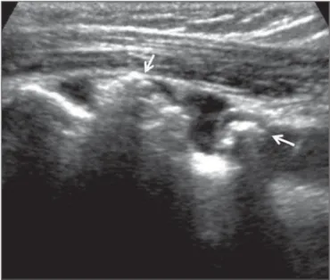
At ultrasonography, cartilaginous nodules are hypoecho-genic (Figure 2), and as they calcify the echohypoecho-genicity in-creases, sometimes determining posterior acoustic shadow-ing (Figure 3).
In the shoulder, ultrasonography is particularly sensi-tive to detect the disease affecting the tendon sheath of the biceps long head, demonstrating nodules attached to the
Figure 1. A:Primary synovial osteochondromatosis. Radiography, anteroposterior view of the shoulder demonstrating multiple, typical, ring-shaped calcifications similar in size and shape, located at the level of the axillary recess of the glenohumeral joint (arrow). B:Secondary synovial osteochondromatosis. Radiography, anteroposterior view of the shoulder demonstrating osteodegenerative changes characterized by the presence of osteophytes in the inferior articular margin of the humeral head (arrowhead) and presence of multiple juxtarticular rounded-shaped calcifications with varied sizes (arrows).
Figure 2.Secondary synovial osteochondromatosis. Sonographic study of the shoulder focusing the tendon of the biceps long head (TBLH) on the transverse plane. Presence of synovial nodules with different levels of echogenicity project-ing toward the interior of the tendinous sheath, easily identifiable by the presence of synovial fluid. The echogenicity depends on the calcification degree, with hypoechoic nodules (black arrow) corresponding to noncalcified chondromas, while calcified nodules present increased echogenicity (arrowhead).
Figure 3.Secondary osteochondromatosis. Sonographic study of the shoulder, focusing the tendon sheath of the biceps long head on the longitudinal plane. The sheath is filled by fluid, and in its interior calcified nodules with posterior acoustic shadow are identified (arrows).
Figure 4. Secondary synovial osteochondromatosis associated with supraspinatus tendinopathy. A: Sonographic study of the shoulder focusing the tendon of the biceps long head on the longitudinal plane, which is ruptured, with only its proximal stump being identified. Multiple small synovial chondromas and effusion within the synovial sheath are observed. B: Longitudinal section of the supraspinatus tendon. Thickened, hypoechoic and heterogeneous tendon, with loss of fibrillar structure, charac-terizing tendinopathy process.
A B
synovial membrane surface, which is facilitated by the pres-ence of fluid within the synovial sheath. Loose bodies are frequently found along the bicipital groove(9). Such patients
frequently have the secondary form of the disease associated with other alterations found upon examination of the shoul-der, such as effusion in the joint and in the subacromial-del-toid bursa, tendinopathies and tendon tear (Figure 4).
Magnetic resonance imaging findings depend on the intensity of synovial proliferation, on the presence of carti-laginous nodules and calcifications(3). Noncalcified lesions
lead to synovial thickening (Figure 5) or to the development
of an isointense intraarticular mass isointense to the muscle on T1-weighted sequences, and hyperintense on T2-weighted sequences(1–3,12).
Such lesions may be differentiated from joint effusion or from other soft tissue tumors by the presence of the typi-cal peripheral and septal contrast uptake of chondral lesions, after intravenous gadolinium injection(1,4). The intravenous
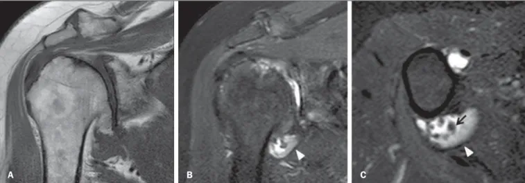
Figure 5.Secondary synovial osteochondromatosis. Magnetic resonance imaging, T1-weighted sequence of the shoulder in the coronal plane (A), coronal T2-weighted fat sat (B), and axial T2weighted fat sat (C) demonstrating marked osteodegenerative glenohumeral alteration characterized by irregularity of the joint surface and marginal osteophytes on the humeral head. Presence of synovial thickening in the axillary joint recess, with intermediate signal on T2-weighted sequence (arrowhead) and small synovial osteochondromas (arrow).
A B C
Intraarticular nodules that undergo endochondral ossi-fication are formed by mature bone, present peripheral cor-tical bone with low signal intensity, and hyperintense yellow bone marrow within the bone on T1-weighted sequences(2,12).
CONCLUSION
In spite of being less common than in other joints of the body, synovial osteochondromatosis may be found in the shoulder at the level of the joint recesses (including the
ten-Figure 6.Primary synovial chondro-matosis. Magnetic resonance imaging of the shoulder, coronal T2-weighted (A) and coronal T1-weighted fat sat se-quence after intravenous gadolinium injection (B). Multiple, noncalcified syn-ovial chondromas in the interior of the subacromial–deltoid bursa, hyperint-ense on T2-weighted sequence and isointense to the muscle on contrast-enhanced T1-weighted sequence, sym-metrical in size and shape. Presence of synovial membrane proliferation thick-ening of the bursa wall, with intermedi-ate signal intensity on T2-weighted se-quence with contrast uptake,
indicat-ing disease activity. A B
Figure 7.Secondary synovial osteo-chondromatosis. Magnetic resonance imaging of the shoulder, sagittal T1-weighted (A) and sagittal T2-weighted fat sat (B) sequences. Presence of calcified nodules (synovial osteochon-dromas) in the subacromial-deltoid bursa (arrows) and in the subscapularis recess (arrowheads) with typical nodu-lar features, with marked hyposignal on
don sheath of the biceps long head) and in the subacromial-deltoid bursa. The primary and secondary presentations of the disease may be suggested by the number, shape and size of the chondral nodules, as well as by the absence or pres-ence of pre-existing joint disease. The diagnosis may be achieved only on the basis of typical imaging findings which, depending on the stage of the disease, may correspond to synovial membrane proliferation, presence of cartilaginous nodules (chondromas) and calcified or ossified nodules (os-teochondromas).
REFERENCES
1. Llauger J, Palmer J, Rosón N, et al. Nonseptic monoarthritis: imag-ing features with clinical and histopathologic correlation. Radiographics. 2000;20 Spec No:S263–78.
2. Murphey MD, Vidal JA, Fanburg-Smith JC, et al. Imaging of syn-ovial chondromatosis with radiologic-pathologic correlation. Radiographics. 2007;27:1465–88.
3. Sheldon PJ, Forrester DM, Learch TJ. Imaging of intraarticular masses. Radiographics. 2005;25:105–19.
4. Walker EA, Murphey MD, Fetsch JF. Imaging characteristics of
tenosynovial and bursal chondromatosis. Skeletal Radiol. 2011; 40:317–25.
5. Kransdorf MJ, Meis JM. Fom the archives of the AFIP. Extraskeletal osseous and cartilaginous tumors of the extremities. Radiographics. 1993;13:853–84.
6. Mohana-Borges AV, Chung CB, Resnick D. Monoarticular arthri-tis. Radiol Clin North Am. 2004;42:135–49.
7. Milgram JW. Synovial osteochondromatosis: a histopatological study of thirty cases. J Bone Joint Surg Am. 1977;59:792–801. 8. Trajkovski T, Mayne IP, Deheshi BM, et al. Synovial chondromatosis
of the shoulder: open synovectomy and insertion of osteoarticular allograft with internal fixation to repair intraoperative glenohumeral joint instability. Am J Orthop (Belle Mead NJ). 2011;40:E154–8. 9. Lunn JV, Castellanos-Rosas J, Walch G. Arthroscopic synovec-tomy, removal of loose bodies and selective biceps tenodesis for synovial chondromatosis of the shoulder. J Bone Joint Surg Br. 2007;89:1329–35.
10. Olsen KM, Chew FS. Tumoral calcinosis: pearls, polemics, and alternative possibilities. Radiographics. 2006;26:871–85. 11. Lasmar NP, Vieira RB, Rosa JO, et al. Condromatose sinovial. Rev
Bras Ortop. 2010;45:490–2.