Letters to the Editor
Radiol Bras. 2016 Jan/Fev;49(1):56–64
62
Endometrial osseous metaplasia: sonographic, radiological and histopathological findings
Metaplasia óssea endometrial: aspecto ultrassonográfico, radiológico e histopatológico
Dear Editor,
A 31-year-old, female patient with previous history of sponta-neous miscarriage with uterine curettage eight years ago, under-going investigation for secondary infertility, increased menstrual flow and pelvic pain.
Transvaginal ultrasonography (US) (Figures 1A and 1B) showed a hyperechoic endometrial, nonspecific, plate-shaped image with posterior acoustic shadowing, and measuring 2.7 × 2.6 cm. Pelvic radiography (Figure 1C) identified a focus of calci-fication at endometrial site.
On the basis of the imaging findings and clinical history, the presumptive diagnosis was endometrial osseous metaplasia, con-firmed by histopathological study revealing the presence of a plate of trabecular bone tissue surrounded by fibrous tissue and prolif-erative endometrium (Figure 2). Cartilage, bone marrow, chronic inflammation and trophoblastic tissue were not present.
Endometrial osseous metaplasia corresponds to the presence of bone-like tissue within the uterine cavity. It is a rare entity, af-fecting only 0.15% of the patients referred to hysteroscopy
clin-ics(1,2). The pathogenesis of such a condition still remains con-troversial. The two most accepted mechanisms involve either the presence of chronic endometrioses with undifferentiated mesen-chymal cells inducing the endometrial stromal cells transforma-tion into osteoblasts, or miscarriage with dystrophic ossificatransforma-tion of the residual ovular tissues(3). Such hypotheses are reinforced as one considers that more than 80% of cases occur after pregnan-cies that evolved to miscarriage, particularly those followed by in-fection(4). Symptoms include pelvic pain and menstrual flow al-terations, but the main consequence of the presence of bone tis-sue in the uterine cavity is infertility(5). The association between osseous metaplasia and infertility occurs because of the similarity between the action of the bone tissue and the action of an intrau-terine contraceptive device (IUCD)(6,7).
The main sonographic finding of endometrial osseous meta-plasia is the presence of a strongly echogenic endometrial plate with posterior acoustic shadowing, assuming the presence of an IUD as main differential diagnosis. Other possible diagnoses in-clude: presence of foreign bodies, Asherman’s syndrome, calci-fied submucosal fibrosis and Müllerian tumor(2,5,6). However, the suspicion of endometrial osseous metaplasia should be taken into consideration by the sonographist in cases where strongly echo-genic endometrial plates are detected in patients with history of miscarriage and chronic endometriosis.
In the presently reported case, the correlation between trans-vaginal US and pelvic radiography has allowed for the diagnosis of endometrial calcification. The previous history of miscarriage with curettage has corroborated the hypothesis of calcification corre-sponding to osseous metaplasia induced by chronic endometritis, which later was confirmed by histopathological analysis of bone fragments collected by means of hysteroscopy.
Transvaginal US is the best imaging method in such cases, since hysterosalpingography and magnetic resonance imaging may miss the findings. In such cases, the investigator must de-scribe the location and the dimensions of the echogenic plate, rule out the presence of an IUCD, and reinforce the history of miscarriage with chronic endometritis, corroborating the hypoth-esis of metaplastic endometrial ossification. Such informations are important for the hysterocopist who will make the resection of the osseous plate with subsequent histopathological analysis.
The treatment for this condition should be performed by means of hysteroscopic removal of osseous fragments to be sub-mitted to histopathological analysis or, as a second option by uter-ine curettage(8). In the present case, the first alternative was Figure 2. Photomicrography with low and medium magnification showing
os-seous trabeculae intermingled with endometrial tissue. Observe the endometrial glands at the lower right corner. Hematoxylin-eosin staining.
Figure 1.A,B: Transvaginal ultrasonography demonstrating hyperechoic image with posterior acoustic shadowing in the endometrium, compatible with calcification.
C: Hip radiography, oblique view showing an image with calcific density corresponding to the one found at ultrasonography, strengthening the suggested hypothesis.
Letters to the Editor
Radiol Bras. 2016 Jan/Fev;49(1):56–64
63
Luiz Felipe Alves Guerra1, Laís Bastos Pessanha1, Gabriel Antonio de Oliveira1, Adriana Maria Fonseca de Melo1, Flavia Silva Braga1, Rodrigo Stênio Moll de Souza1
1. Universidade Federal do Espírito Santo (UFES), Vitória, ES, Brazil. Mailing Address: Dr. Luiz Felipe Alves Guerra. Avenida Marechal Campos, 1468, Maruípe. Vitória, ES, Brazil, 29043-900. E-mail: l.felipeguerra@ hotmail.com.
adopted and the patient had her fertility restored and her men-strual flow reduced.
REFERENCES
1. Parente RCM, Freitas V, Moura Neto RS, et al. Metaplasia óssea en-dometrial: quadro clínico e seguimento após tratamento. Rev Bras Ginecol Obstet. 2010;32:33–8.
2. Shalev J, Meizner I, Bar-Hava I, et al. Predictive value of transvaginal sonography performed before routine diagnostic hysteroscopy for evalua-tion of infertility. Fertil Steril. 2000;73:412–7.
3. Shroff CP, Kudterkar NG, Badhwar VR. Endometrial ossification – re-port of three cases with literature review. Indian J Pathol Microbiol. 1985; 28:71–4.
4. Pinto AP, Guedes GB, Tuon FFB. Metaplasia óssea do endométrio: relato de caso. J Bras Patol Med Lab. 2005;41:287–9.
5. Umashankar T, Patted S, Handigund R. Endometrial osseous metapla-sia: clinicopathological study of a case and literature review. J Hum Reprod
Sci. 2010;3:102–4. http://dx.doi.org/10.1590/0100-3984.2015.0032
6. Basu M, Mammen C, Owen E. Bony fragments in the uterus: an associa-tion with secondary subfertility. Ultrasound Obstet Gynecol. 2003;22: 402–6.
7. Onderoglu LS, Yarali H, Gultekin M, et al. Endometrial osseous meta-plasia; an evolving cause of secondary infertility. Fertil Steril. 2008;90: 2013.e9–11.
8. Cayuela E, Perez-Medina T, Vilanova J, et al. True osseous metaplasia of the endometrium: the bone is not from a fetus. Fertil Steril. 2009;91: 1293.e1–4.
Mesothelioma of the tunica vaginalis in a patient with giant hydrocele
Mesotelioma da túnica vaginal em um paciente com hidrocele gigante
Dear Editor,
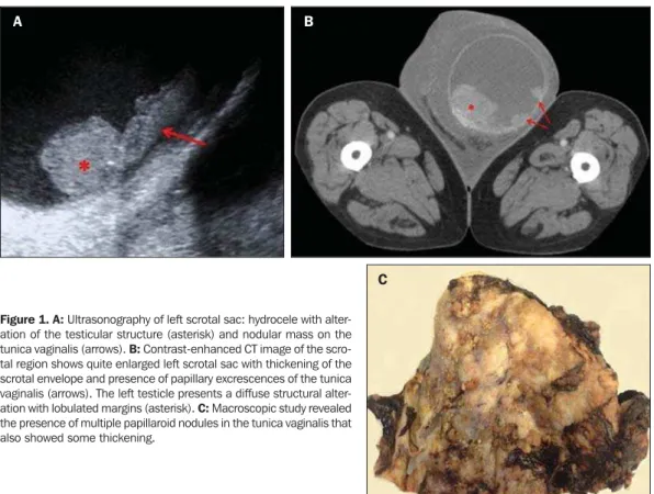
We present the case of an 82-year-old male patient who at-tended our hospital emergency department complaining of pain and enlargement of his left scrotal sac. The patient reported pro-gressive scrotal enlargement evolving over more than 20 years. He did not report any exposure to asbestos. Physical examination revealed an enlarged scrotal sac with increased temperature and testicles not easily palpable. Complete blood count revealed leu-kocytosis with neutrophilia (11.37 × 10³
/µL).
Testicular ultrasonography was performed, showing left hy-drocele with approximately 1100 mL in volume and dense echoes inside, making it difficult to evaluate the testis. The right testis
was displaced upwards, toward the inguinal canal. Given the im-possibility of performing an adequate examination of the left tes-tis, we performed a CT scan of the testicular region. The scan revealed a large hydrocele with dense contents. The left testis showed a diffuse alteration of its structure with a lobulated mar-gin and nodular thickening of the tunica vamar-ginalis. During his hospital stay, the patient presented with cardiovascular instability (arterial pressure of 85/53 mmHg) and was submitted to emer-gency left orchiectomy with surgical drainage of the abscess, causing a complicated hydrocele.
Anatomopathological analysis led to the diagnosis of malig-nant mesothelioma of the tunica vaginalis testis that largely infil-trated the tunica vaginalis, testicular parenchyma and the rest of paratesticular structures (epididymis and rete testis). Immunohis-tochemical study showed: calretinin (+), WT1 (+), CK7 (+), EMA (+) and p53 (+).
Figure 1.A: Ultrasonography of left scrotal sac: hydrocele with alter-ation of the testicular structure (asterisk) and nodular mass on the tunica vaginalis (arrows). B: Contrast-enhanced CT image of the scro-tal region shows quite enlarged left scroscro-tal sac with thickening of the scrotal envelope and presence of papillary excrescences of the tunica vaginalis (arrows). The left testicle presents a diffuse structural alter-ation with lobulated margins (asterisk). C: Macroscopic study revealed the presence of multiple papillaroid nodules in the tunica vaginalis that also showed some thickening.
A B