Imaging diagnosis and clinical findings of cerebral
venous thrombosis in full-term neonates without brain
damage: a ten-year review*
Diagnóstico por imagem e aspectos clínicos da trombose venosa cerebral em recém-natos a termo sem dano cerebral: revisão em 10 anos
Alexandra Maria Vieira Monteiro1, Claudio Marcio Amaral de Oliveira Lima2, Érica Barreiros Ribeiro3, Maria Cristina Lins4, Silvia Miranda5, Luis Eduardo Miranda6
OBJECTIVE: To describe and compare imaging methods and clinical findings of cerebral venous thrombosis in four full-term neonates without brain damage, admitted to a neonatal intensive care unit. MATERIALS AND METHODS: Ten-year review of four cases diagnosed with cerebral venous thrombosis by transfontanellar ultrasonography associated with Doppler fluxometry and confirmed by magnetic resonance imaging/magnetic resonance angiography in correlation with clinical findings and neurological progression. RESULTS: Ultrasonography presented normal results in 75% of cases and magnetic resonance imaging in 100%. Doppler fluxometry and magnetic resonance angiography were abnormal in 100% of cases. Hypoxia (100%) and early seizures (100%) were predominant among clinical findings with evoked potential changes in 50% of cases. In the assessment of the neurodevelopment all the areas remained within normality parameters up to the conclusion of the present study. CONCLUSION: Ultrasonography in association with Doppler can identify changes related to cerebral venous thrombosis and should be complemented with magnetic resonance imaging that is the gold standard for diagnosis in these cases.
Keywords: Cerebral venous thrombosis; Full-term neonates; Magnetic resonance imaging; Magnetic resonance
angiography; Ultrasonography and Doppler.
OBJETIVO: Descrever e comparar os métodos de imagem e os aspectos clínicos em quatro recém-natos a termo diagnosticados como trombose venosa cerebral, sem dano encefálico, adscritos a uma unidade de terapia intensiva neonatal. MATERIAIS E MÉTODOS: Revisão em 10 anos com quatro casos diagnosticados como trombose venosa cerebral por meio de ultrassonografia transfontanela com Doppler e confirmados por ressonância magnética/angiorressonância, correlacionados aos aspectos clínicos e evolução neurológica. RESULTADOS: A ultrassonografia foi normal em 75% dos casos e a ressonância magnética, em 100%. No caso alterado, a dilatação venosa foi identificada. O Doppler e a angiorressonância estavam alterados em 100% dos casos. Dos aspectos clínicos, a hipóxia (100%) e a convulsão precoce (100%) predominaram, com potencial evocado alterado em 50% dos casos. Na avaliação do neurodesenvolvimento, todas as áreas estiveram dentro da normalidade até a última avaliação. CONCLUSÃO: A ultrassonografia associada ao Doppler é capaz de identificar as alterações da trombose venosa cerebral, devendo ser complementada com a resso-nância magnética, que é o padrão ouro de diagnóstico.
Unitermos: Trombose venosa cerebral; Recém-nascidos a termo; Imagem por ressonância magnética;
Angior-ressonância; Ultrassonografia e Doppler. Abstract
Resumo
* Study developed at Casa de Saúde São José (CSSJ-RJ), Rio de Janeiro, RJ, Brazil.
1. PhD, Associate Professor of Radiology at Faculdade de Ciências Médicas da Universidade do Estado do Rio de Janeiro (FCM-UERJ), MD, Radiologist at Casa de Saúde São José (CSSJ-RJ), Rio de Janeiro, RJ, Brazil.
2. MD, Radiologist at Centro de Diagnóstico por Imagem Fá-tima Digittal, Nova Iguaçu, RJ, Brazil.
3. MD, Radiologist at Unit of Magnetic Resonance Imaging of Hospital São José, Criciúma, SC, Brazil.
4. MD, Neonatologist at the Neonatal Intensive Care Unit of Casa de Saúde São José (CSSJ-RJ), Rio de Janeiro, RJ, Brazil. 5. MD, Pediatric Neurologist at the Neonatal Intensive Care Unit of Casa de Saúde São José (CSSJ-RJ), Rio de Janeiro, RJ, Brazil.
with an incidence of less than one case for every 100 thousand newborns per year(1,2).
This condition has been increasingly diag-nosed mainly because the huge develop-ments in diagnostic imaging methods. The most frequent predisposing factor for the development of CVT is hypoxic-ischemic encephalopathy (HIE), with a considerable morbimortality rate(3–6). Clinically, CVT
signs and symptoms are nonspecific and may determine delay or failure in the
diag-Monteiro AMV, Lima CMAO, Ribeiro EB, Lins MC, Miranda S, Miranda LE. Imaging diagnosis and clinical findings of cerebral venous thrombosis in full-term neonates without brain damage: a ten-year review. Radiol Bras. 2010;43(3):149–153.
6. MD, Neonatologist, Head for the Neonatal Intensive Care Unit of Casa de Saúde São José (CSSJ-RJ), Rio de Janeiro, RJ, Brazil.
Mailing address: Dra. Alexandra Maria Vieira Monteiro. HUPE-UERJ. Avenida Vinte e Oito de Setembro, 77, térreo, sala 126, Vila Isabel. Rio de Janeiro, RJ, Brazil, 20551-030. E-mail: [email protected] / [email protected]
Received March 16, 2010. Accepted after revision April 5, 2010.
INTRODUCTION
nosis(7). In approximately 85% of cases, the
thrombosis affects either the superior sag-ittal sinus or the transverse sinus(8–10).
Among the imaging methods, transfon-tanellar ultrasonography (TFUS) associ-ated with Doppler fluxometry is the initial evaluation method, particularly because of the wide availability and bed-side capabil-ity, with low cost, and non-necessity of sedation or ionizing radiation exposure, but magnetic resonance imaging (MRI) re-mains as the gold standard for the diagno-sis of CVT(5,6,9,11,12).
The present study was aimed at describ-ing the imagdescrib-ing finddescrib-ings, compardescrib-ing the methods used in the neonatal period, as well as describing the clinical findings and the neurological progression in four term neonates diagnosed with CVT, with no brain damage, admitted to a neonatal inten-sive care unit over a 10-year period.
MATERIALS AND METHODS
In the period from May 1999 to May 2009, 2,865 neonates were admitted to the Neonatal Intensive Care Unit (ICU) of Casa de Saúde São José (CSSJ) in Rio de Janeiro, RJ, Brazil. Among these neonates, 2,547 were submitted to TFUS as a routine procedure of the ICU, or for different clini-cal indications, with 10% of such studies being associated with Doppler fluxometry
due to a precise indication. In this group, nine full-term neonates were diagnosed with CVT. Two cases of CVT associated with bacterial meningitis, and three asso-ciated with the presence of a cerebral lesion were excluded. All of the four selected cases were submitted to magnetic reso-nance angiography (MR angiography) for diagnosis confirmation. All the neonates underwent evoked auditory potential study to evaluate their degree of auditory loss associated with CVT.
All the neonates were male, with gesta-tional age ranging between 37 and 41 weeks (mean, 39 weeks), weight ranging between 3,010 and 4,085 g (mean weight, 3,548 g), and Apgar score at the first minute between 2 and 8 (mean, 5), and at the fifth minute between 7 and 9 (mean, 8).
Also, risk factors for CVT were evalu-ated according to the following classifica-tion: neonatal risks (perinatal hypoxia and sepsis), and maternal risks (diabetes and in-fection, including infections of any etiol-ogy, in any region or organ, during the pre-natal period). The clinical evaluation of the neonates was based on the presence, or not, of early (up to 24 hours of life) or late on-set seizures, and on the presence of respi-ratory disorders. All the neonates were evaluated by a single pediatric neurologist from their admission to the ICU up to dis-charge or follow-up abandonment.
A Sonoline Versa-Pro ultrasonography unit (Siemens Medical Systems; Erlangen, Germany), with a convex, multifrequency transducer was utilized. Transfontanellar US with Doppler fluxometry was per-formed through the anterior fontanelle, with acquisition of images in the coronal and sagittal planes. Resistance index (RI) was evaluated for both the right and left middle cerebral artery, anterior cerebral artery, and basilar artery, and was consid-ered as altconsid-ered in the setting of increased RI in at least two vessels. The venous flow was mapped with color Doppler. MR an-giography was performed in a Harmony, 1 tesla unit (Siemens Medical Systems; Erlangen, Germany). The neonates re-ceived sedation with chloral hydrate as necessary, and in none of the cases, para-magnetic contrast agent was intravenously administered. The imaging evaluation was aimed at identifying and localizing the thrombosis, its association with parenchy-mal lesions, and the presence, or not, of hydrocephalus.
RESULTS
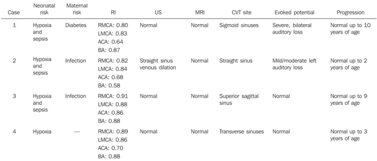
Table 1 consolidates the clinical data and imaging findings.
The neonatal risk factors observed were associated hypoxia and sepsis in three cases (75%) and hypoxia alone as a single risk
Table 1 Clinical and imaging findings.
Case
1
2
3
4
Neonatal risk
Hypoxia and sepsis
Hypoxia and sepsis
Hypoxia and sepsis
Hypoxia
Maternal risk
Diabetes
Infection
Infection
—
RI
RMCA: 0.80 LMCA: 0.83 ACA: 0.64 BA: 0.87
RMCA: 0.82 LMCA: 0.84 ACA: 0.68 BA: 0.58
RMCA: 0.91 LMCA: 0.88 ACA: 0.86 BA: 0.88
RMCA: 0.89 LMCA: 0.86 ACA: 0.70 BA: 0.88
US
Normal
Straight sinus venous dilation
Normal
Normal
MRI
Normal
Normal
Normal
Normal
CVT site
Sigmoid sinuses
Straight sinus
Superior sagittal sinus
Transverse sinuses
Evoked potential
Severe, bilateral auditory loss
Mild/moderate left auditory loss
Normal
Normal
Progression
Normal up to 10 years of age
Normal up to 2 years of age
Normal up to 9 years of age
Normal up to 3 years of age
factor in one case. As regards maternal risks, the most frequent one was infection in two cases (50%) and diabetes in one (25%). In one case no maternal risk factor was identified.
The predominant clinical sign was early seizure onset observed in 100% of cases. Respiratory failure ocurred in the cases 1, 2 and 4, and apnea in one case (25%).
The response to evoked auditory poten-tial ranged from normal in two cases (50%), mild/moderate, unilateral auditory loss in one case (25%) and severe, bilateral loss in another case (25%).
TFUS was normal in three cases (75%). In the case 2, TFUS identified hypoechoic tubular dilatation diagnosed as venous di-lation and absent flow at color Doppler
mapping of the straight sinus (Figure 1). Also, the venous Doppler mapping identi-fied flow absence in the superior sagittal sinus, confirmed by MR angiography in the case 3 (Figure 2). In case 1, ectatic ventricu-lar system was observed (Figure 3).
Altered RI in at least two vessels was observed in all the cases, with values rang-ing between 0.80 and 0.91 (mean, 0.85) for
Figure 1.Case 2. US-Dop-pler, sagittal image (A,B) demonstrating venous dila-tion and failure in vessels filling in the straight sinus (arrows). On C, sagittal MRI T1-weighted image demon-strating hypersignal in the superior sagittal sinus and straight sinus (arrows).
Figure 3. Case 1. US coro-nal image (A) demonstrat-ing ectatic ventricular sys-tem (white arrows), also demonstrated at axial MRI T1-weighted sequence (B), besides hypersignal at the sigmoid sinuses topography (black arrows). On C, MR angiography TOF sequence demonstrating failure in ves-sels filling in the right sig-moid sinus (arrow).
the right middle cerebral artery; between 0.83 and 0.88 (mean, 0.85) for the left middle cerebral artery; between 0.64 and 0.86 (mean, 0.75) for the anterior cerebral artery; and between 0.58 and 0.88 (mean, 0.73) for the basilar artery. The cases with low values for RI were attributed to a mechanism of cerebral self-regulation as a function of arterial redistribution resulting from venous congestion secondary to the thrombosis.
Morphological brain MRI was normal in four cases (100%). Cerebral venous thrombosis was diagnosed at T1-weighted sequences complemented by MR angiog-raphy.
All the sinuses were isolatedly involved. As regards their neurodevelopment, all the neonates progressed within the normal-ity parameters until their discharge or fol-low-up abandonment.
DISCUSSION
The global CVT incidence in children is of 0.67/100,000 children per year, and neonates constitute the most affected group(2,13,14). A retrospective study
devel-oped from 1999 to 2006 by Teksam et al.(13)
with 71 children divided into three groups (neonates, 0–28 days; infants, 29 days–1 year; children/teenagers, 1–18 years) and diagnosed by computed tomography (CT), CT angiography MRI and MR angiogra-phy, reported 48% incidence of CVT in neonates, confirming that this group is more prone to CVT than any other pediat-ric age range, in spite of the fact that the true incidence rate is still unknown because of the scarcity of reliable epidemiological data(13). It is believed that neonates
propen-sity to CVT is a result from a failure of protective mechanisms caused by cerebral immaturity, besides the presence of physi-ological prothrombotic factors and peri-and/or postnatal diseases(14,15).
According to the literature, CVT is most frequent in male neonates(7,16), in a studied
population of 32 neonates(16), similarly to
the results of the present study.
Risk factors for CVT can be classified into neonatal and maternal. deVeber et al.(2), in a study with 160 children (60
neo-nates and 91 non-neoneo-nates) with ages rang-ing from NN (gestational age > 36 weeks)
to 18 years, have reported the presence of any acute systemic disease in 84% of neo-nates with CVT and, among them, about 51% and 30% present, respectively, perina-tal complications and dehydration as a risk factor for CVT. The most frequent compli-cation was hypoxia in 30 cases, besides premature membrane rupture, maternal in-fection and gestational diabetes(2). The
re-sults of the present study are in agreement with the literature, both in terms of neona-tal and maternal risks. In 25% of cases, no maternal risk factor for CVT was observed. It is interesting to note that all the patients were diagnosed with perinatal asphyxia and precociously admitted to the neonatal ICU where, probably, the associated risk factor has been hypoxia and not infection. There-fore, perinatal complications inexorably constitute the most frequent and relevant risk factor for CVT in neonates, probably resulting from the lack of protective mechanisms as a function of immaturity(13).
The clinical presentation is highly vari-able and nonspecific in this age range, and the diversity of associated predisposing factors make the diagnosis difficult and fre-quently delayed(13). Additionally, the
clini-cal condition is influenced by the child age, extent and site of the thrombosis. Also, ac-cording to deVeber et al.(2), 72% of
neo-nates in their study presented some kind of seizure resulting from (HIE) and/or CVT. Besides seizures, most neonates also present some diffuse neurological sign such as agitation or lethargy. This same study shows that 67% of neonates have not presented any focal neurological deficit; on the other hand, hemiparesis, as a clinical sign, has occurred in only 6% of cases, and cranial nerve palsy in 7%. The absence or scarcity of focal signs is explained by the cerebral immaturity. In agreement with the literature, the present study observed the presence of convulsive seizures in all the patients.
In the setting of thrombosis, the intralu-minal venous pressure increases, reducing the blood flow and the cerebral perfusion pressure, which induces the hematoence-phalic barrier disruption and frequently results in vasogenic edema and hydroceph-alus(17). In the multicentric study developed
by Wasay et al.(1), from 1992 to 2001, with
25 neonates with at least two imaging
methods for diagnosis of CVT, hydroceph-alus was present in 10% of cases. In the present study, the inclusion criterion was exactly the absence of associated encepha-lic lesion.
In approximately 85% of cases, throm-bosis affects the superior sagittal sinus or the transverse sinus(1,17) and in the other
15%, the other sinuses of the dura mater and deep veins of the Galen’s system(2,14,18).
The involvement of multiple venous si-nuses may be present in more than 70% of cases(1,19). The results of the present study
presented isolate involvement of venous sinuses.
The brainstem auditory evoked re-sponse test allows the evaluation of the functional integrity of the auditory tract with several clinical applications, particu-larly as a method for auditory screening in high-risk neonates, because of its objectiv-ity, rapidobjectiv-ity, noninvasiveness, bed-side ca-pabilities and applicability to any age range(20,21). It is estimated that, in Brazil,
three to four in every 1,000 children have congenital deafness. This ratio increases to two–four in every 100 neonates when these children come from an ICU. Deafness as-sociated with the occurrence of thrombo-sis was described by Perlman et al.(22), one
of the first authors to demonstrate the co-chlear sensitivity to a vascular event. Cur-rently, many studies suggest the existence of a cochlear vessels spasm or increase in blood viscosity associated to vascular events, like in thrombosis, to explain the temporary deafness(21,23). In the present
study, in 50% of cases there was an audi-tory loss ranging from mild to severe.
TFUS with Doppler fluxometry pre-sents great advantages, principally the noninvasiveness to measure blood flow velocity in intracranial vessels and to evalu-ate physiopathological changes in the ce-rebral hemodynamics, besides the bed-side capability, low cost and non-necessity of sedation and of ionizing radiation expo-sure(24–27). Several studies on TFUS
Dop-pler fluxometry have demonstrated changes of RI in pathological conditions as, for example, hypoxia with presence of re-versed diastolic flow, inclusive, reflecting the increased vascular resistance as a result from edema(17,24). The results of the present
considering that the increased RI values were observed in all the studied vessels.
MR angiography is the most sensitive and specific method for the diagnosis of CVT and, as combined with MRI is con-sidered as gold standard(12,28,29). In
agree-ment with the literature, MR angiography was diagnostic for CVT in 100% of cases in the present study.
According to Teksam et al.(13), in a study
with 34 neonates with CVT diagnosed by CT, CT angiography, MRI and MR angiog-raphy, 62% of neonates with CVT pre-sented an association with parenchymal lesion, with variable location and a pre-dominantly hemorrhagic pattern (76% of cases), but with smaller extent as compared with lesions in older children, and with higher frequency in the frontal and parietal lobes. Additionally, the parenchymal lesion is, in general, correlated with the involved territory of venous drainage (76% of cases). In the present study, association with paren-chymal lesion was not observed.
The literature reports a survival rate of more than 90% among NN with associated cerebral lesion(14). A Canadian longitudinal
study covering an eight-year period reports 8% survival with presence of neurological deficit in 61%, seizures in 21%, and recur-rent cerebral thrombosis in 9%(14). Yet,
some authors report that the best estimate was observed after 2,1 years, as 77% of neonates diagnosed with CVT were neuro-logically normal, although the absence of association with parenchymal lesion has not been highlighted(2).
As regards the evaluation of the neuro-logical development, all the areas remained within the normality parameters up to the abandonment of the follow-up or contact with the clinician.
CONCLUSION
In the neonatal period, combined TFUS and Doppler fluxometry allows the early
diagnosis of CVT, which can be confirmed by MRI/ MR angiography, that is the gold standard for the diagnosis of this severe disease. The absence of associated paren-chymal lesion may, prospectively, represent the absence of a progressive cerebral dam-age.
REFERENCES
1. Wasay M, Dai AI, Ansari M, et al. Cerebral venous sinus thrombosis in children: a multicenter cohort from the United States. J Child Neurol. 2008; 23:26–31.
2. deVeber G, Andrew M, Adams C, et al. Cerebral sinovenous thrombosis in children. N Engl J Med. 2001;345:417–23.
3. De Lorenzi DRS, Tanaka ACd’A, Bozzetti MC, et al. A natimortalidade como indicador de saúde perinatal. Cad Saúde Pública. 2001;17:141–6.
4. deVeber G, Chan A, Monagle P, et al. Anticoagu-lation therapy in pediatric patients with sino-venous thrombosis: a cohort study. Arch Neurol. 1998;55:1533–7.
5. Rivkin MJ. Hypoxic-ischemic brain injury in the term newborn. Neuropathology, clinical aspects, and neuroimaging. Clin Perinatol. 1997;24:607– 25.
6. Silva GS, Almeida CMO, Félix EPV, et al. Trom-bose venosa cerebral e homocistinúria: relato de caso. Arq Neuropsiquiatr. 2001;59:815–6. 7. Sébire G, Tabarki B, Saunders DE, et al. Cerebral
venous sinus thrombosis in children: risk factors, presentation, diagnosis and outcome. Brain. 2005;128:477–89.
8. Khurana DS, Buonanno F, Ebb D, et al. The role of anticoagulation in idiopathic cerebral venous thrombosis. J Child Neurol. 1996;11:248–50.
9. Marques MCP, Pires LA, Damasceno CA, et al. Trombose da veia de Galeno: relato de caso. Arq Neuropsiquiatr. 2003;61:285–7.
10. McMurdo SK Jr, Brant-Zawadzki M, Bradley WG Jr, et al. Dural sinus thrombosis: study us-ing intermediate field strength MR imagus-ing. Ra-diology. 1986;161:83–6.
11. Lee BCP, Park TS, Kaufman BA. MR angiogra-phy in pediatric neurological disorders. Pediatr Radiol. 1995;25:409–19.
12. Brito AR, Vasconcelos MM, Domingues RC, et al. Pseudotumor cerebral secundário a trombose venosa dural: relato de caso pediátrico. Arq Neu-ropsiquiatr. 2005;63:697–700.
13. Teksam M, Moharir M, deVeber G, et al. Fre-quency and topographic distribution of brain le-sions in pediatric cerebral venous thrombosis. AJNR Am J Neuroradiol. 2008;29:1961–5. 14. Andrew ME, Monagle P, deVeber G, et al.
Throm-boembolic disease and antithrombotic therapy in
newborns. Hematology Am Soc Hematol Educ Program. 2001:358–74.
15. Klein L, Bhardwaj V, Gebara B. Cerebral venous sinus thrombosis in a neonate with homozygous prothrombin G20210A genotype. J Perinatol. 2004;24:797–9.
16. Golomb MR, Dick PT, MacGregor DL, et al. Neonatal arterial ischemic stroke and cerebral sinovenous thrombosis are more commonly diag-nosed in boys. J Child Neurol. 2004;19:493–7.
17. Heinz ER, Provenzale JM. Imaging findings in neonatal hypoxia: a practical review. AJR Am J Roentgenol 2009;192:41–7.
18. Ferreira CS, Pellini M, Boasquevisque E, et al. Alterações parenquimatosas na trombose venosa cerebral: aspectos da ressonância magnética e da angiorressonância. Radiol Bras. 2006;39:315–21.
19. Huang BY, Castilllo M. Hypoxic-ischemic brain injury: imaging findings from birth to adulthood. Radiographics. 2008;28:417–39.
20. Lourenço EA, Oliveira MH, Umemura A, et al. Audiometria de resposta evocada de acordo com sexo e idade: achados e aplicabilidade. Rev Bras Otorrinolaringol. 2008;74:545–51.
21. Maia RA, Cahali S. Surdez súbita. Rev Bras Otor-rinolaringol. 2004;70:238–48.
22. Perlman HB, Kimura R, Fernandez C. Experi-ments on temporary obstruction of the internal auditory artery. Laryngoscope. 1959;69:591–613.
23. Barreira-Nielsen C, Futuro Neto HA, Gattaz G. Processo de implantação de programa de saúde auditiva em duas maternidades públicas. Rev Soc Bras Fonoaudiol. 2007;12:99–105.
24. Garcia MHM, Monteiro AMV, Freire SM. Rela-ção entre o índice de resistência obtido pela ul-tra-sonografia Doppler transfontanela e o neuro-desenvolvimento até o primeiro ano de vida em recém-nascidos a termo com encefalopatia hipó-xico-isquêmica leve e moderada. Arq Neuropsi-quiatr. 2007;65:1206–10.
25. Mello RR, Meio MDBB, Morsch DS, et al. Ul-tra-sonografia cerebral neonatal normal no prema-turo – é possível tranqüilizar os pais? J Pediatr (Rio J). 1999;75:45–9.
26. Taylor GA. Recent advances in neonatal cranial ultrasound and Doppler techniques. Clin Perinatol. 1997;24:677–91.
27. Ipsiroglu OS, Eichler F, Stoeckler-Ipsiroglu S. Cerebral Doppler sonography of the neonate. A résumé after 20 years and future aspects. Clin Perinatol. 1999;26:905–46.
28. Gasparetto EL. Trombose venosa cerebral [edi-torial]. Radiol Bras. 2006;39(5):iii.