Preeclampsia
Pré-eclâmpsia
José Geraldo Lopes Ramos
1Nelson Sass
2Sérgio Hofmeister Martins Costa
11Universidade Federal do Rio Grande do Sul, Porto Alegre, RS, Brazil 2Universidade Federal de São Paulo, São Paulo, SP, Brazil
Rev Bras Ginecol Obstet 2017;39:496–512.
Address for correspondence José Geraldo Lopes Ramos, MD,
Professor, Universidade Federal do Rio Grande do Sul, Ramiro Barcelos, 2400, Santa Cecília, Porto Alegre, RS, Brazil (e-mail: [email protected]).
Highlights
• New concepts of diagnosis and risk for preeclampsia; • Guidelines for preeclampsia prevention treatment; • Prediction model of severe maternal outcomes in
pre-eclampsia (fullPIERS) 12.
Introduction
The hypertensive syndromes that occur during pre-gnancy, especially preeclampsia (PE), result in real risk and significant impact on indicators related to maternal and child health. These syndromes are causal factors related to maternal and perinatal death, and they cause definitive limitations to maternal health and serious problems resulting from associated elective pre-maturity. In Brazil, PE is the main cause of elective prematurity.
Keywords
►
pregnancy arterial
hypertension
►
preeclampsia
►
HELLP syndrome
►
high risk pregnancy
►
pregnancy
complications
Abstract
The authors review hypertensive disease during pregnancy with an academic and practical
view, and using the best evidence available. This disease, which is the most important
clinical disease in Brazilian pregnant women, may have its incidence reduced with
prevention through the use of calcium and aspirin in pregnant women at risk. Previously,
it was a disease that presented with
hypertension with proteinuria,
but it has now been
classi
fi
ed with new clinical parameters besides proteinuria. Morbidity and mortality should
be reduced in a continental country such as Brazil using protocols for the early treatment of
complications by calculating severe outcomes in preeclampsia. The early treatment of
acute hypertension, use of magnesium sulfate and early hospitalization in cases of
preeclampsia are concepts to pursue the reduction of our pregnant women
’
s mortality.
Palavras-chave
►
hipertensão arterial
na gestação
►
pré-eclâmpsia
►
síndrome HELLP
►
gestação de alto risco
►
complicações na
gravidez
Resumo
Os autores revisam a doença hipertensiva na gestação com uma visão acadêmica e
prática, utilizando as melhores evidências disponíveis. A doença clínica mais
impor-tante na gesimpor-tante brasileira pode ter sua incidência diminuída com a prevenção por
meio do uso de cálcio e aspirina em gestantes de risco. Antes uma doença que
apresentava hipertensão
arterial com proteinúria,
agora vem sendo classi
fi
cada com
novos parâmetros clínicos além da proteinúria. A morbidade e mortalidade devem ser
diminuídas, em um país continental como o Brasil, utilizando-se protocolos para o
tratamento precoce de suas complicações mediante o cálculo de desfechos graves em
pré-eclâmpsia. O tratamento precoce da hipertensão arterial, o uso do sulfato de
magnésio e a internação precoce em casos de pré-eclâmpsia são conceitos para
perseguirmos a diminuição da mortalidade de nossas gestantes.
This review is part of the Series, Guidelines and Recommendations of the Federação Brasileira das Associações de Ginecologia e Obstetrícia– FEBRASGO, and was prepared by the National Specialized Commission for the Hypertension in Pregnancy.
received
December 5, 2016
accepted
March 29, 2017
published online
August 9, 2017
DOI https://doi.org/ 10.1055/s-0037-1604471.
ISSN 0100-7203.
Copyright © 2017 by Thieme Revinter Publicações Ltda, Rio de Janeiro, Brazil
There is no accurate information on the incidence of preeclampsia worldwide, but it is estimated to occur in 3–5% of pregnancies. Specifically in Brazil, a systematic review identified an incidence of 1.5% for PE and of 0.6% for eclampsia.1Certainly, information concerning Brazil is still underestimated, and definitely varies according to the country’s regions. A Brazilian study2 reports that the estimated prevalence of eclampsia is of 0.2% in the most developed areas, with a maternal death rate of 0.8%, whereas in less favored regions this prevalence rises to 8.1%, with a maternal mortality rate corresponding to 22.0%.
The aim of this text is to sensitize health providers about the magnitude of the problem, recognize local specificities, and adopt interventions based on the best scientific evi-dence available to develop strategies for prevention, early detection of the disease, and reduction of maternal and perinatal harm.
Pathophysiological Foundations
Some evidence supports the hypothesis of maternal im-mune system involvement in the disease. In case there are problems of immunological adaptation to the trophoblast, there will be problems in trophoblast perfusion, with consequent hypoxia. These primary alterations would trig-ger a series of local hypoxia phenomena, and reoxygenation could amplify the local effects, such as the formation of oxygen-reactive species, activation of the maternal infl am-matory system, and acceleration of cellular apoptosis pro-cesses that would limit the establishment of normal placentation and imbalance between pro-angiogenic fac-tors, such as the vascular endothelial growth factor (VEGF) and the placental growth factor (PlGF), and soluble anti-angiogenic factors such as the soluble fms-like tyrosine kinase-1 (sFLT-1), with predominance of the latter, result-ing in generalized activation of the maternal inflammatory system, universal endothelial dysfunction, and limited pla-cental vascularization.3,4
Universal arteriolar spasm due to endothelial activation results in an insidious and progressive process, culminating in multiple organ insufficiency. Preeclampsia should be interpreted as a chronic disease with potential for progres-sive multiple organ failure. This evolutionary character must be taken into account, as well as its unpredictability and clinical instability in decisions. Endothelial activation basi-cally determines: vasoconstriction and consequent increase in peripheral resistance; changes in capillary permeability, which are responsible for edema; and activation of the coagulation system.
The kidneys suffer from anatomopathological patterns (glomerular endotheliosis and focal sclerosis), with conse-quent proteinuria and impairment of the glomerularfi ltra-tion. In the liver, ischemia occurs with varying intensity, leading to dysfunction with elevated levels of transaminases. Focal or confluent edema and/or hemorrhage distend the capsule, and may result in hepatic rupture with massive bleeding.
Vasospasm hinders the uteroplacental bloodflow with varying intensity, depending on the moment of the process and on the existence of a chronic pre-existing injury. Regarding coagulation, there is activation and consumption of platelets with progressive consumption and disseminat-ed coagulation. The brain can be affectdisseminat-ed by ischemia aggravated by diffuse edema, resulting in seizure (eclamp-sia) or stroke. Patients presenting severe conditions, par-ticularly eclampsia, should receive differentiated care, given the progressive functional limitation of multiple organs.
De
fi
nitions of Hypertensive States during
Pregnancy
The expansion of pathophysiological knowledge has resulted in the expansion of clinical possibilities to define PE. How-ever, the recommendations adopted by the International Society for the Study of Hypertension in Pregnancy (ISSHP)5 remain. According to such recommendations, arterial hyper-tension is characterized when the systolic blood pressure (SBP) is140 mm Hg and/or the diastolic blood pressure (DBP) is
90 mm Hg, considering thefifth Korotkoff sound (silence). For the measurement, the patient should sit and place one of the forearms at the height of the atrium (half of the external bone); the measurement should be repeated in one or two fi ve-minute intervals. The usually available cuffs are used for readings in arms with perimeter around 30 cm. Obese patients need appropriate cuffs or correction tables according to their brachial perimeter.
Protein loss of 300 mg or more in 24-hour urine specimen collection should be considered for the definition of protein-uria. For more agility in the diagnosis, evaluations in an isolated sample of urine with proteinuria/creatininuria (P/C) ratio (both in mg/dL)0.3 are considered adequate. In the
absence of such diagnostic possibilities, proteinuria with at least 1þ reagent tape may be considered as long as the
quality of the method is assured. Differently from previous recommendations, the intensity of the proteinuria should no longer be associated with the maternal prognosis, nor be the only aspect to guide decisions.
Preeclampsia is defined as arterial hypertension identi-fied for thefirst time after the 20th week associated with proteinuria, and it may overlap with another hypertensive state. Taking into account the current concept of PE syn-drome, rigid concepts have been abandoned.6Thus, in the absence of proteinuria, the diagnosis of PE may be based on the presence of headache, visual turbidity, abdominal pain or altered laboratory tests, such as thrombocytopenia (less than 100,000/mm3), hepatic enzyme elevation (double the basal), renal impairment (>1.1 mg/dL or double the baseline), or
pulmonary edema and visual or brain disorders such as headache, scotomas, or convulsions.
preeclampsia is considered hypertension after the twentieth week and one of the following criteria:
1. Significant proteinuria (P/C ratio>0.3;>1.0 g/L on
re-agent tape);
2. Maternal organic dysfunctions;
• Loss of renal function (creatinine>1.02 mg/dL);
• Hepatic dysfunction (increase of transaminases by
>2 times the normal upper limit; epigastralgia);
• Neurologic complications (altered mental state; blind-ness; hyperreflexia with clonus, scotomas, visual blur-ring, diplopia, Doppler of maternal ophthalmic artery with peak ratio>0.78);
• Hematologic complications (thrombocytopenia, dissem-inated intravascular coagulation [DIC]<hemolysis);
• Antiangiogenesis status (PlGF<36 pg/mL or sFlt-1/
PlGF ratio>85).
3. Uteroplacental dysfunction (asymmetric intrauterine growth restriction [IUGR]; altered umbilical Doppler, especially if the Doppler is altered in both maternal uterine arteries).
When PE occurs in pregnant women with chronic hy-pertension, it is considered overlapping preeclampsia. Se-vere preeclampsia is defined as PE associated with severe enough maternal-fetal complications to pose imminent risk of maternal-fetal impairment. Persistent SBP160 mm
Hg or DBP110 mm Hg or presence of any of the criteria
listed in►Table 1characterize a pregnant woman as having
severe PE. In general, pregnant women with signs or symptoms of severe PE have a decompensated disease that may rapidly progress to maternal and perinatal mor-bidity and mortality. Proteinuria levels should not be considered criteria of severity in PE.7,8 The presence of PE, regardless of its severity, entails increased fetal and maternal risk. Eclampsia is the occurrence of generalized motor seizures (grand mal seizures) in pregnant women with PE that are not caused by coincident neurological disease and may occur in the prepartum period (50%), during delivery (20%), and in the postpartum period (be-tween 11 and 44%).
Classi
fi
cation
There are several classifications described for hypertensive disorders in pregnancy. In 2014, the ISSHP reviewed the classification of hypertensive disorders during pregnancy (►Table 2).5
Significant Proteinuria
It is the excretion of 300 mg or more of proteins in a 24-hour urine collection. The 24-hour collection is subject to many collection and storage errors, and should not be used for clinical purposes unless 24-hour creatinine clearance is also
Table 1 Severe complications of preeclampsia
Affected organic system Adverse conditions Severe complications indicating termination
of pregnancy
Central nervous system Headache; visual symptoms
Eclampsia; PRES; cortical blindness; Retinal de-tachment; Glasgow scale<13; TIA; stroke;
RND.
Cardiorespiratory Chest pain; dyspnea; saturation O2<97%
Severe uncontrolled hypertension (for a 12-hour period despite maximum doses of hypotensive agents); SO2<90%, need for O2>50%
for>1 hour, intubation, support with
vasoac-tive drugs; pulmonary edema; myocardial ischemia or infarction.
Hematological Leukocytosis; thrombocytopenia; high INR PTT
Platelets<50.000/dL;need for transfusion of
any blood product.
Renal Elevated creatinine and uric acid ARF (creatinine>1.5 mg/dL without previous
renal disease);
need for dialysis (without previous CRF).
Hepatic Nausea; vomiting epigastralgia;
URQ pain; SGOT; SGTP; LDH; elevated bilir-ubin; low plasma albumin
Hepatic impairment (INR>2 in absence of DIC
or use of warfarin); hepatic hematoma with or without rupture.
Fetoplacental Non-reactive CTG; Oligohydramnios; IUGR; Doppler of umbilical artery with absent or reversed diastolicflow
PA; reversed a-wave in ductus venous; fetal death.
Abbreviations: ARF, acute renal failure; CRF, chronic renal failure; CTG, cardiotocography; DIC, disseminated intravascular coagulation; INR; IUGR, intrauterine growth restriction; LDH, lactate dehydrogenase; PA, placental abruption; PPT, prothrombin time; PRES, posterior reversible encephalopathy syndrome; RND, reversible neurological deficit; SGOT, serum glutamic oxaloacetic transaminase; SGTP, serum glutamic pyruvic transaminase; TIA, transient ischemic attack; URQ, upper right quadrant of the abdomen.
Note:Platelets
<100.000 are considered as indication of interruption of pregnancy.
measured to assess the adequacy of the collection.5 The measurement of the P/C ratio in the urine sample has been of clinical utility, and values0.3 demonstrate a good
correlation with significant proteinuria. A P/C ratio in an isolated sample of urine0.3 corresponds to significant
proteinuria 92% of times, and a ratio0.5 corresponds to
significant proteinuria 100% of times.9The presence of 1.0 g/l or more of proteins on the reagent tape strongly suggests significant proteinuria.
Chronic Arterial Hypertension
Chronic arterial hypertension in pregnancy is the occurrence of systemic arterial hypertension (SAH) preceding pregnancy. As there often are no records of BP measurements before gestation, SAH is considered chronic when observed in thefirst trimester of gestation or, at most, up to the 20th week. In most cases, chronic hypertension refers to essential hypertension, usually associated with family history of hypertension, and often accompanied by overweightness or obesity. More rarely, secondary hypertension may occur. Given the age range of the pregnant women, the presence of secondary hypertension is usually due to underlying parenchymal renal diseases, such as glomerulonephritis and reflux nephropathy.
Gestational Hypertension
Gestational hypertension is defined as arterial hypertension arising for the first time after the 20th week of gestation without being accompanied by any signs, symptoms or laboratory abnormalities that characterize preeclampsia.
White Coat Hypertension (Syndrome)
About 25% of people with increased BP measurements in medical consultations have white coat hypertension. The diagnosis can be confirmed by serial measurements (prefer-ably taken by nurses) or ambulatory BP monitoring (ABPM). There are few studies on the repercussion of this type of disorder in pregnancy, some suggesting that up to 50% of these cases evolve to gestational hypertension or PE.5
Preeclampsia diagnosis should be presumed in pregnant women with arterial hypertension and significant protein-uria occurring after the 20th week of gestation (except in cases of hydatidiform mole, when PE can occur before the 20th week). If the increase in BP and proteinuria occurs after the 20th week in a primigravida with family history (mainly sister or mother) of PE or eclampsia, the probability of correct PE diagnosis will be greater than 90%.
Even in the absence of significant proteinuria, the occur-rence of hypertension after the 20th week should translate into a PE diagnosis if there are signs of maternal or placental dysfunction (sFLT-1/PlGF ratio>85, PlGF<36 pg/mL,
creati-nine>1.02 mg/dL; increased transaminase levels by>2
times the upper limit of normal; epigastralgia; altered mental status; blindness; hyperreflexia with clonus, scotomas, visual disturbance, diplopia, maternal ophthalmic artery Doppler with peak ratio>0.78; thrombocytopenia<150,000/dL,
DIC, hemolysis; asymmetric IUGR, umbilical Doppler with decrease or absence of diastolic flow, reverse diastolicflow in umbilical, especially if it is a Doppler with a protodiastolic notch in both maternal uterine arteries).
Serum uric acid increases early in PE, and has a positive correlation with placental bed atheromatosis injuries, lower birth weight infants,10degree of hemoconcentration11and severity of glomerular endotheliosis.12Uric acid levels>4.5 mg/dL are abnormal in gestation.13
The decreased activity of antithrombin III (AT III,<70%)
correlates with renal glomerular endotheliosis, and its mea-surement may be important in the differential diagnosis with chronic hypertension.14Calciuria is decreased in PE, and may also be useful in the differential diagnosis with chronic hyper-tension. A 24-hour calciuria below 100 mg suggests PE.15
In patients at high risk for PE (►Table 1), it is prudent to
perform baseline tests at the beginning of pregnancy for further comparison. This evaluation should be restricted to the measurement of platelets, creatinine, uric acid, and a search for basal proteinuria (that is, a P/C ratio in the urine sample). In these patients, a precise dating of the gestational age (GA) through ultrasonographic examination in thefirst trimester is fundamental. A Doppler evaluation of the uter-ine arteries after the 23rd week of GA is useful to evaluate the presence of an adequate placental implantation or not. Uterine arteries with normal resistance indices indicate low probability of occurrence of PE during pregnancy (high negative predictive value).16,17However, pulsatility indices above the 95th percentile for GA and presence of bilateral protodiastolic notch beyond 27 weeks are signs of deficient trophoblastic invasion and consequent increased risk of PE and/or IUGR.
Differential Diagnosis between
Preeclampsia and Chronic Systemic
Arterial Hypertension
Thefirst onset of hypertension and proteinuria in a primi-gravida after the 20th week of gestation easily leads to the diagnosis of PE. Likewise, pregnant women with high BP levels before the 20th week or even before the beginning of pregnancy should be diagnosed as having chronic hyperten-sion. However, the differential diagnosis can become difficult when the pregnant woman is seen for thefirst time after the 20th week with arterial hypertension and cannot inform her previous blood pressure levels accurately. If the pregnant woman is not a primigravida, her serum uric acid level is<4.5 mg/dL, and the 24-hour calciuria>100 mg, the
diagnosis of chronic hypertension is more likely.
Table 2 Classification of hypertensive disorders of pregnancy
Classification
1. Chronic hypertension
2. Gestational hypertension
3. Preeclampsia with or without overlapping chronic hypertension
4. White coat hypertension
Prediction of Preeclampsia
Advances in the knowledge of the pathophysiology of PE have resulted in the adoption of prediction methods. Through epidemiological data, it is possible to recognize women more likely to develop the disease18 (
►Table 1), and develop a
differentiated prenatal follow-up strategy. In addition to the clinical features, the literature is rich in publications suggest-ing prediction methods. Among the several alternatives, the use of Doppler of the uterine arteries and detection of plasmatic substances, such as proteins of placental origin or resulting from angiogenic imbalance, stand out.
The uterine artery Doppler performed in thefirst or second trimesters has limited accuracy, presents difficulties in assur-ing the standardization and qualifications in its measurement, and the equipment is costly. The various alternatives of plasma markers also lack the accuracy that justifies their adoption in the clinical practice. The use of plasma markers related to angiogenesis/antiangiogenesis imbalance has been described in the literature as a promising tool for the early detection of PE. However, additional studies are needed to define uniform methods of quantification and evaluate their accuracy before recommending the use in the clinical practice. Despite the large number of ‘predictive factors,’ there is no consistent evidence identifying the impact of these methods on maternal and perinatal prognosis. Thus, there is no consistent evidence to adopt universal screening in the clinical practice besides the identification of clinical risk.
Due to the high incidence and severity of PE, several attempts have been made to identify the patients at greatest risk of developing it. Preeclampsia in a previous pregnancy poses an average risk of around 15% for PE recurrence, and of 22% for gestational hypertension. Recurrence is more likely if the previous PE had early onset, was severe, or complicated by eclampsia or HELLP syndrome. A high BMI during the previous PE increases the risk of recurrence.8 Among the several tests proposed to predict the occurrence of PE, the most used currently is the Dopplerflowmetry of the uterine arteries.
The Doppler study of the uterine arteries in patients at risk for PE showing persistent protodiastolic incisions beyond the 23rd week of gestation identifies high-resistance placental circulation that usually results from this deficiency of vascu-lar invasion by the trophoblast and consequent increased risk of PE and/or IUGR in the current pregnancy. In a systematic review including 74 studies with 79,547 patients, it was concluded that the 24-week uterine artery Doppler study is the best predictor of PE. The Doppler should be considered positive in the presence of an altered pulsatility index (above the 95th percentile for GA) in combination or not with the persistence of a bilateral protodiastolic notch in the uterine arteries.19The presence of these alterations in the velocimetry test is not a diagnosis of PE, but in patients with clinical risk, it shows a greater chance of having pregnancy-specific hypertensive disease and/or IUGR in the current gestation. The greatest usefulness of this Doppler evaluation is its high negative predictive value. Thus, if a patient at high clinical risk for PE (that is, mother and sister
with positive history of PE) has a Dopplerflowmetry test indicating good diastolicflow in the uterine arteries after the 25th week, her risk of developing PE decreases. In pregnant women at low clinical risk for PE and IUGR, there is no use for a Doppler evaluation of the uterine arteries, since this test cannot identify an increased risk in this population of pregnant women.
Prevention
Only the use ofcalcium and low-dose aspirin are recommended and considered effective in the clinical practice. Calcium sup-plementation (calcium carbonate, 1,000–2,000 mg/day) and the use of small daily doses (50–170 mg) of aspirin for at-risk groups are the only alternatives that have shown some degree of effectiveness in randomized clinical trials (Grade A of recommendation).
Antiplatelet Agents
Since 1985, several studies have been published analyzing the effects of using low doses of aspirin for PE prevention. A systematic review published in the Cochrane Library20 in-cluded 37,560 pregnant women at moderate and high risk for preeclampsia. The authors concluded that low-dose aspirin (50–150 mg/day) reduces by 17% the risk of developing PE (risk ratio [RR]: 0.83) with a number needed to treat (NNT) of 72 pregnant women.
Roberge et al21 reviewed 42 randomized clinical trials (27,222 women) comparing groups using acetylsalicylic acid (ASA, 50–150 mg once a day) with controls. When compared with controls, the groups using ASA initiated before 16 weeks compared with initiation after the 16th week was associated with: a large reduction in perinatal mortality (RR: 0.41, 95% confidence interval [95%CI]: 0.9–0.92 versus RR: 0.93, 95%CI: 0.73–1.19); PE (RR: 0.47, 95%CI: 0.36–0.62 versus RR¼0.78,
95%CI: 0.61–0.99); severe PE (RR: 0.18, 95%CI: 0.08–0.41 versus RR: 0.65, 95%CI: 0.40–1.07); IUGR (RR: 0.46, 95%CI: 0.33–0.64 versus RR: 0.98, 95%CI: 0.88–1.08); and preterm birth (RR: 0.35, 95%CI: 0.22–0.57 versus RR¼0.90, 95%CI:
0.83–0.97).
A critical analysis of the various studies enables the conclusion that although there is no benefit in prescribing aspirin for patients at low risk for PE, its use in the high-risk population can bring benefits. For pregnant women at risk (of PE, eclampsia or hemolysis, elevated liver enzymes and low platelet count [HELLP] syndrome in the previous gesta-tion, recurrent fetal loss or antiphospholipid antibody syn-drome), aspirin should be administered prophylactically at low doses (75–170 mg) once a day in the evening (before going to sleep), and initiated before the 16th week. Although it can be maintained until delivery, suspension after the 36th week is rational, as it would avoid potential risks of increased bleeding during delivery.
Supplementation with Calcium
The use of calcium is based on the relationship between a diet with little calcium and increased incidence of eclampsia. Furthermore, in low-income populations with calcium-rich diets, there is lower incidence of PE and eclampsia. There are several studies correlating calcium supplementation and the ingested amounts of calcium in the diet with BP levels and PE. According to a Cochrane Library review,22in 12 studies involving 15,206 pregnant women, calcium supplementa-tion reduced the risk of PE (RR: 0.7) and hypertension (RR: 0.48). This effect is higher among pregnant women at high risk for PE and among those on a low-calcium diet. There was no increase in maternal or fetal adverse events in the population studied. The largest study on calcium supple-mentation performed with low-risk pregnant women did not show decreased PE frequency,23while the majority of ran-domized controlled trials with pregnant women at high risk for PE have shown a significant decrease of the disorder.24 The use of calcium (1 g/day) is recommended from the 12th
week of gestation, and only for pregnant women at high risk for PE development, especially those on a low-calcium diet.
Screening
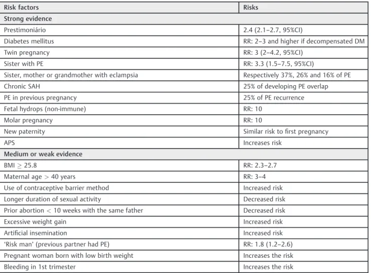
The main risk factors for the development of PE are first pregnancy, previous or family history of PE, chronic hyper-tension, diabetes, collagenosis, black ethnicity, obesity and thrombophilia (►Table 3).25,26 Special attention must be
paid during the prenatal care of these patients to perform the diagnosis of preeclampsia as early as possible.
The evaluation of biomarkers for PE has been the subject of numerous studies, and may be useful in the early diagnosis of PE. Ideally, the biomarker evaluation should be easy to perform, low-cost, and enable the detection of pregnancy-specific hypertensive disease as early as possible, preferably in the first trimester of pregnancy, before the onset of hypertension. Recent reviews show that, to date, none of the available clinical trials has achieved an ideal sensitivity level (> 90%) for the prediction of PE. Only the Doppler
Table 3 Risk factors for PE
Risk factors Risks
Strong evidence
Prestimoniário 2.4 (2.1–2.7, 95%CI)
Diabetes mellitus RR: 2–3 and higher if decompensated DM
Twin pregnancy RR: 3 (2–4.2, 95%CI)
Sister with PE RR: 3.3 (1.5–7.5, 95%CI)
Sister, mother or grandmother with eclampsia Respectively 37%, 26% and 16% of PE
Chronic SAH 25% of developing PE overlap
PE in previous pregnancy 25% of PE recurrence
Fetal hydrops (non-immune) RR: 10
Molar pregnancy RR: 10
New paternity Similar risk tofirst pregnancy
APS Increases risk
Medium or weak evidence
BMI25.8 RR: 2.3–2.7
Maternal age>40 years RR: 3–4
Use of contraceptive barrier method Increased risk
Longer duration of sexual activity Decreased risk
Prior abortion<10 weeks with the same father Decreased risk
Excessive weight gain Increased risk
Artificial insemination Increased risk
‘Risk man’(previous partner had PE) RR: 1.8 (1.2–2.6) Pregnant woman born with low birth weight Increases the risk
Bleeding in 1st trimester Increases the risk
Abbreviations: APS, antiphospholipid syndrome; BMI, body mass index; PE, preeclampsia; RR, relative risk; PA, placental abruption; SAH, systemic arterial hypertension.
Notes: Medium or weak evidence, some studies have demonstrated the association; strong evidence, several studies have shown risk.
Adapted from:Magee et al & Canadian Hypertensive Disorders of Pregnancy (HDP) Working Group (2014)8, Corrêa Júnior et al (2009)25and Sibai
performed between 20–24 weeks showed sensitivity>60%
for PE detection, particularly if performed in pregnant wom-en at increased risk in the 2nd trimester, and to predict severe PE of early onset.19,27–30
Using a mathematical model and taking into account the relative risk regarding the maternal age, nuchal translucency procedure, beta-human chorionic gonadotrophin (β-HCG) and pregnancy-associated plasma protein A (PAPP-A) dosing, Nicolaides classifies pregnant women as being at high risk (>
1/50), intermediate risk (1/51–1,000) and low risk (<1/1000)
of having preeclampsia. Therefore, low-risk pregnant women are advised to undergo only three prenatal consultations, while high-risk pregnant women are advised to undergo more visits. This structure of prenatal care has been criti-cized because when classified as low-risk, many pregnant women could have a delayed PE diagnosis, especially those with later onset of PE. This prenatal care model to predict preeclampsia must be effectively tested.31
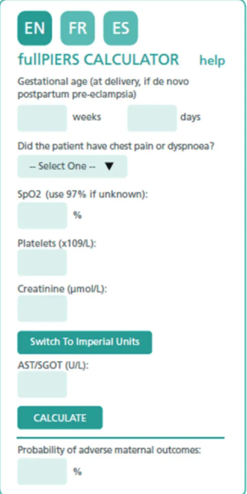
Model to Predict Severe Maternal Outcomes
Von Dadelszen et al32 have developed an interesting and practical model to predict severe maternal outcomes (►Fig. 1). This model was developed in four countries
(Canada, New Zealand, Australia and the United Kingdom) and externally validated.33It can assist clinicians to assess the patients’percentage of risk of having a fatal outcome or a severe complication within the following seven days. For its use, simply access the fullPIERS calculator website (available in four languages) andfind a risk calculator (►Fig. 1), insert
the data on GA, presence or absence of dyspnea or chest pain, O2saturation, dosage of creatinine, platelets, serum glutamic oxaloacetic transaminase (SGOT) or serum glutamic pyruvic transaminase (SGPT), and obtain the percentage of occur-rence of severe complications.
Complications
Systemic arterial hypertension during pregnancy can gener-ate several complications (►Table 4) that will invariably
require careful evaluation and management by the medical staff.
Renal Insufficiency
Renal capillary glomerular endotheliosis was considered the characteristic injury of PE for many years. Some authors only considered the PE diagnosis to be accurate in the presence of this renal injury. Damage to the glomerular membrane causes renal dysfunction, and the glomerularfiltration rate and renal plasmaflow are decreased in relation to healthy pregnant women. There is hyperuricemia in PE, but the elevation of uric acid plasma is transient (dependent on the contraction of plasma volume), and the levels return to Fig. 1 Risk calculator.Source:von Dadelszen, Payne (2011).32
Table 4 Complications of SAH in pregnancy
Affected system Disorder
Cardiovascular Severe SAH, pulmonary edema, pulmonary embolism, vascular accidents
Renal Oliguria, ARF
Hematological Hemolysis, thrombocytopenia, DIC
Neurological Eclampsia, cerebral edema, stroke, PRES
Ophthalmologic Amaurosis, retinal hemorrhages, exudates, papilledema
Hepatic Dysfunction, ischemia, hematoma, capsular rupture
Placental Ischemia, thrombosis, PA, fetal hypoperfusion
normalfigures after childbirth. Acute renal failure (ARF) is an uncommon event in PE. In general, bilateral cortical necrosis is associated with bleeding and excessive hypotension.34
Oliguria in PE has a pre-renal cause most of the times. Therefore, when the urine output drops<25 mL/h, 1,000 mL
of saline solution should be administered within 30 minutes. If the urinary output does not normalize, central hemody-namic monitoring is indicated. Normal or increased pulmo-nary capillary pressure (PCP) and increased uripulmo-nary concentration mean that oliguria is caused by intrinsic renal arteriolar spasm caused by angiospasm. At other times, oliguria may be a consequence of decreased ventricular function. In general, these patients have very high PCP and incipient pulmonary edema.
Pulmonary Edema
Most pulmonary edema cases in pregnant women are asso-ciated with difficult-to-control hypertension. In PE, pulmo-nary edema occurs more frequently after delivery, associated with excessivefluid infusion.
The etiology of pulmonary edema in PE appears to be multifactorial. The reduction in colloid osmotic pressure (COP), increase in capillary permeability, and elevation in vascular hydrostatic pressure produce extravasation offluids in the interstitium and alveolar space. In non-pregnant patients, the decrease in COP/PCP gradient has been corre-lated with pulmonary edema development. Gestation indu-ces decreased COP, and this decrease is accentuated in PE.
The diagnosis and treatment of pulmonary edema in PE is similar to those of non-pregnant patients: oxygen therapy, water restriction, intravenous (IV) furosemide (80 mg ini-tially) and central hemodynamic monitoring. Reduction in afterload is obtained with the use of vasodilators (hydral-azine, nifedipine).
Coagulopathy
Patients with PE frequently have abnormalities in the coagulation system. Reduction in AT III activity (< 70%),
increase in factor VIII consumption, and elevation of platelet factor IV can be detected before the clinical manifesta-tions.13 Although there are changes in the coagulation system since the onset of the disease, in patients with PE, most blood coagulability changes occur due to HELLP syn-drome (thrombocytopenia and hepatic dysfunction) and not to DIC.
Management of Preeclampsia
Regardless of the severity of the clinical picture, every patient diagnosed with PE should be hospitalized for fol-low-up in a high-risk gestational unit. Any patient with PE apparently with a benign condition may suddenly develop complications severe enough to result in maternal and/or fetal death.
The fetuses of mothers with PE who remain hospitalized have half the risk of death compared with fetuses of mothers who are not hospitalized. In addition, hospital-based patients with PE have newborns with more advanced GA at delivery and greater birthweight.35
Antihypertensive Therapy in Preeclampsia
Severe systolic hypertension is an independent factor for stroke in pregnancy.36The goal of the antihypertensive treat-ment is to protect the pregnant women from stroke (stroke, rupture of hepatic hematoma). In 2011, the World Health Organization (WHO) strongly recommended the antihyper-tensive treatment for severe preeclampsia with the aim of reducing maternal morbidity and mortality.37Moderate hy-pertensive pregnant women with long-term SAH and those with secondary SAH and/or repercussion in target organs should be treated with antihypertensive medication to remain normotensive. The CHIPS study demonstrated that strict con-trol of arterial hypertensionwith initiation of antihypertensive treatment from pressure levels of 140/90 mm Hg improves fetal weight, decreases prematurity rates, the diagnosis of severe SAH, and cases of thrombocytopenia and transfusion. This study advises the initiation of the hypertension treatment earlier than we had previously indicated.38
Acute Hypertension
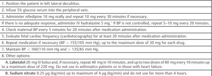
Nifedipine administered orally is thefirst drug of choice for the treatment of a hypertensive crisis (►Table 5).
Alterna-tively, hydralazine can be used intravenously or intramus-cularly with similar success as nifedipine.39 However, the meta-analysis of Magee et al40 showed that the use of hydralazine for hypertensive crisis control presented disad-vantages compared with nifedipine and labetalol, demon-strating increased risk of maternal hypotension (RR: 3.29), placental abruption (PA; RR: 4.17), fetal adverse events and fetal bradycardia (RR: 2.04). Labetalol is an effective alter-native for the treatment of acute hypertension during pregnancy, even though it is not commercially available in Brazil. Sodium nitroprusside should be reserved for cases of hypertensive encephalopathy or hypertensive crisis not responsive to other treatments, and the dose should always be>4μg/kg/min per infusion pump.16,39,41,42
Angiotensin-converting-enzyme inhibitors, angiotensin inhibitors or blockers, diazoxide and propranolol should not be used in PE because they pose too much risk to the health of the fetuses.40,43
Anticonvulsive Preventive Therapy
Magnesium sulfate (MgSO4) is the drug of choice for pre-venting eclampsia, and the only drug with proven preventive effects against eclamptic seizures. Randomized clinical trials demonstrate that MgSO4is superior to hydantoin, diazepam, and placebo for the prevention of eclampsia and its recurrent seizures. The treatment with MgSO4should be used during labor, prior to cesarean section, or whenever there are signs/ symptoms consistent with imminent eclampsia. Magnesium sulfate reduces the risk of eclampsia by 57%, and decreases the risk (RR: 0.55) of maternal death without deleterious effects on the fetus.44
depression and respiratory arrest due to overdosage should be used.
Although MgSO4 therapy has been more effective than placebo for the prevention of eclampsia, even in mild PE cases, and its use has not been associated with unfavorable maternal fetal outcomes,44,45the use in patients with mild PE is controversial, given the low incidence (0.6%) of eclamp-sia in these patients. In patients with mild PE, the NNT for the prevention of 1 case is 129, while in patients with severe PE it is 36. The rational use of MgSO4, avoiding routine use in the group known to have mild PE, has a lower cost.
The use of a low-dose MgSO4infusion (0.6 g/h) after a standard 4 g IV attack dose was as effective as the traditional 4 g intramuscular (IM) regimen of 4/4 hours, with 3.3% recurrence in patients with IM MgSO4, and 2% in patients
with IV infusion of 0.6 g/h.46 Therefore, continuous IV infusion at a low dose (0.6 g/h) may be an alternative, especially in patients with higher incidence of side effects or even impaired renal function. The preferred treatment is IV therapy in infusion pump at a concentration of 1 g/h. Schemes for the use of MgSO4are shown in►Tables 6and7.
The degree of maternal and fetal impairment should be assessed simultaneously with the treatment of severe hy-pertension and prevention of eclampsia. If there is intense and persistent epigastralgia, mainly associated with very high BP levels, there may be distension of the hepatic capsule by subcapsular hemorrhage. In this situation, it is important to evaluate the liver with an ultrasound or tomography. The confirmation of a hematoma implies the necessity of strict BP control and the indication of cesarean section, because there
Table 5 Treatment of acute hypertension (BP>160/110 mm Hg)
1. Position the patient in left lateral decubitus.
2. Infuse 5% glucose serum into the peripheral vein.
3. Administer nifedipine 10 mg orally and repeat 10 mg every 30 minutes if necessary.
If there is no adequate response, administer IV hydralazine 5 mg.If BP is not controlled, repeat 5
–10 mg every 20 minutes. 4. Check maternal BP every 5 minutes for 20 minutes after medication administration.
5. Evaluate fetal cardiac frequency (cardiotocography) for at least 20 minutes after medication administration.
6. Repeat medication if necessary (BP>155/105 mm Hg), up to the maximum dose of 30 mg for each drug.
7. Maintain BP<160/110 mm Hg and>135/85 mm Hg.
8. Other options:
A. Labetalol20 mg IV bolus and, if necessary, repeat 40 mg in 10 minutes, and up to two doses of 80 mg every 10 minutes up to a maximum dose of 220 mg. Do not use in asthmatics patients or in those with heart failure.
B. Sodium nitrate0.25μg (kg/min) up to maximum of 4μg (kg/min) and do not use for more than 4 hours.
Abbreviations: BP, blood pressure; IV, intravenous.
Note:Dilute 1 ampoule (20 mg 2 mL) in 3 mL of distilled water: each milliliter will have 5 mg of hydralazine.
Adapted from:Report of the National High Blood Pressure Education Program (2000).15
Table 6 Prevention of convulsions with magnesium sulfate heptahydrate (MgSO47H2O)
I. Attack dose:4 g of MgSO4(8 mL of 50% MgSO47H2O diluted in 12 mL of distilled water) IV in 5–10 minutes.
II. Maintenance dose IV:0.6–2 g/h IV (dilute 10 mL of 50% MgSO47H2O in 240 mL of saline solution and infuse at a rate of 50 mL/hour (1 g/hour) or 100 mL/hour (2 g/hour) continuously. Every 120 minutes, check if diuresis is preserved (>25 mL/hour)
and if tendon reflexes are present.
III. Maintenance dose IM:10 mL at 50% in the upper outer quadrant of the buttock every 4 hours (alternating buttocks). Evaluate diuresis (>25 mL/hour) and patellar reflexes before each application.
Abbreviations: IM, intramuscular; IV, intravenous.
Note:Especially useful for transporting patients in ambulance and in ambulatories, situations in which IV infusion control is precarious.
Table 7 Magnesium sulfate therapy: special situations
I. If there is a lapse6 hours between maintenance doses and diuresis is25 mL/hour, restart treatment with the attack dose.
II. If renal function is impaired (serum creatinine1.3 mg/dL):Apply half the maintenance dose. Measure the serum magnesium level before each new dose 4–7 mEq/L: therapeutic levels 8–10 mEq/L: inhibition of tendon reflexes>10 mEq/L:
risk of cardiorespiratory arrest.
may be hepatic rupture during the expulsive period. In addition, laboratory tests should be requested to evaluate renal and hepatic functions and possible hematological changes (►Table 8).
Management in Pregnancy at Gestational
Age
>
36 Weeks or with Proven Fetal Lung
Maturity
The cure of PE occurs only after the removal of the placenta; thus, the clinical management depends basically on a balance between the severity of the disease and the GA. Aimed at reducing maternal and fetal complications, patients should be referred to tertiary services where pre-established pro-tocols are followed. These measures lead to a reduction from 5.1% to 0.7% in the occurrence of combined maternal adverse events.47In addition, delivery before 37 weeks is an inde-pendent factor that protects against the recurrence of PE in the next gestation.48 Koopmans et al49 randomized 756 patients with mild PE or gestational hypertension for expec-tant management (watchful waiting) or induction of labor from the 36th week. In the induction group, fewer maternal complications occurred, with no difference in the rates of cesarean or perinatal complications. The planned induction in PE with mature fetuses significantly reduces the morbidity of PE with a significant decrease in care costs.
The existence of a mature fetus is sufficient reason for the definitive treatment of the disease (birth). Therefore, the management of pregnant women with fetuses close to term (GA36 weeks) and PE (even mild PE) should be based on
the following parameters:
a. Patient hospitalization in an obstetric center.
b. Treatment of acute arterial hypertension episodes (►Table 5).
c. Prevention of severe forms of convulsions with MgSO4 (►Tables 6and7).
d. Evaluation of the degree of maternal and fetal impairment. e. Interruption of the gestation, preferably by inducing labor.
Management in Pregnancy at Gestational
Age
>
33 Weeks and
<
36 Weeks
Pregnant women with PE and a preterm fetus should be admitted to a hospital obstetrical center with neonatal and maternal intensive care unit (ICU) facilities for evaluation and treatment. The goal of the management is to reach a GA closer to term without this posing too much risk for the pregnant woman and the concept.
Initially, antihypertensive and anticonvulsant therapies should be used as described before (►Tables 5,6and7). The
MgSO4treatment will be discontinued if the conservative management is adopted. The use of hypotensive drugs (methyldopa) is reserved for cases in which the BP exceeds safe levels (SBP>160 mm Hg or DBP>110 mm Hg) and in
the presence of other risk components indicating immediate cessation of pregnancy.
The assessment of the maternal involvement by physical examination (BP, diuresis, state of consciousness, O2 satura-tion), laboratory evaluation (►Table 8), and fetal impairment
screening are indicated.
After thefirst 24 hours of observation and evaluation, it is necessary to decide for conservative conduct or interruption of gestation. The definition of the best moment to interrupt the pregnancy depends on several individual factors, neona-tal ICU conditions, and the degree of maternal and/or feneona-tal impairment. As a general rule: 1) if the PE is classified as mild, that is, without imminent risk to maternal and fetal health, the interruption should be postponed, if possible, up to the 36th week; and 2) if the PE is classified as severe (►Table 9), the pregnancy should be interrupted.
By adopting the conservative approach, pregnant women should remain hospitalized with restricted physical activity (avoid resting restricted to the bed because it does not contribute to the stabilization of the clinical picture and increases the risk of thrombosis). The diet can be unrestrict-ed and normosodic. The pregnant woman’s weight should be recorded every two days, and the vital signs should be evaluated only during the waking period, avoiding waking the patient up during sleep. Weekly or in a shorter term, in case of clinical necessity, a laboratory evaluation should be performed (►Table 8). The fetus should be auscultated every
day, with observation of the daily rate of fetal movement. In patients with mild PE, it is advisable to evaluate the fetal well-being once a week, and whenever any changes in the maternal state occur. Ultrasonography to check fetal devel-opment and assessment of fetal-maternal hemodynamics (Dopplerflowmetry) should be performed at the time of PE diagnosis.
To monitor fetal development, an ultrasound should be repeated at least in ten-day intervals due to the high inci-dence of IUGR. The evaluation of placental circulation by the Doppler study of the umbilical arteries is the only fetal evaluation test with level 1 of evidence that has proven to decrease perinatal mortality in pregnant women with SAH and IUGR.16Therefore, ideally, patients with PE in conserva-tive management should undergo at least one weekly
Table 8 Laboratory evaluation in PE
Suspected diagnosis Initial evaluation Follow-up
Proteinuria/creatininuria ratio or proteinuria in reagent tape
Pulse oximetry Hemogram Creatinine Platelets
Serum glutamic oxaloacetic transaminase or lactate dehydrogenase
Platelets
Doppler evaluation. Antepartum cardiotocography and fetal biophysical profile may be used complementarily when the Doppler examination is altered in preterm gestations, and when there is need or possibility of prolonging gestation. During labor, cardiotocography with continuous or intermit-tent monitoring of the fetal heart rate is the test of choice for fetal surveillance.
The induction of fetal lung maturity with corticosteroids can be performed in pregnancies<34 weeks in which the birth is
predicted for the next the 24 or 48 hours.16 If an elective cesarean is indicated (without labor) for a pregnant woman at<39 weeks, the use of corticosteroids for pulmonary
matu-ration brings benefits by reducing the need for hospitalization in the neonatal ICU for the newborn’s mechanical ventila-tion.50,51When pregnancy interruption is indicated and the fetus is<36 weeks of GA, the patient has to be hospitalized or
transferred to a tertiary-level healthcare hospital.
Management in Pregnancy at Gestational
Age
<
33 Weeks
In pregnant women at GA<33 weeks and stable fetal
maternal condition, we can opt for conservative manage-ment with assiduous managemanage-ment of all parameters of maternal and fetal well-being. By choosing the expectant management, one should be alert to any signs of clinical decompensation. Particular attention should be paid to the degree of maternal thrombocytopenia, which is an impor-tant indicator of morbidity and mortality. Patients with PE and platelets between 150,000 and 100,000 cells/mm3 al-ready have an increase in fetal and maternal morbidity and mortality, which will be greater the lower the platelet count.
Conservative Management of Severe
Preeclampsia
The prevalence of severe PE is of1% of pregnancies, and is
associated with progressive deterioration of the fetal-mater-nal picture.52,53All pregnant women with severe PE should be hospitalized, and the initial management should include administration of MgSO4and antihypertensive drugs (SBP
160 mm Hg or DBP110 mm Hg).52In the presence of
eclampsia, pulmonary edema, coagulopathy and
non-reac-tive fetal evaluation, labor should be performed even before the completion of the corticosteroid therapy for fetal matu-rity.►Table 4shows the main parameters for the
interrup-tion of gestainterrup-tion.
Several studies52,53 describe the complications in the conservative management of severe PE<34 weeks, namely:
PA (16–39%); perinatal death (up to 17%); small fetuses for GA (up to 70%); presence of nonreactive fetal tests (26–74%); pulmonary edema (up to 8%); eclampsia (up to 5.6%); HELLP syndrome (4–27%); and renal failure (up to 17%). The main reason for gestational discontinuation in this group of preg-nant women is the worsening of the fetal status; therefore, fetal and maternal evaluation should be performed daily, using the various methods available. If the pregnancy is32
weeks, but there is risk of maternal and/or fetal death, PA, HELLP syndrome, DIC, eclampsia, severe uncontrollable hy-pertension (160/110 mm Hg) or hepatic hematoma, the
choice should be interruption of pregnancy.
The prospective fullPIERS study32assessed the occurrence of severe maternal outcomes (maternal death and life-threatening complications) in 2,023 pregnant women with PE admitted to tertiary-level hospitals for follow-up in four countries (Canada, New Zealand, Australia and the United Kingdom) in. There were severe complications in 261 women (5%). The predictors for these complications were: GA<34
weeks, chest pain, dyspnea, low O2saturation, thrombocy-topenia, increased serum creatinine and altered hepatic transaminases (SGOT). The authors also showed that requir-ing lactate dehydrogenase (LDH) measurement when the liver enzymes are normal is redundant and should be avoided. It is only necessary to titrate one of the liver enzymes (SGOT or SGPT), and it is not necessary to request coagulation tests.
Some authors recommend trying the conservative man-agement in women with severe PE who received betame-thasone only up to the 32nd week on the grounds that the risk of serious maternal complications is not compensated by the additional gain in fetal maturity.54
HELLP Syndrome
The acronym HELLP stands for hemolysis, elevated liver enzymes and low platelet count (►Table 10). The
Table 9 Maternal and fetal indications of termination of pregnancy in severe preeclampsia<34 weeks39
Maternal Fetal
HELLP syndrome Fetal growth below percentile 5
Eclampsia Repeated late fetal decelerations on cardiotocography
Pulmonary edema or O2saturation<94% Vein Doppler with pathological a-wave
BP without control despite medications Fetal death
Serum creatinine>1.5 mg/dL or oliguria (<500 mL/ mL/24 hours) Suspected PA, ROM or onset of labor
Suspected PA, ROM or onset of labor
Abbreviations: BP, blood pressure; HELLP, hemolysis, elevated liver enzymes and low platelet count; PA, placental abruption; ROM, rupture of membranes.
pathophysiology of this disease is unclear, but the hepatic hematologic involvement of PE can be considered. Hemoly-sis, elevated liver enzymes and low platelet count syndrome develops in 0.1 to 0.8 of all pregnancies, and in 10–20% of pregnant women with severe PE/eclampsia. About a third of HELLP syndrome diagnoses are performed in the postpartum period. In patients with antepartum diagnosis, 10% of diag-noses were performed before the 27th week, 20% after the 37th week, and 70% between the 27th and 37th weeks.55,56 Hemolysis, elevated liver enzymes and low platelet count syndrome is related to microangiopathic hemolytic anemia and vasospasm in the maternal liver. The symptomatology is usually poor, and may include malaise, epigastralgia, nausea and headache. The degree of clinical suspicion of HELLP syndrome cases is very important. In the presence of throm-bocytopenia in a patient with PE, HELLP syndrome should be strongly considered. Many cases go through days with a vague symptom of malaise and the patient reporting non-specific symptoms, similar to a cold, with generalized pain, nausea and epigastric pain. Some studies point to a varying prevalence of the main symptoms, such as malaise (50 to 90%), pain in the right hypochondrium or epigastralgia (30 to 90%), and nausea and vomiting (20 to 50%); proteinuria may be absent.57,58
The diagnostic confirmation of HELLP syndrome is by laboratory tests (►Table 10), using the laboratory parameters
described by Sibai.55 Thrombocytopenia is the main and earliest laboratorial modification found. The appearance of coagulation abnormalities, such as change in prothrombin time, partial thromboplastin time, andfibrinogen, is uncom-mon. When thrombocytopenia is severe (< 50,000/mm3),
products offibrin degradation and activation of AT III appear, indicating the initiation of an intravascular coagulation pro-cess. Eventually, patients with HELLP syndrome have hemor-rhagic diastasis with bleeding at multiple sites (hematuria, hematemesis, surgical wound bleeding). Red cell fragmenta-tion is present in HELLP syndrome, and although the amount of fragmentation is not associated with the severity of multiple organ dysfunction, it represents the involvement of the endo-thelial system in the microcirculation. Fragmentation is a result of the passage of red blood cells through small damaged vessels. Hepatic dysfunction can be measured by various parameters, such as increased LDH and transaminases (SGOT
and SGPT). Renal dysfunction will depend on the severity of the condition, and it can be diagnosed in up to 46% of HELLP syndrome cases.59 After hepatic and renal dysfunction, the patient may present pulmonary damage with DIC, character-izing a multiple organ dysfunction. In less than 2% of HELLP syndrome cases, a hepatic hematoma is formed. The diagnosis can be made by ultrasonography, and the treatment varies from conservative therapy to surgical management in cases of hepatic rupture.60 If there is hepatic hematoma without rupture, a cesarean section is indicated, and surgical explora-tion should not be performed given the risk of rupture at that time.
Differential Diagnosis
Differential diagnosis between HELLP syndrome and other pathologies (especially hemorrhagic and hepatic patholo-gies) that may occur in the puerperal cycle is fundamental. Among the main pathologies, the following stand out: acute hepatitis, cholecystitis, pancreatitis, lupus, fatty liver of pregnancy, thrombocytopenic purpura, hemolytic-uremic syndrome, and septic or hemorrhagic shock, among others. Severe complications of HELLP syndrome occur with hemorrhage (central nervous system, liver, operative wound, PA).
Thrombocytopenia<50,000/mm3is associated with the
occurrence of DIC and a strong indicator of hemorrhagic complications. The presence of headache, visual changes and epigastralgia significantly increases the risk of eclampsia. In a Brazilian study61 performed with 105 patients with HELLP syndrome, the main complications found were bleeding (34%), oliguria (47%), acute renal failure (20%), acute pulmonary edema (7%), need for blood transfusion (33%), and maternal death (4%). These data confirm the severity of this syndrome and the importance of the management at a tertiary center with experienced teams. The most important factor for the reduction of maternal morbidity and mortality is the early diagnosis, which should be made in the asymptomatic phase through laboratory investigation of thrombocytopenia, hemo-lysis and hepatic alterations in all patients with PE. Although the main cause of jaundice in pregnancy is hepatitis, if it occurs, the presence of HELLP syndrome with advanced he-molysis should always be ruled out.
Table 10 Diagnosis of HELLP syndrome
Exam Parameter
Hemolysis
Peripheral blood smear
(schistocytosis, anisocytosis, echinocytosis, poikilocytosis)
Bilirubin >1.2 mg/dL
Lactate dehydrogenase >600 U/L
Hepatic impairment Serum glutamic oxaloacetic transaminase >70 U/L
Thrombocytopenia Platelets <100,000/mm3
Abbreviation: HELLP, hemolysis, elevated liver enzymes and low platelet count.
Management in HELLP Syndrome
As it happens with eclampsia, HELLP syndrome should be regarded as an obstetric emergency requiring immediate care. The treatment is based on the prevention of hemor-rhagic complications and eclampsia, control of SAH and the onset of labor.
The timing of interruption can be programmed depending on the severity of each case and the GA. In pregnancies>34
weeks, labor induction should start immediately, with si-multaneous control of the hypertensive crisis by using MgSO4 and blood products when indicated. In pregnant women at GA<34 weeks, in the absence of serious
compli-cations, such as hepatic hematoma, severe thrombocytope-nia and eclampsia, corticosteroid therapy should be performed for pulmonary maturation before the interrup-tion of pregnancy. O’Brien et al62propose fundamental steps for the care of HELLP syndrome, as follows:
1. Have high diagnostic suspicion in pregnant women with PE;
2. Perform laboratory tests and differential diagnosis; 3. Evaluate maternal and fetal conditions;
4. Control blood pressure;
5. Stabilize the clinical picture: venous access; administra-tion of MgSO4and antihypertensive drugs;
6. Consider the use of corticosteroids for fetal maturity; 7. Hemotherapy if necessary;
8. Check if there is need for hepatic imaging (epigastralgia); 9. If cesarean section is indicated, evaluate with the
anes-thesiologist the technique to be adopted;
10. Actively manage labor or plan the cesarean section with the proper technique;
11. Plan for care in maternal and neonatal ICUs if necessary; 12. Perform laboratory evaluation every 6–24 hours, depending on the severity of the condition, until stabilization;
13. Maintain the use of antihypertensive and MgSO4in the puerperal period; and
14. Counseling for future pregnancies.
As the management of patients with HELLP syndrome should be performed in tertiary centers with maternal and neonatal ICUs, suspected cases should be transferred imme-diately in an adequate ambulance in the presence of a life-saving physician after contact with the reference maternity. The patient should be on IV MgSO4, and if an infusion pump is not available, the attack dose should be administered intra-venously, avoiding IM administration if thrombocytopenia
<100,000/mm3due to the risk of gluteal hematoma.
Mag-nesium sulfate should be started immediately, and main-tained for up to 24 hours postpartum, with control of diuresis, tendon reflexes, and respiratory rate (►Table 10).
Fetal conditions, GA and uterine cervix (Bishop score) are fundamental in deciding the route of birth. If<30 weeks, in
absence of labor, and Bishop score<5, elective caesarean
section is recommended after initiating MgSO4.6In pregnant women with<32 weeks and fetuses with restricted growth,
and alteration of the umbilical artery Doppler, it is preferable
to perform a cesarean section, except in cases already in labor.61 The other patients may be submitted to labor induction. Anesthesia of the pudendal nerve should be avoided due to the risk of hematoma. Caesarean sections should be performed by experienced professionals using the best surgical technique and with attention to intra-operative hemostasis. In the presence of thrombocytopenia (<100,000/mm3), infraumbilical median laparotomy is
rec-ommended to reduce the risk of hematomas in the aponeu-rotic detachment. If thrombocytopenia is<75,000/mm3,
epidural or subdural anesthesia should be avoided, and general anesthesia should be performed. The use of an aspiration drain is recommended in the most severe patients, especially in those with DIC, facilitating postoperative con-trol. The Portovac (Howmedica, Toronto, Ontario, Canada) drain (polyethylene with closed drainage system) or the Blake (Ethicon, Somerville, NJ, US) drain (silicone, soft, continuous drainage) can be used. The latter has the advan-tage of continuous drainage, and since it does not have a closed drainage system, it causes less obstruction problems due to small clots. These should be removed 24 to 48 hours after the cesarean section, depending on the evolution of the patient’s surgical clinical status and the amount of drainage. Care should be taken with puerperal blood loss and the risk of uterine hypotonia. Thus, the prophylactic use of IV oxytocin and misoprostol (rectal or intrauterine) is extremely valuable.
Use of Corticosteroids for
Thrombocytopenia Rescue
Corticosteroids have been used for the treatment of women with HELLP syndrome, especially those with platelets
<50,000/mm3. The mechanism of action includes reduction of
platelet adhesion, reduction in platelet removal by the spleen, and increase in platelet activation. Currently, a Brazilian study (COHELLP) is underway to verify the efficacy of dexamethasone in patients with HELLP and thrombocytopenia<50,000.
Some centers use dexamethasone 10 mg intravenously every 12 hours before delivery and after birth until labora-tory recovery. Some studies have demonstrated an im-provement in thrombocytopenia and other laboratory tests with this practice, as well as a decrease in the need for transfusions, hypertension and the use of antihyperten-sive drugs, presenting a postpartum recovery with lower morbidity.63However, thisfinding has not been reported in other studies.64We still lack more consistent evidence on the benefit of corticosteroid therapy in maternal morbidity and mortality. In a recent systematic review of the Cochrane Library, the conclusion is that there is insufficient evidence for the routine use of steroids in HELLP syndrome, and that their use may be justified in special situations in which platelet increase is important.65 Intravenous dexa-methasone may be used if platelets are<50,000/dL. This
Blood and Platelet Transfusion
In the presence of abnormal bleeding and HELLP syndrome, or in the presence of severe thrombocytopenia (<20,000
platelets), even without bleeding, transfusion of platelet concentrate is always indicated. If the patient underwent a cesarean section, the transfusion of platelets is recom-mended when the count is<50,000/mm3. Each platelet
concentrate unit elevates the platelets by5,000 mm3to
10,000 mm3in an adult weighing 70 kg.62
Postpartum Management
The postpartum period remains extremely critical. In gener-al, in thefirst 24 hours of the puerperal period, there is a transient worsening of the clinical picture due to consump-tion of platelets and coagulaconsump-tion factors. This worsening is more pronounced when the birth occurs by caesarean sec-tion. Therefore, we should not base on the postoperative process of preeclampsia. Many maternal deaths have oc-curred in the postpartum period because of hemorrhagic complications and some degree of little importance given to care in that period. Even if the patient does not have clinical parameters for an ICU admission yet, she must be admitted to this type of unit for immediate control of any kind of postpartum change. Laboratory control will be performed using the same parameters of diagnosis (platelets, LDH, SGOT, bilirubin). Diuresis should be controlled and tained above 25 mL/hour. Hypertension should be main-tained below 160/100 mm Hg. If there is spontaneous diuresis above 25 mL/hour, normal creatinine, LDH decrease, improvement in platelet levels and hepatic transaminases, we can consider the disease entered remission.
Preeclampsia Delivery Route
The preferred route of delivery in PE is vaginal, with no contraindication for cervical maturation procedures (Foley catheter, prostaglandin analogues), and cesarean section is reserved for usual obstetric indications. There should be constant monitoring of the fetal heart rate (FHR) during the first or second periods of childbirth. The presence of uterine hyperactivity, increased uterine tone, vaginal bleed-ing or pathological decelerations of the fetal heart rate should be seen as signs of possible PA.
For the cesarean section, epidural or subdural anesthesia may be used. In this situation, the patient should be hydrated with an infusion of 1,000 mL lactated ringer or saline before sympathetic block to avoid severe hypotension with de-creased tissue perfusion of vital organs (kidneys and placenta).
In addition, while the patient remains supine during cesarean section, a cushion should be placed under the pregnant woman’s rightflank, thereby reducing the com-pression of the uterus on the large vessels of the abdomen. If severe hypotension still occurs, liquid infusion will be nec-essary tofill the dilated vascular space, avoiding the use of vasopressor substances. In emergency situations or when
there is a complicated pregnancy-specific hypertensive dis-ease (eclampsia, HELLP syndrome, DIC), general anesthesia is the preferred option. In this eventuality, it is important to alert the anesthesiologist about the use of MgSO4, because its sedative action may be dangerous in conjunction with succinylcholine.
In general, the hypertensive picture disappears or improves substantially in thefirst 24 hours of the puerperal period, although the symptoms may remain up to six weeks after childbirth. If BP is<150/100 mm Hg, the patient may
be discharged without antihypertensive therapy and under-go a weekly evaluation in an outpatient setting until PE signs disappear.
Management in Gestational
Age
<
24 Weeks
The presence of severe PE in the second trimester, and especially<25 weeks, is associated with high rates of
peri-natal mortality (up to 83%) and maternal complications (27 to 71%), including maternal death.52,66Immediate delivery is associated with a lower chance of fetal survival, while prolongation of the pregnancy may somewhat increase the chance of fetal survival, but it adds an important risk of maternal morbidity and mortality. In these cases, the ideal management is not established yet, and is the reason for numerous studies and discussions in the literature. Some authors52–54recommend the interruption of pregnancy in these cases after discussing with the couple and obtaining signed informed consent. When the option is for expectant management, fetal and maternal evaluations should be performed daily, controlled in centers with obstetricians, neonatologists and intensivists experienced in high-risk obstetrics.
Persistent Postpartum Hypertension
Chronic hypertensive patients may develop hypertensive encephalopathy, pulmonary edema and cardiac insufficiency in the puerperal period. These events are more frequent in patients with overlapping PE, previous cardiac or renal disease, PA, or with difficult to control BP. In patients who remain hypertensive, drugs for its control should be admin-istered orally. In the other patients, BP can be controlled weekly for a month, then at intervals of three to six months for one year.
When prescribing antihypertensive medication, it is nec-essary to bear in mind that the vast majority is excreted in human milk and can be absorbed by the newborn. Although there is a lack of good studies on the use of antihypertensive drugs in lactation, the recommendation to avoid diuretics seems reasonable, given their potential to suppress lactation. Neonatal exposure to methyldopa, labetalol, captopril and nifedipine is considered safe, and, therefore, a good option in the breastfeeding period.