EZH2 Protein Expression and Tumor Response to
Neoadjuvant Chemotherapy in Locally
Advanced Breast Cancer
Expressão da proteína EZH2 e resposta tumoral à
quimioterapia neoadjuvante no câncer de mama
localmente avançado
Lucienne Pereira Del Grossi Neusquen, MSc
1José Roberto Filassi, PhD
1Carlos Elias Fristachi, PhD
2Kátia Candido Carvalho, PhD
3Maíra Teixeira Dória, MD
1José Maria Soares Júnior, PhD
1,3José Roberto Morales Piato, PhD
11Department of Obstetrics and Gynecology, Universidade de São Paulo, São Paulo, São Paulo, Brazil
2Instituto Arnaldo Vieira de Carvalho, São Paulo, São Paulo, Brazil 3Laboratory of Structural and Molecular Gynecology (LIM-58), Faculdade
de Medicina da Universidade de São Paulo, São Paulo, São Paulo, Brazil Rev Bras Ginecol Obstet 2016;38:280–286.
Address for correspondence Maíra Teixeira Dória, MD, Department of Obstetrics and Gynecology, Universidade de São Paulo, Av. Dr. Enéas de Carvalho Aguiar, 255, 10° andar, Cerqueira Cesar, 05403-000, São Paulo, Brazil (e-mail: [email protected]).
Keywords
►
EZH2
►
breast neoplasms
►
tumor markers
►
neoadjuvant therapy
►
gene expression
Abstract
Introduction
Neoadjuvant chemotherapy (NAC) is the standard treatment for locally
advanced breast cancer. However, some tumors will not respond to this treatment due
to histological and molecular features. The protein EZH2 (enhancer of zest homolog 2)
is a histone methyltransferase that is correlated with poorly differentiated breast
carcinomas and aggressive tumor behavior.
Purpose
The present study evaluated the association between EZH2 expression and
response to NAC, and its correlation with HER2 overexpression, estrogen and
proges-terone receptors (ER, PR) and Ki-67 proliferation index.
Methods
A total of 60 patients with locally advanced breast cancer treated with NAC
were selected for this study. Twenty-three paraf
fi
n blocks had not enough material for
tissue resection, and were not evaluated. A tissue microarray based in
immunohis-tochemistry (IHC) analysis of EZH2 was performed for the remaining 37 specimens.
Patients were divided into two groups based on response to NAC.
Results
EZH2 expression was signi
fi
cantly associated with markers of poor prognosis
such as ER negativity (
p
¼
0.001), PR negativity (
p
¼
0.042) and high K
i-67
prolifera-tion index (
p
¼
0.002). High EZH2 expression was not correlated with the response to
NAC.
received January 23, 2016 accepted May 18, 2016
DOI http://dx.doi.org/ 10.1055/s-0036-1584954. ISSN 0100-7203.
Copyright © 2016 by Thieme Publicações Ltda, Rio de Janeiro, Brazil
Introduction
Neoadjuvant chemotherapy (NAC) is the standard of care for
patients with locally advanced breast cancer.1–6It provides
local control and reduction in tumor size, increasing breast-preserving rates. Neoadjuvant treatment also provides an opportunity to test tumor sensitivity to chemotherapy in vivo. Approximately 15% of patients will achieve a complete response to NAC, whereas another 15% will display minimal
changes or a progressive disease.7Patients who achieve a
pathologically complete response (pCR) after NAC have
bet-ter disease-free survival and overall survival rates.8 The
identification of tumor markers that predict response to
therapy, distinguishing responders from non-responders, can improve therapeutic decisions.
Clinical and pathological features considered to be predic-tors of disease progression with NAC include negative expres-sion of estrogen receptor (ER) and progesterone receptor (PR),
high score of Ki-67, and high nuclear grade of tumor cells.7
These markers have also been associated with a complete pathologic response to neoadjuvant chemotherapy, suggesting that although morphologically similar, highly proliferative tumors can be divided into two subpopulations, namely those that are highly sensitive and those that are highly resistant to
chemotherapeutics.9There remains a need for the discovery of
new molecular markers that can differentiate these subpopu-lations of highly proliferative tumors.
The protein EZH2 (enhancer of zest homolog 2) encodes a histone methyltransferase, which is the catalytic core protein of
the polycomb repressor complex 2 (PCR2).10PCR2 are epigenetic
chromatin modifiers that regulate transcription through
nucleo-some modification, chromatin remodeling, and interaction with
other transcription factors.11These proteins are involved in cell
regulation and cell division, and are well known for initiating target gene silencing by promoting H3K27me3 trimethylation,
which is catalyzed by EZH2.12Several studies showed that the
EZH2 and the signal transducer and activator of transcription 3 (STAT3) are involved in the proliferation, self-renewal and
pluripotency of cancer stem cells.13
Overexpression of EZH2 has been described in prostate,
breast, bladder, gastric, lung and ovarian cancers.10In breast
cancer, elevated EZH2 expression has been correlated with poorly differentiated tumors and aggressive tumor
beha-vior.14–17 Holm et al18 tested the EZH2 and H3K27me3
abundance in more than 400 tumors using
immunohisto-chemistry (IHC). They found significantly high expression of
EZH2 in triple-negative, basal-like and HER2-enriched tu-mors, and high H3K27me3 in luminal A, HER2-enriched and
normal-like tumors.18 Therefore, high abundance of EZH2
was associated with poor distant free-survival.
Conclusions
Our data suggested that EZH2 protein expression may not correlate with
the clinical response to NAC. Other studies with more patients are needed to con
fi
rm
this observation.
Resumo
Introdução
A quimioterapia neoadjuvante é o tratamento padrão para os cânceres de
mama localmente avançados. Entretanto, apenas uma porcentagem desses tumores
irá responder ao tratamento, devido a características histológicas e moleculares. A
proteína EZH2 (enhancer of zest homolog 2) é uma histona metiltransferase associada
a tumores mal diferenciados e de comportamento agressivo.
Objetivo
O presente estudo teve como objetivo avaliar a associação entre a
expressão da proteína EZH2 e a resposta à quimioterapia neoadjuvante, além da
correlação dessa proteína com hiper-expressão de HER2, receptores de estrogênio e
progesterona, e o marcador de proliferação Ki-67.
Métodos
Um total de 60 pacientes com câncer de mama localmente avançado
tratadas com quimioterapia neoadjuvante foram selecionadas para esse estudo. Vinte e
três blocos de para
fi
na não continham material su
fi
ciente para ressecção e não foram
avaliados. Foi realizado
microarray
baseado em análise imuno-histoquímica da proteína
EZH2 para as 36 pacientes restantes. As pacientes foram divididas em dois grupos
baseado na resposta à quimioterapia neoadjuvante.
Resultados
A expressão da proteína EZH2 foi signi
fi
cativamente associada com
marcadores de pior prognóstico, como negatividade para receptor de estrogênio
(
p
¼
0,001) e progesterona (
p
¼
0,042), além de alto K
i-67 (
p
¼
0,002). Entretanto, a
alta expressão da EZH2 não se correlacionou com a resposta à quimioterapia
neoadjuvante.
Conclusões
Nossos dados sugerem que a expressão da proteína EZH2 pode não estar
relacionada com a resposta clínica à quimioterapia neoadjuvante. Outros estudos com
maior número de pacientes são necessários para con
fi
rmar esses achados.
Palavras-chave
►
EZH2
►
neoplasias da mama
►
marcadores
tumorais
►
terapia
neoadjuvante
EZH2 is also overexpressed in inflammatory breast cancer (IBC), the most metastatic variant of breast cancer with the
poorest survival.11,19,20An analysis of 88 tissue microarrays
from IBC demonstrated that EZH2 is highly expressed in
75.7% of them, and was associated significantly with
unfa-vorable prognostic factors, such as higher tumor grade and
triple-negative status.11
To date, there are no studies in the literature evaluating if there is a clear correlation between EZH2 protein expression and the response of breast cancer tumors to NAC. Therefore, the aim of this study was to determine if EZH2 protein expression can predict tumor response to NAC in locally
advanced breast cancers. One predefined secondary aim of
this study was to evaluate the association of EZH2 expression and other pathological features, such as ER and PR
expres-sion, Ki-67 proliferation index and HER2 overexpression.
Materials and Methods
Patient Selection
This was a case-control pilot study developed at Faculdade de Medicina da Universidade de São Paulo with the analysis of breast cancer patients treated at Instituto de Oncologia e Mastologia Arnaldo Vieira de Carvalho. Primary tumor speci-mens with local advanced breast cancer (stage IIB or IIIA) from 60 patients were selected. The patients received antra-cycline-based neoadjuvant chemotherapy, and were treated at Instituto de Oncologia e Mastologia Arnaldo Vieira de Carvalho. The tumor samples were obtained in accordance with protocols approved by the Institutional Ethics Commit-tee at the Instituto de Oncologia Arnaldo Vieira de Carvalho and at Faculdade de Medicina da Universidade de São Paulo. Exclusion criteria were bilateral disease and pregnancy con-comitant with the diagnosis of breast cancer.
Pathological Assessment
After analysis of the tumor specimens, 23 paraffin blocks had
not enough material for tissue resection, and were not
evaluated. Thirty-seven paraffin-embedded tumor specimen
blocks were selected for this study and cut into 5-µm slices prior to being stained with hematoxylin and eosin. The morphologically representative areas (only tumor cells, no
necrosis or normal breast cells) were defined as the site for
replicate core extraction from the master block.
Tissue microarrays were constructed from these tumor
samples, and IHC was performed for EZH2 (clone NCL–
L-EXH2, 1:200 dilution, Novocastra) and the usual prognostic markers: ER (clone SP1; 1: 200 dilution, Neomark); PR (clone pgR636, 1: 600 dilution, DAKO); HER-2 (1: 400 dilution,
Spring); and Ki-67 (clone MIB-1, 1: 4800 dilution, DAKO).
Evaluation of Staining Results
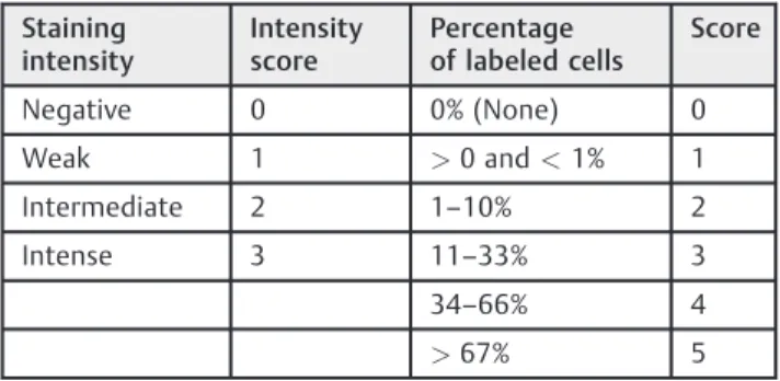
The expression of the EZH2 protein was based on the IHC
labeling in accordance with Allred’s criteria, which are
commonly used for the evaluation of hormone receptor
expression (►Fig. 1). Thus, the observed intensity of
cellular staining was classified as negative, weak,
inter-mediate, or intense. The percentage of stained cells was
assigned a proportion score as follows: 0%;>0 and<1%;
1–10%; 11–33%; 34–66%; and>67% cells positively
la-beled. The intensity scores (range, 0–3) and proportional
scores (range, 1–5) were summed, resulting in a total
score between 0 and 8 to enable comparison with other
biomarkers (►Table 1).
Assessment of Clinical Response
Clinical assessment of tumor response was performed before initiating NAC and pre-operatively using calipers (Sanny Professional Skinfold caliper, Ann Arbor, USA). The response criteria set forth by the International Union for Cancer
Control (UICC) were employed.21 According to clinical
re-sponse, patients were divided into two groups: Group 1 (the
responders) consisted of patients who achieved 50%
reduction in tumor size; and Group 2 (the non-responders) consisted of patients in whom the aforementioned objective response was not observed.
For data analysis, patients were grouped according to the UICC response criteria: the responder group included
patients with complete or partial clinical response to NAC, and the non-responder group included patients who had no change in tumor size or disease progression.
Statistical Analysis
Statistical analysis was performed using a linear correlation
analysis by Spearman’s test for non-parametric correlations.
The means and distributions of the quantitative variables of
both groups were determined with the Student’st-test or the
Mann-Whitney test for non-parametric data, when
appro-priate. Fisher’s exact test was performed to compare the
proportions. Mean values are presented with standard
de-viations (SDs) in the results. Statistical significance was
determined asp<0.05 with a two-sided test.
Results
A reduction of 50% or more in tumor size was achieved in 19
of 37 cases (51.3%) – Group 1. The other 18 cases were
classified as non-responders (Group 2). The pathological
characteristics of Group 1 are summarized in ►Table 2,
and ►Table 3 presents the data from Group 2. The mean
age was 4611 years for responders and 5611years for
non-responders (p¼0.008). It was not possible to perform
IHC for Ki-67 proliferation index in 6 tumor specimens, EZH2
expression in 5, HER2 in 1, ER status in 2, and PR status in 1 specimen.
Of all tumor specimens evaluated for EZH2, the lowest amount of expression was 11%. The majority of patients (62.5%) had more than 66% of cells positive for EZH2 (►Table 4).
To evaluate the association between EZH2 expression and the other pathological factors, EZH2 expression was divided into two categories according to percentage of cells positive
for EZH2: 34–66% and67%. High EZH2 expression was
significantly associated with markers of poor prognosis, such
as ER negativity (p¼0.001), PR negativity (p¼0.042), and
Table 1 The evaluation of EZH2 protein expression on the tumor tissue
Staining intensity
Intensity score
Percentage of labeled cells
Score
Negative 0 0% (None) 0
Weak 1 >0 and<1% 1
Intermediate 2 1–10% 2
Intense 3 11–33% 3
34–66% 4
>67% 5
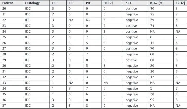
Table 2 Pathological characteristics of Group 1
Patient Histology HG ER PR HER2† p53 Ki-67 (%) EZH2‡
1 IDC 2 7 7 0 negative 40 8
2 IDC 3 0 0 0 negative 90 8
3 IDC 2 8 8 0 negative 10 6
4 IDC 3 0 0 0 positive 50 7
5 IDC 2 6 8 3 negative 60 8
6 IDC 2 8 8 1 negative 50 7
7 IDC 1 5 7 0 negative 5 5
8 IDC 2 7 5 0 positive 30 7
9 IDC 2 7 4 0 negative 60 7
10 IDC 3 0 0 0 negative 90 8
11 IDC 3 0 0 0 positive 90 8
12 IDC 3 0 0 0 positive 80 8
13 ILC 1 6 7 0 negative 30 7
14 IDC 1 6 7 1 negative NA 8
15 IDC 2 0 0 0 negative 40 8
16 IDC 2 6 8 3 negative 35 7
17 IDC 1 7 5 0 negative 25 7
18 IDC 3 6 3 0 negative NA NA
19 IDC 2 4 6 2 positive NA NA
Abbreviations: ER, estrogen receptor; IDC, invasive ductal carcinoma; ILC, invasive lobular carcinoma; HG, histological grade; NA, not available; p53, protein p53; PR, progesterone receptor.
Allred score for ER and PR;†HER2: 0 and 1, negative; 2, undetermined; 3, positive;‡Score for EZH2 as presented in
high Ki-67 proliferation index (p¼0.002). There was no association between EZH2 expression and either age or HER2 status.
To assess if there was an association between EZH2 expression and response to NAC, we also divided patients
into the two categories of EZH2 expression (34–66% and
67%). As reported in►Table 5, our analysis revealed no
association between responder status and EZH2 protein
expression (p¼0.135).
Discussion
The heterogeneity of breast cancers in their evolution and response to standard treatments compels us to identify markers that allow individualized therapies. Despite the improvement in breast cancer treatment in the last decades,
there is still a significant percentage of patients that do not
respond to therapy. Antibody targeted immunotherapy for HER2 protein represented an important step toward tailored treatment for patients who express that protein. The dis-covery of other antigen targets is a great challenge, and will allow the development of novel therapeutic strategies for
breast cancer.8,22–24 Our study focused on EZH2 protein
expression and its correlation with the response to neoadju-vant chemotherapy.
In our study, all of the tumors analyzed were positive for EZH2 protein expression in at least 11% of invasive tumor
cells. Thisfinding is in accordance with a previous report
from Bachmann et al, which shows EZH2 expression in
locally advanced breast tumors.15 The variability of EZH2
protein expression detected by IHC allowed us to classify EZH2 presence by the percentage of cells stained and the intensity of staining, producing a composite score ranging from 0 to 8. This score allowed the data to be compared with other parameters that had a history of being evaluated in this manner, such as ER and PR statuses.
Our findings regarding the association between EZH2
protein expression and HER2 and Ki-67 proteins expression
was significantly positive for the correlation between EZH2
and Ki-67 expression, while the association between EZH2
and HER2 did not reach statistical significance. This
correla-tion between EZH2 and Ki-67 is consistent with prior
pub-lications.15,25,26The potential association between EZH2 and
HER2 remains controversial. Collett et al25and Holm et al18
Table 3 Pathological characteristics of Group 2
Patient Histology HG ER PR HER2† p53 Ki-67 (%) EZH2‡
20 IDC 3 0 0 0 positive 10 8
21 IDC 2 5 8 0 negative 75 8
22 IDC 3 NA NA 3 negative 39 8
23 IDC 3 0 0 2 positive 74 8
24 IDC 3 0 0 3 positive NA NA
25 IDC 2 8 7 0 negative 8 7
26 IDC 2 3 5 0 negative 11 8
27 IDC 3 0 0 0 positive 70 8
28 IDC 2 7 7 0 negative 60 8
29 IDC 3 0 0 3 positive 80 8
30 IDC 2 6 5 3 negative 80 8
31 IDC 2 6 0 0 negative 30 7
32 IDC 2 5 3 0 negative 12 6
33 IDC 1 NA 0 NA negative NA NA
34 IDC 1 8 7 0 negative 5 7
35 IDC 1 6 6 0 negative 30 8
36 IDC 3 0 0 0 negative 95 8
37 IDC 2 8 8 0 negative NA NA
Abbreviations: ER, estrogen receptor; IDC, invasive ductal carcinoma; ILC, invasive lobular carcinoma; HG, histological grade; NA, not available; p53, protein p53; PR, progesterone receptor.
Allred score for ER and PR;†HER2: 0 and 1, negative; 2, undetermined; 3, positive;‡Score for EZH2 as presented in►Table 1.
Table 4 Percentage of EZH2þcells in all specimens analyzed
EZH2 (%) No. of patients %
11–33% 1 3.1
34–66% 11 34.4
67% 20 62.5
found high abundance of EZH2 in HER2-enriched tumors,
whereas our data and Kleer et al27did not demonstrate this
association. Our study also demonstrated an association between EZH2 high expression and negativity for ER and PR, which are also markers of poor prognosis.
Interestingly, high EZH2 expression was not correlated with the response to NAC. These results may suggest that EZH2 is a marker for aggressive subtypes of breast cancer, but
not a predictor of response to NAC. However, thesefindings
may be due to the small number of specimens analyzed, not
reflecting the real predictive value of EZH2.
The current study has limitations that include the small number of cases analyzed, which is prone to selection bias. The loss of some cases, either due to loss of tissue cores during processing, or due to lack of material, might constitute another limitation. Nonetheless, the strength of our study was the evaluation of a series of breast cancers from a single
institution, analyzing EZH2 expression, Ki-67 proliferation
index, HER2 overexpression, ER and PR statuses, all by immunohistochemistry. To the best of our knowledge, this
is thefirst study to evaluate the association of EZH2 and the
response to neoadjuvant chemotherapy.
Chemo resistance is the principal obstacle in successful therapy for patients with breast cancer. Evidences suggest that EZH2 may regulate cancer stem/initiating cell
equili-brium in advanced breast cancer.19Therefore, EZH2
expres-sion might be useful not only as a marker of poor prognosis, but also as a target for new therapies. Mu et al demonstrated
that the EZH2 knockdown significantly suppressed tumor
growth in vivo in a new human inflammatory breast cancer
model.19
Clinical tools capable of determining which types of
tumors would be expected to benefit from particular
thera-pies are still in development. Our results demonstrate that EZH2 expression is frequent in locally advanced tumors, and suggest that EZH2 presence may not be associated with the response to neoadjuvant treatment with anthracyclines. However, other studies with more patients are needed to
confirm this observation.
References
1 Schwartz GF, Hortobagyi GN, Masood S, Palazzo J, Holland R, Page D; Consensus Conference Committee. Proceedings of the con-sensus conference on neoadjuvant chemotherapy in carcinoma of
the breast, April 26-28, 2003, Philadelphia, PA. Hum Pathol 2004; 35(7):781–784
2 Charfare H, Limongelli S, Purushotham AD. Neoadjuvant che-motherapy in breast cancer. Br J Surg 2005;92(1):14–23 3 Mathew J, Asgeirsson KS, Agrawal A, et al. Neoadjuvant
che-motherapy in locally advanced primary breast cancers: the Nottingham experience. Eur J Surg Oncol 2007;33(8):972–976 4 Specht J, Gralow JR. Neoadjuvant chemotherapy for locally
ad-vanced breast cancer. Semin Radiat Oncol 2009;19(4):222–228 5 Costa SD, Loibl S, Kaufmann M, et al. Neoadjuvant chemotherapy
shows similar response in patients with inflammatory or locally advanced breast cancer when compared with operable breast cancer: a secondary analysis of the GeparTrio trial data. J Clin Oncol 2010;28(1):83–91
6 Liu SV, Melstrom L, Yao K, Russell CA, Sener SF. Neoadjuvant therapy for breast cancer. J Surg Oncol 2010;101(4):283–291 7 Fernández-Sánchez M, Gamboa-Dominguez A, Uribe N, et al.
Clinical and pathological predictors of the response to neoadju-vant anthracycline chemotherapy in locally advanced breast cancer. Med Oncol 2006;23(2):171–183
8 Rastogi P, Anderson SJ, Bear HD, et al. Preoperative chemotherapy: updates of National Surgical Adjuvant Breast and Bowel Project Protocols B-18 and B-27. J Clin Oncol 2008;26(5):778–785 9 Caudle AS, Gonzalez-Angulo AM, Hunt KK, et al. Predictors of
tumor progression during neoadjuvant chemotherapy in breast cancer. J Clin Oncol 2010;28(11):1821–1828
10 Chase A, Cross NC. Aberrations of EZH2 in cancer. Clin Cancer Res 2011;17(9):2613–2618
11 Gong Y, Huo L, Liu P, et al. Polycomb group protein EZH2 is frequently expressed in inflammatory breast cancer and is pre-dictive of worse clinical outcome. Cancer 2011;117(24): 5476–5484
12 Shen L, Cui J, Liang S, Pang Y, Liu P. Update of research on the role of EZH2 in cancer progression. Onco Targets Ther 2013;6:321–324 13 Chang CJ, Hung MC. The role of EZH2 in tumour progression. Br J
Cancer 2012;106(2):243–247
14 Raaphorst FM, Meijer CJ, Fieret E, et al. Poorly differentiated breast carcinoma is associated with increased expression of the human polycomb group EZH2 gene. Neoplasia 2003;5(6): 481–488
15 Bachmann IM, Halvorsen OJ, Collett K, et al. EZH2 expression is associated with high proliferation rate and aggressive tumor subgroups in cutaneous melanoma and cancers of the endome-trium, prostate, and breast. J Clin Oncol 2006;24(2):268–273 16 Jiang T, Wang Y, Zhou F, Gao G, Ren S, Zhou C. Prognostic value of
high EZH2 expression in patients with different types of cancer: a systematic review with meta-analysis. Oncotarget 2016;7(4): 4584–4597
17 Guo S, Li X, Rohr J, et al. EZH2 overexpression in different immunophenotypes of breast carcinoma and association with clinicopathologic features. Diagn Pathol 2016;11:41
18 Holm K, Grabau D, Lövgren K, et al. Global H3K27 trimethylation and EZH2 abundance in breast tumor subtypes. Mol Oncol 2012; 6(5):494–506
Table 5 Percentage of EZH2þcells and response to chemotherapy among samples with11% EZH2þcells
EZH2 (%) Responders Non-responders Total p
n % n % N %
34–66% 8 50.0 3 20.0 11 34.4
67% 8 50.0 12 80.0 20 62.5 0.135
TOTAL 16 100.0 15 100.0 31 100.0
Abbreviation: n, number of patients.
19 Mu Z, Li H, Fernandez SV, Alpaugh KR, Zhang R, Cristofanilli M. EZH2 knockdown suppresses the growth and invasion of human infl am-matory breast cancer cells. J Exp Clin Cancer Res 2013;32:70 20 Debeb BG, Gong Y, Atkinson RL, et al. EZH2 expression correlates
with locoregional recurrence after radiation in inflammatory breast cancer. J Exp Clin Cancer Res 2014;33:58
21 Hayward JL, Carbone PP, Heusen JC, Kumaoka S, Segaloff A, Rubens RD. Assessment of response to therapy in advanced breast cancer. Br J Cancer 1977;35(3):292–298
22 Fisher B, Brown A, Mamounas E, et al. Effect of preoperative chemotherapy on local-regional disease in women with operable breast cancer:findings from National Surgical Adjuvant Breast and Bowel Project B-18. J Clin Oncol 1997;15(7):2483–2493 23 Fisher B, Bryant J, Wolmark N, et al. Effect of preoperative
chemotherapy on the outcome of women with operable breast cancer. J Clin Oncol 1998;16(8):2672–2685
24 Wolmark N, Wang J, Mamounas E, Bryant J, Fisher B. Preoperative chemotherapy in patients with operable breast cancer: nine-year results from National Surgical Adjuvant Breast and Bowel Project B-18. J Natl Cancer Inst Monogr 2001; ((30):96–102
25 Collett K, Eide GE, Arnes J, et al. Expression of enhancer of zeste homologue 2 is significantly associated with increased tumor cell proliferation and is a marker of aggressive breast cancer. Clin Cancer Res 2006;12(4):1168–1174
26 Athanassiadou AM, Tsipis A, Patsouris E, et al. Enhancer of zeste homologue 2 expression in breast carcinoma smears in relation-ship with p53, Ki-67 and other prognostic parameters. Acta Cytol 2011;55(2):180–186