* Corresponding author:Kayako Isohashi,2-2, Yamadaoka, Suita City, Osaka, 565-0871, Japan. Tel: +81-6-6879-3461; Fax: +81-6-6879-3469; E-mail: [email protected]
© 2015 mums.ac.ir All rights reserved.
This is an Open Access article distributed under the terms of the Creative Commons Attribution License (http:// creativecommons.org/licenses/by/3.0), which permits unrestricted use, distribution, and reproduction in any medium, provided the original work is properly cited.
Prognostic Value of FDG-PET, Based on the Revised Response
Criteria, in Patients with Malignant Lymphoma: A Comparison with
CT/MRI Evaluations, Based on the International Working Group/
Cotswolds Meeting Criteria
Kayako Isohashi
1*, Mitsuaki Tatsumi
2, Hiroki Kato
1, Kentaro Fukushima
3, Tetsuo
Maeda
3, Tadashi Watabe
1, Eku Shimosegawa
4, Yuzuru Kanakura
3, Jun Hatazawa
1,2,51 Department of Nuclear Medicine and Tracer Kinetics, Osaka University Graduate School of Medicine, Osaka, Japan 2 Department of Radiology, Osaka University Graduate School of Medicine, Osaka, Japan
3 Department of Hematology and Oncology, Osaka University Graduate School of Medicine, Osaka, Japan 4 Department of Molecular Imaging in Medicine, Osaka University Graduate School of Medicine, Osaka, Japan 5 Immunology Frontier Research Center, Osaka University, Osaka, Japan
A R T I C L E I N F O A B S T R A C T
Article type:
Original article
Objective(s): Post-treatment evaluations by CT/MRI (based on the International Working Group/ Cotswolds meeting guidelines) and PET (based on Revised Response Criteria), were examined in terms of progression-free survival (PFS) in patients with malignant lymphoma (ML).
Methods: 79 patients, undergoing CT/MRI for the examination of suspected lesions and whole-body PET/CT before and after therapy, were included in the study during April 2007-January 2013. The relationship between post-treatment evaluations (CT/MRI and PET) and PFS during the follow-up period was examined, using Kaplan-Meier survival analysis. The patients were grouped according to the histological type into Hodgkin’s lymphoma (HL), diffuse large B-cell lymphoma (DLBCL), and other histological types. The association between post-treatment evaluations (PET or PET combined with CT/ MRI) and PFS was examined separately. Moreover, the relationship between disease recurrence and serum soluble interleukin-2 receptor, lactic dehydrogenase, and C-reactive protein levels was evaluated before and after the treatment.
Results: Patients with incomplete remission on both CT/MRI and PET had a significantly shorter
PFS, compared to patients with complete remission on both CT/MRI and PET and those exhibiting incomplete remission on CT/MRI and complete remission on PET (P<0.001). Post-treatment PET evaluations were strongly correlated with patient outcomes in cases with HL or DLBCL (P<0.01) and other histological types (P<0.001). In patients with HL or DLBCL, incomplete remission on both CT/MRI
and PET was associated with a significantly shorter PFS, compared to patients with complete remission
on both CT/MRI and PET (P<0.05) and those showing incomplete remission on CT/MRI and complete remission on PET (P<0.01). In patients with other histological types, incomplete remission on both
CT/MRI and PET was associated with a significantly shorter PFS, compared to cases with complete remission on both CT/MRI and PET (P<0.001). None of the serum parameters differed significantly
between recurrent and non-recurrent cases.
Conclusion: Post-treatment PET evaluations were well correlated with the outcomes of patients with ML, exhibiting FDG uptake. Among patients with HL or DLBCL, a post-treatment complete remission on PET was predictive of a relatively long PFS. For predicting the prognosis of patients with other histological types, a combination of CT/MRI and PET, rather than PET alone, is recommended.
Article history:
Received: 25 Jan 2015 Revised: 16 Mar 2015 Accepted: 21 Mar 2015
Keywords:
FDG-PET CT MRI
Malignant Lymphoma Prognosis
Please cite this paper as:
Isohashi k, Tatsumi M, Kato H, Fukushima K, Meada T, Watabe T, Shimosegawa E, Kanakura Y, Hatazawa J. Prognostic Value of FDG-PET, Based on the Revised Response Criteria, in Patients with Malignant Lymphoma : A Comparison with CT/MRI Evaluations, Based on the International Working Group/Cotswolds Meeting Criteria. Asia Oceania J Nucl Med Biol. 2015; 3(2): 91-98.
Introduction
The International Working Group (IWC) guidelines for response assessment in patients
Table 1. Staging of patients and histological characteristics of the disease
Initial stage (Ann Arbor) I: 13, II: 17, III: 14, IV: 35
Histology
Hodgkin’s lymphoma 14
Diffuse large B-cell lymphoma 31
Follicular lymphoma 21
Mucosa-associated lymphoid tissue lymphoma 4
Lymphoplasmacytic lymphoma 3
Marginal zone B-cell lymphoma 1
Anaplastic large cell lymphoma 1
Mantle cell lymphoma 1
Natural killer T-cell lymphoma 1
Intravascular lymphoma 1
Burkitt lymphoma 1
with Hodgkin’s lymphoma (HL) are widely used (1, 2). Based on these guidelines, the size of malignant lymphoma (ML) lesions, which are visible on computed tomography (CT) scans, is the main basis for evaluations.
However, it is often difficult to differentiate
post-treatment tumor tissues from the surrounding tissues on CT images due to the presence of
necrotic and fibrous tissues. Consequently, the
ability of CT scan to evaluate tumor response is somewhat limited and there is a possibility of underestimation (3).
The common types of ML including diffuse large B-cell lymphoma (DLBCL), follicular NHL,
mantle cell lymphoma, and HL exhibit significant
FDG uptakes. FDG-positron emission tomography (PET) is a well-established modality for the staging
and monitoring of ML (4-13). FDG-PET frequently
detects nodal and extranodal ML lesions that are missed using conventional imaging methods, and this may result in a more accurate staging of ML (4, 5, 12, 14). FDG-PET also allows viable tumor tissue
to be distinguished from areas of necrosis or fibrosis
in the residual mass after therapy; thus, FDG-PET is likely to provide more accurate and earlier evaluations compared to CT scan (3, 8, 15-18).
The Revised Response Criteria (RRC), which are mainly used to assess patient response in aggressive NHL and HL by incorporating FDG-PET
findings, were first published in 2007 (15). Based
on RRC, ML patients with negative FDG-PET results after the completion of therapy are considered to have entered the complete remission (CR).
In curable subtypes of lymphoma such as DLBCL and HL, accurate information on tumor status after treatment is essential (19). For incurable subtypes of lymphoma, objective response rate, CR rate (in particular) or progression-free survival (PFS) period are usually the primary endpoints of clinical trials, evaluating patient response to treatment (19, 20).
In this study, we retrospectively investigated post-treatment CT/magnetic resonance imaging (MRI) evaluations, based on IWC/CMC guidelines and PET evaluations, based on RRC in ML patients. We also examined the relationship between these evaluations and PFS during the follow-up period.
Moreover, serum soluble interleukin-2 receptor (sIL-2R), serum lactic dehydrogenase (LDH), and C-reactive protein (CRP) have been recently used as markers of tumor burden and disease activity
in ML. In fact, sIL-2R and LDH are significant
prognostic indicators in NHL (21-23), and serum sIL-2R and CRP levels are helpful prognostic indices in patients with HL (24). Therefore, we
also examined the association between these tumor markers (i.e., sIL-2R, LDH, and CRP) before and after treatment and disease recurrence.
Methods
Patients
A total of 89 patients with ML underwent whole-body FDG-PET before and after treatment and were evaluated, based on RRC during April 2007-January 2013 at our institute.
In total, 10 out of 89 patients were excluded from the study due to the following reasons:
not being an adult (≥20 years old) (one patient), absence of a significantly abnormal
FDG accumulation on PET evaluation before the treatment (one patient), primary cancer accompanied by ML (three patients), diabetes mellitus (one patient), high blood glucose level (>150 mg/dl) immediately before FDG injection (two patients), and change of hospital without follow-up (for at least 6 months) after treatment at our hospital (two patients). Young patients were excluded since pediatric lymphoma differs from adult lymphoma in terms of form, treatment response, and patient prognosis (25, 26).
The remaining 79 patients (42 men and 37 women; median age: 58 years; range: 20-83 years) were analyzed. Overall, 14 patients had HL and 65
cases had NHL. The diagnosis of ML was confirmed
histopathologically in all patients. The patients’ characteristics are listed in (Table 1 and 2).
Imaging protocols
All patients underwent whole-body FDG-PET/CT and CT/MRI examinations for suspected lesions before and after therapy. Post-treatment FDG-PET/CT scans were performed at least three weeks after the completion of chemotherapy and 8 weeks after the completion of radiation or chemoradiotherapy, according to RRC (15). Cases not complying with these criteria were excluded from the study, even if treated.
Whole-body FDG-PET/CT was performed, using a Gemini GXL PET/CT system (Philips, Cleveland, OH, USA) or an Eminence PET/CT system (Shimadzu, Kyoto, Japan) during 2007-2013. After fasting for at least four hours, patients received an intravenous injection of FDG (3.7
MBq/kg for PET/CT) and images were acquired
60 min later. FDG-PET/CT scans were obtained from the parietal to mid-femur level.
Imaging interpretation and analysis
All FDG-PET/CT and CT/MRI scans were performed at the same nuclear medicine/ radiology department and were reviewed by three radiologists and nuclear medicine physicians (K.I., H.K. and J.H.) on the same team; the reviewers
were blinded to patient outcomes. Any equivocal
cases were resolved by consensus.
The size of each nodal mass on CT/MRI images was measured, based on two-dimensional diameters in the transverse plane before and after therapy. The sum of the product of diameters was evaluated for each mass, using IWC guidelines for NHL and CMC guidelines for HL (1, 2).
On PET images, clear foci of increased FDG uptake over the background, not located in areas of normal FDG uptake and/or excretion, were considered positive for tumor cells and were evaluated, based on RRC as follows (15): CR,
defined as no evidence of PET-positive disease before therapy; partial remission (PR), defined as
reduced evidence of PET-positive disease before therapy and absence of new sites; stable disease
(SD), known as failure to attain CR/PR, without
the progressive disease (PD); and PD, defined as
the appearance of new PET-positive lesions or increased abnormal FDG uptake in previously involved site.
Post-treatment CT/MRI (based on IWC/CMC guidelines) and PET evaluations (based on RRC) were investigated in relation to PFS during the follow-up period. PFS was calculated since the completion of therapy until disease progression, the last follow-up session, or hospital transfer. For cases with hospital transfer, follow-up for at least six months after the completion of therapy was performed at our hospital prior to transfer.
PET scans are recommended for response assessment in patients with HL or DLBCL (15, 19). All patients were grouped according to the histological type as follows: HL or DLBCL, and other histological types. PET evaluations and PET combined with CT/ MRI were also investigated regarding PFS during the follow-up period, respectively.
Laboratory data analysis
Most patients underwent routine evaluations including physical examinations and laboratory studies such as the measurement of serum sIL-2R, LDH, and CRP levels as tumor markers within 14 days before the start of therapy (pre-treatment) and after the completion of therapy (post-treatment).
Serum sIL-2R level (normal range: 127-582 U/ml) was measured, using a sandwich enzyme immunoassay. The serum LDH level (normal range: 103-229 U/L) was determined, using a method described by the Japan Society of Clinical Chemistry, using lactic acid as the substrate; the serum CRP level (normal <0.2 mg/dl) was measured, using latex immune nephelometry.
During the follow-up period after the end of treatment, patients diagnosed with disease recurrence or disease progression, based on physical examinations and follow-up images, were
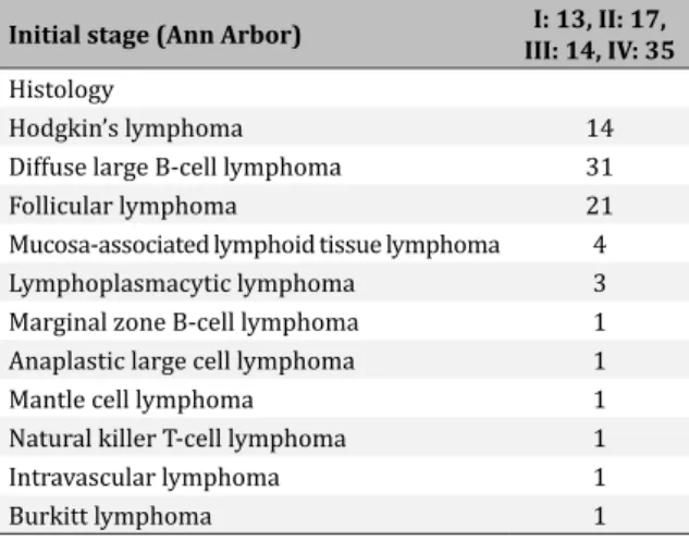
Figure 1. Kaplan-Meier curves for PFS in different settings
Table 2. Treatment characteristics of patients
Therapy Number
Chemotherapy and Rituximab 46
Chemotherapy and radiotherapy 6
Chemotherapy 12
Rituximab 9
Chemotherapy, radiotherapy and Rituximab 4
Surgical therapy and Rituximab 2
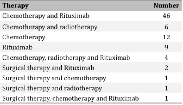
Figure 2. Comparison of PFS curves in patients with CR on PET versus those with incomplete remission on PET in HL or DLBCL (A) and other histological subtypes (B)
Table 3. Post-treatment evaluations, using CT/MRI and PET (n=82)
CR on PET
PR on PET
SD on PET
PD on PET
CR on CT/MRI 51 0 0 0
PR on CT/MRI 17 10 0 0
SD on CT/MRI 0 1 1 0
PD on CT/MRI 0 0 0 2
Abbreviations: CR, complete remission; PR, partial remission; PD, progressive disease; SD, stable disease
included in the recurrent group. The association between tumor marker levels before and after treatment and disease recurrence was also examined.
Statistical analysis
PFS was compared between the groups, using log-rank test. The comparison between each parameter (i.e., sIL-2R, LDH, and CRP) before and after treatment between the recurrent and non-recurrent groupswas performed, using Mann-Whitney U test. For all statistical analyses, P-value < 0.05 was
considered statistically significant. All the analyses
were performed, using Stat Mate IV (ATMS Co., Ltd., Tokyo, Japan).
Results
A total of 82 post-treatment evaluations in 79 ML patients were analyzed. The median follow-up duration was 45 months (range: 4-88 months). The results of post-treatment CT/MRI and PET evaluations were consistent in 78% of cases (64:82). CR on both CT/MRI and PET was the most common status (62%, 51:82). Among the CT/ MRI and PET evaluations with results that were not concordant, 94% (17/18) had incomplete remission on CT/MRI and complete remission on PET. These results are presented in (Table 3).
The 3-year PFS rates in cases with CR on both CT/MRI and PET (n=51), incomplete remission on CT/MRI and CR on PET (n=17), and incomplete remission on both CT/MRI and PET (n=14) were 90%, 94%, and 26%, respectively. Patients with incomplete remission on both CT/MRI and PET had
a significantly shorter PFS, compared to patients
with CR on both CT/MRI and PET (P<0.001) and those exhibiting incomplete remission on CT/MRI and CR on PET (P<0.001) (Figure 1). No significant difference was observed between cases with CR on both CT/MRI and PET and those with incomplete remission on CT/MRI and CR on PET (P=0.984).
As the RRC has recommended, patient response was assessed, using PET in patients with HL and DLBCL. These cases were divided into two groups
(HL or DLBCL, and other histological types) and the following results were obtained. Among patients with HL or DLBCL, those with CR on PET had a longer PFS, compared to patients with incomplete remission on PET (P<0.01) (Figure 2A). Among patients with other histological types, cases with CR on PET also had a longer PFS, compared to those with incomplete remission on PET (P<0.001) (Figure 2B).
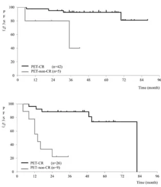
Further evaluation of combined CT/MRI and PET in patients with HL or DLBCL showed that incomplete remission on both CT/MRI and PET
was associated with a significantly shorter PFS,
compared to cases with CR on both CT/MRI and PET (P<0.05) and those exhibiting incomplete remission on CT/MRI and CR on PET (P<0.01)
(Figure 3A). No significant difference was
observed between cases with CR on both CT/MRI and PET and those showing incomplete remission on CT/MRI and CR on PET (P=0.568).
Among patients with other histological types, incomplete remission on both CT/MRI and PET
was associated with a significantly shorter PFS,
Figure 3. Comparison of PFS curves in different settings in HL or DLBCL (A) and other histological subtypes (B)
than the latter (P=0.058). No significant difference was observed between cases with incomplete remission on CT/MRI and CR on PET and those showing incomplete remission on both CT/MRI and PET (P=0.617).
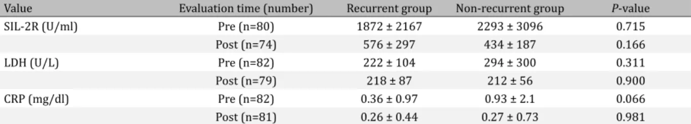
Disease recurrence occurred in 18 out of 82 cases, who were evaluated during the follow-up period. None of serum parameters (sIL-2R, LDH,
and CRP) differed significantly between recurrent
and non-recurrent cases (Table 4).
Discussion
This study showed that cases with post-treatment CR on both CT/MRI and PET and subjects with incomplete remission on CT/MRI and CR on PET had a longer PFS, compared to ML cases with incomplete remission on both CT/ MRI and PET. This study also showed that post-treatment PET evaluations were well correlated with the outcomes of patients with HL or NHL, exhibiting FDG uptake.
Even if post-treatment CT/MRI and PET results differed, post-treatment CR on PET was predictive of a long PFS among patients with HL or DLBCL. Among patients with other histological types, an evaluation consisting of a combination of CT/MRI and PET, rather than PET alone, is recommended.
Many ML subtypes have high cell density and exhibit high FDG uptake. In particular, aggressive types such as DLBCL and lymphoblastic lymphoma exhibit relatively high FDG uptakes (27). HL is composed of a few scattered neoplastic cells, referred to as Hodgkin and Reed-Sternberg cells, which are surrounded by mononuclear cells with a high metabolic activity (13). To evaluate patient response to treatment, FDG-PET is recommended for patients with aggressive NHL or HL, given the consistent FDG avidity of these lesions and their potential curability (11, 15).
Unlike CT and MRI, FDG-PET can detect metabolic changes earlier than morphological changes after treatment induction and can distinguish the remaining viable tumor tissues from inactive scar tissues after treatment (11, 14, 27, 28). A meta-analysis by Zijlstra et al. showed
that the pooled sensitivities and specificities of FDG-PET for evaluating patient response to
first-line therapy were 72% and 100% for NHL and 84% and 90% for HL, respectively (29). Our study also showed that regardless of CT/MRI evaluation, post-treatment PET evaluation is well correlated with the outcomes of patients with HL or DLBCL.
However, PET is limited in its ability to detect microscopic lesions, considering the partial volume
effect (PVE). Since PVE is influenced by factors
including tumor size and shape, tumor shrinkage after treatment tends to be underestimated in some cases (30, 31). Therefore, patient response
evaluations, based on PET findings, are associated
with a risk of failure to predict late relapse (29). About two-thirds of patients with HL and one-third of patients with NHL have residual masses visible on CT scan. They have negative PET results after the completion of therapy and clinical or other biochemical signs of relapse are absent in these cases; however, relapse occurs in less than 10% of patients with HL and 15-20% of patients with NHL (27).
In this study, 17 cases with CR on PET exhibited a residual mass lesion, based on CT/
MRI examinations; recurrence was confirmed in
three cases during the follow-up after the end of
treatment. Although no statistically significant
difference was found between patients with CR on both CT/MRI and PET and those exhibiting incomplete remission on CT/MRI and CR on PET, subjects with morphological changes, accompanied by metabolic alterations after therapy, tended to have a longer PFS; this tendency was particularly observed in patients with histological types other than HL and DLBCL.
Table 4. Comparison of pre- and post-treatment serum parameters and disease recurrence
Value Evaluation time (number) Recurrent group Non-recurrent group P-value
SIL-2R (U/ml) Pre (n=80) 1872 ± 2167 2293 ± 3096 0.715
Post (n=74) 576 ± 297 434 ± 187 0.166
LDH (U/L) Pre (n=82) 222 ± 104 294 ± 300 0.311
Post (n=79) 218 ± 87 212 ± 56 0.900
CRP (mg/dl) Pre (n=82) 0.36 ± 0.97 0.93 ± 2.1 0.066
Post (n=81) 0.26 ± 0.44 0.27 ± 0.73 0.981
Abbreviations: Pre = pre-treatment, Post= post-treatment, SD = standard deviation Data are shown as mean±SD.
histological types, the most frequent diagnosis
was follicular NHL (21 cases). Among studies focusing on PET evaluations, several reports have indicated that a negative PET status at the end of the treatment is associated with a longer PFS, compared to a PET-positive status in patients with follicular NHL (32-34). The present study also showed that post-treatment CR on PET was predictive of a prolonged CR duration among patients with other histological types.
On the other hand, although a statistically
significant difference was not observed, patients
with CR on both CT/MRI and PET tended to have a longer PFS, compared to those with incomplete remission on CT/MRI and CR on PET.
Follicular lymphoma is an incurable lymphoma subtype. The relapse rate among patients with follicular lymphoma is higher than that of patients with other more aggressive lymphomas (35). A reduction in the number of viable tumor cells can lead to a decrease in the anatomical tumor size (36). Therefore, the combination of CT/MRI and PET is likely to be more advantageous than PET alone for predicting the prognosis of patients with other histological types.
In the current study, none of the examined serum markers were capable of predicting disease recurrence. Standardized uptake values on PET images may be a better marker of disease status in patients with NHL, compared to sIL-2R level (37). The advantage of imaging for the prediction of disease recurrence was also evident in the current study, which examined a relatively large number of cases and histological types and had a median follow-up duration of 45 months.
The present study had a few limitations. Firstly, the study population was comprised of cases with different HL and NHL subtypes, and the number of cases with some subtypes was low. Secondly, not all lesions with abnormal FDG uptake were histopathologically examined.
Conclusion
In conclusion, post-treatment CR on both
CT/MRI and PET was predictive of a long PFS in patients with ML. Post-treatment PET evaluations were well correlated with the outcomes of patients with ML, exhibiting FDG uptake. Among patients with HL or DLBCL, post-treatment CR on PET was predictive of a relatively long PFS. When predicting the prognosis of patients with other histological types, a combination of CT/MRI and PET, rather than PET alone, is recommended.
Acknowledgments
The authors would like to thank the staff of the Department of Nuclear Medicine at Osaka University Hospital for their assistance. This study was supported in part by
Grants-in-Aid for Scientific Research (S) (24229008), for Scientific Research (25461822,
26462204, 24591758), and for Young Scientists (B) (25861099, 25861095) from the Ministries of Education, Culture, Sports, Science and Technology, Japan. This study won a second-place poster prize in the tumor section of the 52nd Annual Scientific Meeting by the Japanese Society
of Nuclear Medicine.
References
1. Cheson BD, Horning SJ, Coiffier B, Shipp MA, Fisher RI, Connors JM, et al. Report of an international workshop to standardize response criteria for non-Hodgkin's lymphomas. NCI Sponsored International Working Group. J Clin Oncol. 1999;17(4):1244-53. 2. Lister TA, Crowther D, Sutcliffe SB, Glatstein E,
Canellos GP, Young RC, et al. Report of a committee convened to discuss the evaluation and staging of patients with Hodgkin’s disease: Cotswolds meeting. J Clin Oncol. 1989;7(11):1630–6.
3. Weber WA. Assessing tumor response to therapy. J Nucl Med. 2009;50(Suppl 1):1-10.
4. Hutchings M, Barrington SF. PET/CT for therapy response assessment in lymphoma. J Nucl Med. 2009;50:(Suppl 1):21-30.
5. Moog F, Bangerter M, Diederichs CG, Guhlmann A, Merkle E, Frickhofen N, et al. Extranodal malignant lymphoma: detection with FDG PET versus CT. Radiology. 1998;206(2):475-81.
Buck A, von Schulthess GK. Whole-body positron
emission tomography using fluorodeoxyglucose
for staging of lymphoma: effectiveness and comparison with computed tomography. Eur J Nucl Med. 1998;25(7):721-8.
7. Newman JS, Francis IR, Kaminski MS, Wahl RL. Imaging of lymphoma with PET with 2-[F-18]-
fluoro-2-deoxy-D-glucose: correlation with CT.
Radiology. 1994;190(1):111-6.
8. Buchmann I, Reinhardt M, Elsner K, Bunjes D, Altehoefer C, Finke J, et al. 2-(fluorine-18) fluoro-2-deoxy-D-glucose positron emission tomography in the detection and staging of malignant lymphoma. A bicentertrial.Cancer. 2001;91(5):889-99.
9. Rhodes MM, Delbeke D, Whitlock JA, Martin W, Kuttesch JF, Frangoul HA, et al. Utility of FDG-PET/ CT in follow-up of children treated for Hodgkin and non-Hodgkin lymphoma. J Pediatr Hematol Oncol.2006;28(5):300-6.
10. Tatsumi M, Cohade C, Nakamoto Y, Fishman EK, Wahl RL. Direct comparison of FDG PET and
CT findings in patients with lymphoma: initial
experience. Radiology.2005;237(3):1038-45. 11. Juweid ME, Wiseman GA, Vose JM, Ritchie JM,
Menda Y, Wooldridge JE, et al. Response assessment of aggressive non-Hodgkin's lymphoma by integrated International Workshop Criteria and
fluorine-18-fluorodeoxyglucose positron emission
tomography.J Clin Oncol. 2005;23(21):4652-61. 12. Isasi CR, Lu P, Blaufox MD. A metaanalysis of
18F-2-deoxy-2-fluoro-D-glucose positron emission
tomography in the staging and restaging of patients with lymphoma. Cancer. 2005;104(5):1066-74. 13. Gallamini A. Positron emission tomography
scanning: a new paradigm for the management of Hodgkin's lymphoma. Haematologica. 2010;95(7):1046-8.
14. Isohashi K, Tatsumi M, Higuchi I, Inoue A, Nakajo K, Ishikawa J, et al. 18F-FDG-PET in patients with
malignant lymphoma having long-term follow-up: staging and restaging, and evaluation of treatment response and recurrence. Ann Nucl Med. 2008;22(9):795-802.
15. Cheson BD, Pfistner B, Juweid ME, Gascoyne RD, Specht L, Horning SJ, et al. Revised ResponseCriteria for Malignant Lymphoma. J Clin Oncol. 2007;25(5):579-86.
16. Lavely WC, Delbeke D, Greer JP, Morgan DS, Byrne DW, Price RR, et al. FDG PET in the follow-up management of patients with newly diagnosed
Hodgkin and non-Hodgkin lymphoma after
first-line chemotherapy. Int J Radiat Oncol Biol Phys. 2003;57(2):307-15.
17. Jerusalem G, Warland V, Najjar F, Paulus P, Fassotte MF, Fillet G, et al. Whole-body 18F-FDG PET for the evaluation of patients with Hodgkin's disease and non-Hodgkin's lymphoma. Nucl Med Commun. 1999;20(1):13-20.
18. Zinzani PL, Stefoni V, Tani M, Fanti S, Musuraca G, Castellucci P, et al. Role of [18F] fluorodeoxyglucose positron emission tomography scan in the
follow-up of lymphoma. J Clin Oncol. 2009;27(11):1781-7. 19. Juweid ME, Stroobants S, Hoekstra OS, Mottaghy
FM, Dietlein M, Guermazi A, et al. Use of positron emission tomography for response assessment of lymphoma: consensus of the Imaging Subcommittee of International Harmonization Project in Lymphoma. J Clin Oncol. 2007;25(5):571-8.
20. Pyo J, Won Kim K, Jacene HA, Sakellis CG, Brown JR, Van den Abbeele AD. End-TherapyPositronEmissio nTomography for TreatmentResponseAssessment in FollicularLymphoma: A SystematicReview and Meta-analysis. Clin Cancer Res. 2013;19(23):1–12. 21. Goto N, Tsurumi H, Goto H, Shimomura YI, Kasahara
S, Hara T, et.al. Serum soluble interleukin-2 receptor (sIL-2R) level is associated with the outcome of patients with diffuse large B cell lymphoma treated with R-CHOP regimens. Ann Hematol. 2012;91(5):705-14.
22. Shimomura Y, Tsurumi H, Sawada M, Yamada
T, Hara T, Fukuno K, et.al. Clinical significance
of serum soluble interleukin-2 receptor level in patients with non-Hodgkin's lymphoma. RinshoKetsueki.1999;40(8):639-45.
23. Yoshizato T, Nannya Y, Imai Y, Ichikawa M,
Kurokawa M. Clinical significance of serum-soluble
interleukin-2 receptor in patients with follicular lymphoma. Clin Lymphoma Myeloma Leuk. 2013;13(4):410-6.
24. Wieland A, Kerbi R, Berqhold A, Schwinger W, Mann G, Urban C.C-reactive protein (CRP) as tumor marker in pediatric and adolescent patients with Hodgkin disease. Med Pediatr Oncol. 2003;41(1):21-5.
25. Bakhshi S, Radhakrishnan V, Sharma P, Kumar R, Thulkar S, Vishnubhatla S, et al. Pediatricnonlymphoblastic non-Hodgkin lymphoma: baseline, interim, and posttreatment
PET/CT versus contrast-enhanced CT for evaluation--a prospective study. Radiology. 2012;262(3):956-68.
26. Kelly KM, Hodgson D, Appel B, Chen L, Cole PD, Horton T, et al. Children's Oncology Group's 2013 blueprint for research: Hodgkin lymphoma. Pediatr Blood Cancer. 2013;60(6):972-8.
27. Baba S, Abe K, Isoda T, Maruoka Y, Sasaki M, Honda H. Impact of FDG-PET/CT in the management of lymphoma. Ann Nucl Med. 2011;25(10):701-16. 28. Wahl RL, Zasadny K, Helvie M, Hutchins GD, Weber
B, Cody R. Metabolic monitoring of breast cancer chemohormonotherapy using positron emission tomography: initial evaluation. J Clin Oncol. 1993;11(11):2101–11.
29. Zijlstra JM, Lindauer-van der Werf G, Hoekstra
OS, Hooft L, Riphagen II, Huijgens
PC.18F-fluoro-deoxyglucose positron emission tomography for post-treatment evaluation of malignant lymphoma: a systematic review. Haematologica. 2006;91(4):522-9.
31. Srinivas SM, Dhurairaj T, Basu S, Bural G, Surti S,
Alavi A. A recovery coefficient method for partial
volume correction of PET images. Ann Nucl Med.2009;23(4):341-8.
32. Lu Z, Lin M, Downe P, Chong S, Ling S. The prognostic value of mid- and post-treatment [18F]
fluorodeoxyglucose (FDG) positron emission
tomography (PET) in indolent follicular lymphoma. Ann Nucl Med.2014;28(8):805-11.
33. Lopci E, Zanoni L, Chiti A, Fonti C, Santi I, Zinzani PL, et al. FDGPET/CT predictive role in follicular lymphoma. Eur J Nucl Med Mol Imaging. 2012;39(5):864-71.
34. Alcantara M, Dupuis J, Mareschal S, Julian A, Cottereau AS, Becker S, et al. PET/CT before autologous stem cell transplantation predicts outcome in refractory/relapsed follicular
lymphoma. Eur J Nucl Med Mol Imaging. 2015; 42(2):215-21.
35. Bodet-Milin C, Eugène T, Gastinne T, Frampas E, Le Gouill S, Kraeber-Bodéré F. FDG-PET in Follicular Lymphoma Management.J Oncol. 2012;2012:370272.
36. Wu X, Pertovaara H, Korkola P, Vornanen M, Järvenpää R, Dastidar P, et al. Early interim PET/CT predicts post-treatment response in diffuse large B-cell lymphoma. Acta Oncol. 2014;53(8):1093-9. 37. Tatsumi M, Sugahara H, Higuchi I, Fukunaga H,