ABSTRACT
Assessment of enamel-dentin caries lesions
Marianna Guanaes Gomes TORRES1, Aline da Silva SANTOS2, Frederico Sampaio NEVES3, Marcel Lautenschlager ARRIAGA4, Paulo Sérgio Flores CAMPOS5, Iêda CRUSOÉ-REBELLO5
1- DDS, MSc, PhD student, Department of Oral Radiology, School of Dentistry, Federal University of Bahia, Salvador, BA, Brazil. 2- DDS, Department of Oral Radiology, School of Dentistry, Federal University of Bahia, Salvador, BA, Brazil.
3- DDS, MSc, PhD student, Department of Oral Diagnosis, Piracicaba Dental School, State University of Campinas, Piracicaba, SP, Brazil. 4- DDS, MSc, PhD, Department of Public Health, School of Dentistry, Federal University of Bahia, Salvador, BA, Brazil.
5- DDS, MSc, PhD, Department of Oral Radiology, School of Dentistry, Federal University of Bahia, Salvador, BA, Brazil.
Corresponding address: !""!!"!#$%&&'(")***+!!/ - e-mail: [email protected]
5HFHLYHG-XO\0RGL¿FDWLRQ0D\$FFHSWHG2FWREHU
O
caries using photostimulable phosphor plates. Material and Methods: The ability to detect enamel-dentin occlusal caries in 607 premolars and molars from 47 patients ! " # $ %! %&! ' (! '' ' ) * +, using Digora® (Soredex Medical Systems, Helsinki, Finland) phosphor plates. The plates ' ' ' ) 3 "5 8 9) ! (5) teeth based on the radiographic criteria proposed in a previous study. Descriptive analysis ; '' +< = ) > ( ' ? & = @ B ' @ +B) 3 ! CE ' ! circumscribed, in dentin under occlusal enamel (enamel-dentin caries lesions). Conclusions: I ' =! diagnosed by radiographic images as having enamel-dentin caries, no caries could be detected by clinical examination.Key words: " ) % ') " ')
phosphor.
INTRODUCTION
The detection of caries lesions is still a matter of investigation in Dentistry. Epidemiologic studies prevalent than cavitated lesions3,8. Extensive lesions
can develop in dentin beneath an apparently sound ! '= ! may ultimately undermine the enamel7.
N ! ? caries, is a phenomenon that leads to formation of highly mineralized, strengthened enamel !
progress gradually and the carious lesion might ( ? at the enamel surface. There is no consensus on the cause of enamel-dentin caries. It has been ! ' =! the high remineralization capacity of fluoride ? ! (' Q) * alternative hypothesis is that the phenomenon and dentin7,13,14.
0 Sound (absence of demineralization or color change)
1 #89;9<=>99 ?
2 #8E<9;9<=E9? =>9
3 Dentine lesion
4 Restored
5 Sealed
Figure 1- Weerheijm, Gruythuysen and Van Amerongen19 (1992) criteria for clinical examination
dentin, relatively high concentrations of dentin ' = ' = confer the same degree of protection on the more ) to progress undetected beneath an apparently '= & ) The potential also exists for remineralization of an ! ? & = of detecting the enamel-dentin caries7.
3 ? ( a distinct entity and it has been asserted that to ' ' = () U! (' = under apparently sound enamel7,17,18. Thus, the
detection of enamel-dentin caries, or hidden ! =! diagnosis methods is fundamental to determine the presence of these lesions. The radiographic exam, used as a complement of the clinical visual exam, is a disposable tool for dental researchers and professionals4,5,20.
D i g i t a l i m a g e s y s t e m s d e m o n s t r a t e diagnostic accuracy comparable to conventional radiography4-6,11,15,22. Digital systems present a
= (' 50-90%. Digital receptors are more sensitive to radiation than film, require less exposure to radiation, and chemical processing are not needed5,21.
Digora® (Soredex Medical Systems, Helsinki,
Finland) is one of the several digital systems that ' '! ' 'Q particles that are sensitive to x-rays, forming a latent image. The optic plate is scanned on a Digora®
' ! = ' ' ) =
[ 10,21.
The present study evaluated the detection of enamel-dentin occlusal caries using photostimulable '' ') ' evaluate the frequency of enamel-dentin occlusal caries in relation to gender and age; to compare the occurrence of enamel-dentin occlusal caries in different tooth groups; to compare detection
of enamel-dentin occlusal caries based on digital = the criteria proposed by Weerheijm, Gruythuysen and Van Amerongen19 (1992).
MATERIAL AND METHODS
3 "' I! Pediatric Dentistry, and Dentistry II of the School of Dentistry of the Federal University of Bahia @#{$#%*B! ' !
prevalence of enamel-dentin caries18!
evaluated clinically. The purpose of the research (' ' forms for minors and patient informed consent '| ) '' #{$#%*[ Research Ethics Committee (Protocol number 0006.0.368.000-07).
Before the examination sessions, verbal and ' performed. For the calibration tests, the occlusal surfaces in 10 patients (maxillary and mandibular B proposed by Weerheijm, Gruythuysen and Van
Amerongen19 @~~9B @# B! !
! ( agreement of 0.98 and kappa value of 0.92.
After supervised toothbrushing, the teenagers ( ' '! ' ! ' dry the surfaces to be evaluated. For the visual (! ' to hold back the soft tissue and permit better visualization of the area. The visual examination ' ) + ' residual dental plaque by applying soft pressure. The occlusal surfaces of permanent premolars ! the criteria of Weerheijm, Gruythuysen and Van Amerongen19 (1992). Scores of 1 and 2 represent
0 Absence of any visible radiolucency in dentine under occlusal enamel
3 Presence of visible radiolucency, circumscribed, in dentine under occlusal enamel-enamel-dentine caries
lesions (only high quality pattern radiographic images)
4 Radiopaque image suggesting restoration
6 Impossible to judge and density measurement and histogram tools may be applied
Figure 2- Weerheijm, Gruythuysen and Van Amerongen19 (1992) criteria for radiographic examinations
Figure 3- Radiographic aspects of the occlusal surfaces according to Weerheijm, Gruythuysen and Van Amerongen19
(1992) criteria
' ( ' the treatment protocol in this study for patients ' '' ! '' ' ' )
Using the Digora® digital system, phosphor
' ' U = holders (JON, São Paulo, SP, Brazil), all patients ' Timex 70 C machine (Gnatus, Ribeirão Preto, SP, %&B! =( ' E ?'! *! ) ) % ' )
(' radiologists, at different times, in a dimly lit room, ? ? to the monitor, so that the image seen by the observer corresponded to the real image size, and shielding it from the light emitted by the screen.
3 "® for
8 9) ! (®. The radiologists
& tools for better visualization during the scoring of the teeth based on the aforementioned criteria19
@# 9B) # ' ' of the occlusal surfaces according to the criteria adopted in the study19. In case of disagreements,
both observers evaluated the images again until they came to a consensus.
=
of Weerheijm, Gruythuysen and Van Amerongen19
(1992) to archive the data from the clinical and radiographic examinations of each patient. The ) "' ; '' ' )+ =)
* ! ' visible radiolucency, circumscribed, in dentin under occlusal enamel (enamel-dentin caries lesions)
' evaluated.
RESULTS
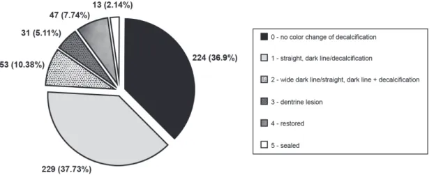
( CE ' evaluated (17 males and 30 females). The mean 9)E 9)+ ) { ,E ! 9 ! E~ = ! C+ ' ,9 = ' @# CB) distributed based on the criteria adopted in the study19 (Figure 5).
? & = @ B ' @99~E)E<B! by sound teeth (scored as 0, 224-36.90%). The ' ' + 9)C< the total sample.
' (' ! high inter-examiner reproducibility value (kappa value 0.89). The results for the radiographic # ,) CE
on Weerheijm, Gruythuysen and Van Amerongen19
@~~9B )
Of the 47 patients examined, 24 did not present dentin occlusal caries, and 23 had enamel-dentin occlusal caries. The distribution of patients 9) ; ' enamel-dentin caries lesions for females and males, = ' @')~,E9B @ 9B)
Figure 4- Distribution of evaluated teeth based on tooth groups (1st premolar, 2nd premolar, 1st molar and 2nd molar)
Criteria Enamel-dentine caries lesions (%)
0 7 (14.89)
1 10 (21.28)
2 24 (51.06)
3 5 (10.64)
4 0
5 1 (2.13)
Table 1- Distribution of teeth with enamel-dentin caries
based on Weerheijm, Gruythuysen and Van Amerongen19
(1992) clinical criteria
Figure 5- Distribution of Weerheijm, Gruythuysen and Van Amerongen19 (1992) clinical criteria on the evaluated teeth
Figure 6- Distribution of Weerheijm, Gruythuysen and Van Amerongen19 (1992) radiographic criteria on the evaluated teeth
= @')EB prevalence of enamel-dentin lesions among these age groups.
When evaluating different dental groups for the presence of enamel-dentin caries, 14.29% (17 teeth) of the second molars presented
! +)9< @9, B = ! 1.47% (2 teeth) of the second premolars and 1.51% @9 B = ') ; in molars (Figure 7).
Enamel-dentine caries
Gender Total (%)
Male (%) Female (%)
Absence 9 (37.50) 15 (62.50) 24 (51.06)
Presence 8 (34.78) 15 (65.21) 23 (48.94)
Total 17 (36.17) 30 (63.83) 47 (100.00)
Table 2- Distribution of patients by gender with and without enamel-dentin caries (p=0.9672)
Clinical criterion Enamel-dentine caries Total (%)
2nd M (%) 1st M (%) 2nd PM (%) 1st PM (%)
0 5 (29.41) 1 (3.85) 0 1 (50.00) 7 (14.89)
1 1 (5.88) 7 (26.92) 1 (50.00) 1 (50.00) 10 (21.28)
2 8 (47.06) 16 (61.54) 0 0 24 (51.06)
3 3 (17.65) 1 (3.85) 1 (50.00) 0 5 (10.64)
4 0 0 0 0 0
5 0 1 (3.85) 0 0 1 (2.13)
Total 17 (36.17) 26 (55.32) 2 (4.25) 2 (4.25) 47 (100)
Table 3- Frequency of different clinical aspects of teeth with enamel-dentin caries related to tooth group (p=0.5)
(M= molar; PM= premolar)
Figure 7- Distribution of Weerheijm, Gruythuysen and Van Amerongen19 (1992) radiographic criteria on the different tooth
groups (M= molar; PM= premolar)
nd st nd st
! = ! ' = 'B) # = ! ' 9 @ ? & =B @')+B)
When evaluating the distribution of enamel-dentin lesions on maxillary and mandibular teeth, ( ' @, teeth) than the mandibular premolars and molars @ B! = @')E+B @ CB)
Of the 47 teeth diagnosed by radiographic ( ! ~
opened for treatment and all of them presented ) ' ; endodontic treatment due to pulp infection caused by an extensive caries lesion. The remaining teeth ' @Q therapy and/or sealant placement) and monitoring, based on a multidisciplinary decision.
=
Weerheijm, Gruythuysen and Van Amerongen19
(1992) as score 6 on radiographic images (Figure 3) had the clinical option of monitoring the lesion.
DISCUSSION
The total number of teeth evaluated by radiographic examination (559) did not correspond to the total number of teeth evaluated clinically @,EB! C ! due to incomplete imaging of some teeth in the radiographs.
Based on tooth eruption chronology, 1st molars
erupt earlier than 2nd molars and then 1st and 2nd
Enamel-dentine caries
Arch Total (%)
Upper (%) Lower (%)
Absence 270 (52.73) 242 (47.27) 512 (91.59)
Presence 15 (31.91) 32 (68.09) 47 (8.41)
Total 285 (50.98) 274 (49.02) 559 (100.00)
Table 4- Distribution of enamel-dentin caries on maxillary and mandibular premolars and molars (p=0.075)
and are also frequently restored2. That can explain
the occurrence of more sealed surfaces on 1st molars
(11 1st molars of 13 sealed teeth) and the occurrence
of restorations in this same group of teeth (27 1st
molars of 47 restored teeth); premolars presented completely sound occlusal surfaces (score 0) in a ' @++ premolars and 69 molars).
In the present study, 32 teeth presented cavities during clinical examination, and 47 teeth presented enamel-dentin occlusal caries, demonstrating a higher occurrence of non-cavitated lesions than cavitated lesions.
The dentin lesions detected on the radiographs ! ' a different pattern regarding age and gender @ 9B! =
of Weerheijm, Gruythuysen and Van Amerongen19
(1992).
3 ' 9 ! ' @9, B! ' + @~ B , @9 B! =) the fact that in the older teenagers, most of the ) 3 9 ! + , ! ,! , ~! E 9 ! respectively.
When considering the prevalence of enamel-dentin caries in maxillary and mandibular molars,
Weerheijm, Gruythuysen and Van Amerongen19
(1992) affirmed that there is no significant ( arches. Our study found a higher occurrence of enamel-dentin caries in mandibular molars than ( ! = @ CB)
Comparing the presence and distribution of enamel-dentin caries lesions in the various tooth '! = ' ' cavity had the highest number of enamel-dentin ) = + CE ! ( = !
second mandibular molars in 13 cases and second ( C ) { enamel-dentin caries detected, only four premolars )
# ! ! = ! @# B (Table 3).
> ( complementary radiographic examinations do not detect the occurrence of caries lesions in some teeth. In many examinations, radiographic images ' caries lesion on proximal surfaces9,11. This situation
'( ' studies involving a larger number of individuals. In these cases, no radiographic images are taken and caries lesions are under-reported3.
According to the guidelines of the American Dental Association1 (2006), radiographic diagnosis
should only be used after clinical examination, considering the dental and general health needs of the patient. Because each patient is different, radiographic examination should be individualized1.
3 ' ! dentin caries could not be detected in clinical examination: 8.41% of the teeth clinically diagnosed as sound presented dentin caries (Figure 7). Thus, ' part of the initial routine dental examination in
) ! )9 (2009) support the
' ' ' the initial routine dental examination of children because the prevalence of hidden occlusal caries detectable only radiographically is approximately 12%.
Civera, et al.5 (2007) also concluded that the use
of radiographic techniques, conventional or digital, increases the number of caries lesions diagnosed compared to clinical examination alone; 3.23 times 9) '! ' to clinical examination.
Digital radiography is a reality, and its reduced ' = patient. Besides having similar accuracy for the detection of caries lesions as conventional radiography4,6,11,15, it also has the advantage of
; = '21. More
studies on the application of density measurement tools and histograms are necessary.
structure, function and esthetics12,16. Non-invasive
@Q ' | sealant placement), by consensual multidisciplinary decision, for 38 of the 47 teeth diagnosed as having enamel-dentin caries.
CONCLUSIONS
It is important to note that radiographic examination, even using digital systems, presents considerable limitations; for example, one cannot determine if the lesion is active or not, or the presence or absence of cavities. Research on detection of enamel-dentin caries is still ongoing and this study permits the implementation of future studies in this area. It is important to have a precise ! ' ' patients that present clinically sound dental units.
ACKNOWLEDGMENTS
We are grateful to Fapesb (Edital Temático 2004) and CNPq for their contributions for this study.
REFERENCES
* " * I = *) use of dental radiographs: update and recommendations. J Am Dent Assoc. 2006;137:1304-12.
2- Alvarez JO, Navia JM. Nutritional status, tooth eruption, and ) * I ) ~~C~CE9,) * *! I! ! #! *I! Ambrosano GMB. Assessment of different methods for diagnosing dental caries in epidemiological surveys. Commun Dent Oral Epidemiol. 2004;32:418-25.
4- Castro VM, Katz JO, Hardman PK, Glaros AG, Spencer P. In vitro ' =
in the detection of approximal caries. Dentomaxillofac Radiol. 2007;36:138-42.
5- Civera VG, Silla JMA, Company JMM, Navarro LF. Clinical and radiographic diagnosis of approximal and occlusal dental caries in a ? '') { { I %) 9E9N9+9E) 6- Ferreira RI, Haiter-Neto F, Tabchoury CPM, Paiva GAN, Bóscolo FN. Assessment of enamel demineralization using conventional, digital, and digitized radiography. Braz Oral Res. 2006;20:114-9. 7- Lynch RJM, Ten Cate JM. The effects of adjacent dentine blocks on the demineralization and remineralization of enamel in vitro.
Caries Res. 2006;40:38-42.
I! * *! ! &? #I! *I! Ambrosano GM. Comparison of diagnostic methods for dental caries. J Dent Child (Chic). 2003;70:115-9.
~ %! 8! & ! # "! U ) I ' ') * " ) 9~+C9)
? N! 8 #) " ' ) Contemp Dent Pract. 2002;3:23-39.
11- Peker I, Toraman Alkurt M, Altunkaynak B. Film tomography ' = ' '( caries detection. Dentomaxillofac Radiol. 2007;36:495-9. 12- Pretty IA. Caries detection and diagnosis: novel technologies. J Dent. 2006;34:727-39.
>? "! N? >! ! N >! * ! ) * ' radiographic assessment and subtraction radiography in detecting demineralization in occlusal surfaces. Caries Res. 2007;41:121-8. C & 8I! I I! & *N! > RP, Corona SAM, Palma-Dibb RG. Validity and reproducibility of different combinations of methods for occlusal caries detection: an in vitro comparison. Caries Res. 2006;40:194-201.
15- Syriopoulos K, Sanderink GCH, Velder XL, Van Der Stelt PF. Radiographic detection of approximal caries: a comparison of = ) "( >) 2000;29:312-8.
16- Thompson VP, Kaim JM. Nonsurgical treatment of incipient and hidden caries. Dent Clin North Am. 2005;49:905-21.
17- Weerheijm KL. Occlusal “hidden caries”. Dent Update. 1997;24:182-4.
18- Weerheijm KL, Groen HJ, Bast AJ, Kieft JA, Eijkman MA, Van Amerongen WE. Clinically undetected occlusal dentine caries: a radiographic comparison. Caries Res. 1992;26:305-9.
19- Weerheijm KL, Gruythuysen RJ, Van Amerongen WE. Prevalence of hidden caries. ASDC J Dent Child. 1992;59:408-12. 9 8& *) % ' detection of caries lesions. J Dent Res. 2004;83:C72-5.
9 8& *) * ' ) "( >) 2006;35:307-14.