Clinicoradiological Session
Case 2/2008 – Twenty Two Year Old Male with Ventricular Septal
Defect of Persistently Small Proportions
Edmar Atik
Clínica do Dr. Edmar Atik - São Paulo, SP - Brazil
Mailing address: Edmar Atik •
InCor - Av. Dr. Enéas Carvalho de Aguiar, 44 - 05403-000 - São Paulo, SP - Brazil E-mail: [email protected]
Key words
Ventricular septal defect, congenital cardiopathy, clinical.
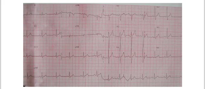
normal electrical potentials. ÂP: + 50o, ÂQRS: +70o, ÂT: +50o (Fig. 1).
Radiographic image
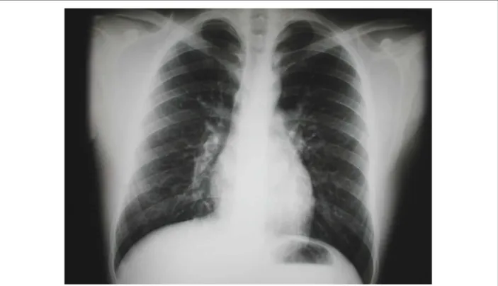
Cardiac area shows normal dimensions and slightly increased vascular network (rete), primarily in the hilar region (Fig. 2).
Diagnostic impression
This image is consistent with a slight shift of blood flow from left to right as in congenital acyanogenic cardiopathies such as ventricular septal defect.
Differential diagnosis
Other cardiopathies should be considered, such as ASD (atrial septal defect), PDA, and AV fistulas, as long as they
Clinical data
Cardiac murmur with early detection in the first days of life which remained unchanged over time. During this period, the patient experienced no symptoms or clinical complications and was able to participate normally in routine physical activities.
Physical examination
Patient was eupneic, had good skin color, and pulses were palpated on all 4 limbs. Weight: 64 Kg, Height: 170cm, BP: 125/70 mm Hg, HR: 76 bpm. Aorta was not palpated at the supraesternal notch. Precordium showed no deformities or impulsions. Ictus cordis was not palpated. Normal heart sounds, with inconsistent splitting of second heart sound. Rough ejection systolic murmur +/++ in 3rd, 2nd, and 4th left intercostal spaces, not accompanied by thrills.
Lungs and abdomen: no abnormalities.
The electrocardiogram showed normal sinus rhythm and
Figure 1 -Normal electrocardiogram.
Clinico Radiological Session
Edmar AtikArq Bras Cardiol 2008; 90(4): 274-275
Figure 2 -Chest X-ray showing cardiac area within limits of normality and slightly increased pulmonary arterial network.
cause minor repercussions.
Diagnostic confirmations
Clinical elements easily point towards the diagnosis of ventricular septal defect with mild hemodynamic repercussions. From the first days of life and to date, the echocardiogram has shown outlet muscular VSD (ventricular septal defect) with 2 to 3 mm diameter and normal cardiac chambers (Fig. 3).
Treatment
Always considered conservative, with close clinical observation in light of the mild clinical and laboratorial manifestations and following rigid prophylactic orientation for infectious endocarditis.
The same orientation continued to be adopted because of the perspective of life expectancy similar to that of the normal population.
Figure 3 -Demonstration of ventricular septal defect on the echocardiogram, color low, in longitudinal parasternal view. LA - left atrium, AO - aorta, RV - right ventricle,
LV - left ventricle.