Clinicoradiological Session
Case 2/2007 – A Two-Year-Old Child with Ventricular Septal Defect
and Right Ventricular Anomalous Muscle Bundle
Edmar Atik
Instituto do Coração do Hospital das Clínicas – FMUSP – São Paulo, SP - Brazil
Mailing address: Edmar Atik •
InCor – Av. Dr. Enéas C. de Aguiar, 44 – 05403-000 – São Paulo, SP - Brazil E-mail: [email protected]
Clinical data
A two-year-old white male child in whom a heart murmur was detected in a routine examination when he was only 12 days old. Since then, it was noted that he tired during breast-feeding, a symptom that improved with digoxin. Physical examination showed dyspnea +, normal pulses, body weight 10.3 kg, body height 88 cm, blood pressure 91/69 - 75 mmHg, and heart rate 100 bpm. No cyanosis was observed. The aorta was not palpable in the suprasternal notch. Chest examination revealed mild impulses along the left sternal border and a valvular and muscular apical impulse ++ located at the 4th
left intercostal space. Heart sounds were loud, and the first sound was more pronounced in the tricuspid area. The second sound was single. A harsh grade II holosystolic murmur was heard in the 2nd, 3rd, and 4th left intercostal spaces, irradiating
to the right sternal border. The liver was palpable 1 cm from the right costal margin.
The electrocardiogram showed sinus rhythm and evidence of overload on both ventricles and the left atrium. There were broad RS waves from V2 to V5. P wave was negative in V1 and V2. P-axis:+70o , QRS-axis:+120o,
T-axis:+40o.
Radiographic examination
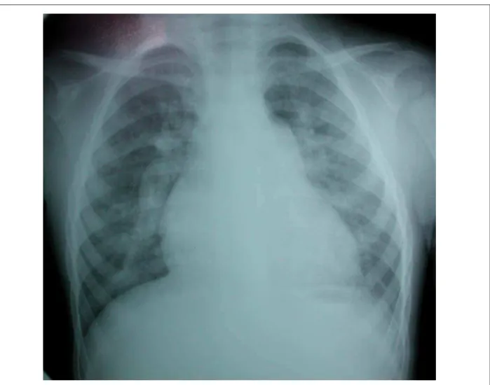
Chest X-ray showing an enlarged cardiac silhouette due to the right atrium and left ventricle. The mid-arch is bulged, and the pulmonary vascularity is increased (Figure 1).
Diagnostic impression
This image suggests the presence of combined ventricular and atrial septal defects, in view of the enlarged right atrium and left ventricle.
Differential diagnosis
Such enlargements also suggest the possibility of high ventricular septal defect associated with anomalous right ventricular muscle bundle. Ductus arteriosus-like heart disease with pulmonary hypertension must also be considered when the pulmonary trunk is enlarged. Another possibility is partial atrioventricular septal defect coupled with atrial septal defect and valvular regurgitation.
Diagnostic confirmation
Clinical findings were consistent with ventricular septal defect with moderate repercussion. The echocardiogram revealed a 10-mm muscular (trabecular) VSD with anomalous right ventricular muscle bundle, in addition to posterior deviation of the interventricular septum and fibrous subaortic ring (Figure 2). Measurements were the following: right ventricle 20 mm, left ventricle 38 mm, left atrium 32 mm, aorta 20 mm, septum 4 mm, posterior wall 5 mm, and myocardial fiber shortening fraction 39%. Angiographic images during cardiac catheterization revealed the presence of high ventricular septal defect and anomalous right ventricular muscle bundle (Figure 2). Hemodynamic data were as follows: right atrium: 5, right ventricle: 60/10, pulmonary trunk: 60/30 - 40, pulmonary wedge pressure: 13, left ventricle: 80/10, aorta: 75/45 - 55 mm Hg. Pulmonary flow was 3.8 l/min/m2
and systemic flow was 1.5 l/min/m2, pulmonary resistance was
2.9 UW and systemic resistance was 13.5 UW.
Management
During surgery, a high muscular ventricular septal defect measuring 10 mm in diameter was found. The anomalous right ventricular muscle bundle, obstructing the RV outflow tract, had to be resected. The patient evolved favorably.
Key words
Ventricular septal defect, right ventricular anomalous muscle bundle.
Clinico Radiological Session
Edmar AtikArq Bras Cardiol 2007; 88(5) : 545-546 Fig. 1 -Chest X-ray showing increased pulmonary arterial vasculature. When the right atrium and left ventricle are enlarged, a communication between chambers must be considered.
Fig. 2 -Cross-sectional echocardiographic (A) and angiographic (B) images showing anomalous right ventricular muscle bundle (A). An obstruction between inflow and outflow tracts (arrow) can be clearly seen, in addition to ventricular septal defect. RA - right atrium; Ao - aorta; VSD - ventricular septal defect; PT - pulmonary trunk; RV - right ventricle; PV - pulmonary valve.
RV
BUNDLE
PV
RA VSD
AO
PT
RV