ARTICLE
Aquaporin 9 in rat brain after severe traumatic
brain injury
Aquaporina 9 em cérebro de rato após traumatismo cerebral grave
Hui Liu1, Mei Yang1, Guo-ping Qiu1, Fei Zhuo1, Wei-hua Yu1, Shan-quan Sun1, Yun Xiu2
Predominantly caused by motor vehicle accidents, the traumatic brain injury(TBI) is the leading cause of death and severe disability amongpeople under the age of 45 in Western industrialized countries1. Almost all patients sufer from severe
disorder of energy metabolism2, ionic dysfunction3, and water
homeostasis4 after TBI. Experimental evidences indicate that
aquaporin 9 (AQP9), which is one member of the water chan-nel family in brain, can facilitate the low of water and perme-ate glycerol, monocarboxylperme-ates, and urea through the plasma membrane and it is involved in water homeostasis and neu-ronal energy metabolism in brain5. In brain ischemia models,
the studies show that AQP9 may be a helper in the regulation
of postischemic edema6. Yet, as far as we know, little is known
about AQP9 in brain after TBI. he present study was per-formed to study whether brain AQP9 expression is altered and what are the possible roles of AQP9 in brain after TBI.
METHODS
Experimental animal
his study had an ethical approval by the Ethics Committee of Chongqing Medical University (CQMU). A total of 480 adult male Sprague Dawley rats supplied by the Experimental
1 Institute of Neuroscience, Chongqing Medical University, Chongqing, China;
2 Institute of Life Science, Chongqing Medical University, Chongqing, China.
Correspondence: Shan-quan Sun; Institute of Neuroscience, Chongqing Medical University; 400016 Chongqing - China; E-mail: [email protected]
Support:This research was supported by the National Natural Science Foundation of China (No. 30470608 and 81000566), Ph.D. Programs Foundation of Ministry of Education of China for New Teacher (No. 20105503120009) and Chongqing Natural Science Foundation (cstc2011jjA10093).
Conflict of interest: There is no conflict of interest to declare.
Received 21 July 2011; Received in final form 17 November 2011; Accepted 25 November 2011
ABSTRACT
Objective: To reveal the expression and possible roles of aquaporin 9 (AQP9)in rat brain, after severe traumatic brain injury (TBI). Methods: Brain water content (BWC), tetrazolium chloride staining, Evans blue staining, immunohistochemistry (IHC), immunofluorescence (IF), western blot, and real-time polymerase chain reaction were used. Results: The BWC reached the first and second (highest) peaks at 6 and 72 hours, and the blood brain barrier (BBB) was severely destroyed at six hours after the TBI. The worst brain ischemia occurred at 72 hours after TBI. Widespread AQP9-positive astrocytes and neurons in the hypothalamus were detected by means of IHC and IF after TBI. The abundance of AQP9 and its mRNA increased after TBI and reached two peaks at 6 and 72 hours, respectively, after TBI. Conclusions: Increased AQP9 might contribute to clearance of excess water and lactate in the early stage of TBI. Widespread AQP9-positive astrocytes might help lactate move into neurons and result in cellular brain edema in the later stage of TBI. AQP9-positive neurons suggest that AQP9 plays a role in energy balance after TBI.
Key words: aquaporin-9, traumatic brain injury, brain edema.
RESUMO
Objetivo: Revelar a expressão e os possíveis papéis da aquaporina 9 (AQP9) no cérebro de ratos após lesão cerebral traumática (LCT) grave. Métodos: Foram utilizados: determinação do conteúdo cerebral de água, corante cloreto de tetrazólio, corante azul de Evans, imunoisto-química (IHQ), imunofluorescência (IF), western blot e PCR em tempo real. Resultados: O conteúdo cerebral de água alcançou o primeiro e o segundo (o mais alto) picos após 6 e 72 horas. A função da barreira hematoencefálica se mostrou muito prejudicada após 6 horas da LCT. A pior isquemia cerebral ocorreu após 72 horas da LCT. Astrócitos AQP9 positivos e neurônios no hipotálamo foram detectados difusamente pela IHQ e IF após LCT. A abundância de AQP9 e de sua mRNA aumentou após LCT e alcançou dois picos após 6 e 72 horas, respectivamente, da LCT. Conclusões: AQP9 aumentada pode contribuir para a eliminação de água e lactato em excesso na fase precoce da LCT. Astrócitos di-fusamente localizados AQP9 positivos podem ajudar a entrada do lactato nos neurônios, promovendo edema cerebral celular na fase tardia da LCT. Neurônios AQP9 positivos sugerem que AQP9 tem um papel no equilíbrio energético após LCT.
Animal Center of CQMU (certiication: 2401115), weighing (250±50 g) was selected for this study. All rats were raised in individually ventilated cages, with abundance water and food, in a warm, dark and quiet room. hey were well anes-thetized by an intraperitoneal injection (IP) of chloral hydrate (350 mg/kg) before surgery, penicillin (PNC) was applied to their wounds and they were given PNC (200,000 UI/kg, IP) and buprenorphine (0.25 mg/kg, per OS) after operation. he animals were randomly divided into Control Group (n=240) and TBI Group (n=240). Five rats were used in each method at each time point in every group. he tissue samples were collected at 1, 3, 6, 12, 24, 48, 72 hours and one week.
Model establishment
According to the methods of Feeney7, a craniectomy was
performed on the skull over the right parietal cortex, leav-ing the dura intact. he 40 g fallleav-ing weight dropped from 40 cm high to induce severe brain injury. he operation to the Control Group was the same as TBI Group, with the excep-tion of the falling weight dropped onto the rat’s head.
Brain water content
Rat brains were removed and weighed immediately af-ter dissection (wet weight) and then dried in a vacuum oven at 120°C for 48 hours. he dried brain was re-weighted. he percentage of the water content was calculated as ([wet weight−dry weight]/wet weight) × 100%.
Evans blue dye extravasation method
he rats were anesthetized and Evans blue (2% in saline; 2 mL/kg) was intravenously administered by internal carotid vein one hour before rats were sacriiced. he rats were per-fused transcardially with saline until the luid from the right atrium became colorless. Rat brains were removed, weighed, and homogenized. 4 mL of 99.5% formamide per gram of tissue were added and placed on a shaker for 48 hours. Supernatants were collected and measured with a spectro-photometer at 620 nm and they were compared with a stan-dard curve. he results were expressed as micrograms of al-bumin-Evans blue/milligram of brain tissue.
Triphenyl tetrazolium chloride staining
Rats were anesthetized and sacriiced. he brains were rapidly removed and sliced coronally at 2-mm intervals. All slices were incubated in 2% 2,3,5-triphenyltetrazolium chlo-ride (TTC) (TaKaRa, China) at 37 °C for 20 minutes, then they were ixed in 4% paraformaldehyde solution for 24 hours.
Reverse transcription real-time PCR
Rat right brain cortices were divided and total RNA was isolated using tissue/cell RNA mini kit. he cDNAs were generated from 1 μg of total RNA by superscript II RNase H-reverse transcriptase with Oligo(dT) primer. Quantity of AQP9mRNA levels was done by real time PCR, using Taqman probe. he primer pairs (Invitrogen) were given in Table 1.
he β-actin primer sets were included as house-keeping control genes. Reactions were carried out in 20 μL volumes consisting of Premix Ex Taq (2×) 10 μL, PCR Forward Primer (10 μm) at 0.4 μL, PCR Reverse Primer (10 μm) at 0.4 μL, Taqman probe (20 μm) at 0.8 μL, Rox reference dye (50×) at 0.4 μL, cDNA at 2 μL and ddH2O at 6 μL (TaKaRa, China). Each run consisted of serial dilution (10×) of standard prepa-ration and rat cDNA samples to generate a standard curve. In each reaction, 2 μL cDNA was ampliied. he ampliica-tion program was as follows: pre-incubaampliica-tion at 95°C for ten seconds, fast start polymerase action at 95°C for ive seconds, followed by 60°C for 31 seconds. Taqman probe luorescence was acquired at 60°C in each ampliication cycle. Changes in AQP9 mRNA expression were examined with ABI7000 Sequence Detection System (Perkin Elmer, USA). A standard curve was used to extrapolate the copy number of target cDNA in rat brain.
Western blot
he rat right brain cortices were divided and homoge-nized into ice-cold lysis bufer (P0013, Beyotime, China) for 15 minutes; they were centrifuged at 12,000 rpm at 4°C for 10 minutes; collected the supernatant and added PMSF to the inal concentration (1 mmol/L). 30 μL of protein and 7 μL 5× bufer were boiled for ive minutes and separated by SDS/ PAGE, then transferred in 55 minutes to PVDF membranes. he membranes were blocked with 5% non-fat dried milk in 0.01 M PBS for two hours, probed with anti-AQP9 antibody (1:300) and β-actin antibody (1:500, Santa Cruz) overnight at 4°C. he blots were incubated with horse radish peroxidase conjugated secondary antibody (1:10000 for β-actin, 1:500 for AQP9, Santa Cruz) for four hours and they were developed in chemiluminescent substrate (Pierce, USA). he bands were quantiied by gel densitometry (Bio-Rad, Hercules, USA). Results were expressed as AQP9/β-actin.
Immunohistochemistry and Immunofluorescence
Anesthetized rats were perfused transcardially with 4% paraformaldehyde in 0.01M PBS. Brains were removed. he frozen serial coronal sections (10 μm) were sliced. he
Table 1. Primer pairs used I reverse transcription real time polymerase chain reaction.
Forward primers Reverse primers TaqMan-probe Length (bp) AQP9 5’-tcccaggctcttcactgca-3’ 5’-acccacgacaggtatccacc-3’ 5’-fttgacctcaacacagttggp-3’ 85
β-actin 5’-ccctggctcctagcaccat-3’ 5’-cacagagtacttgcgctcagga-3’ 5’-faagatcaagatcattgctcp-3’ 186
sections were blocked in 0.01M PBS, containing 10% horse serum for six hours at 4°C, and incubated with primary an-tibodies for one day at 4°C, then with secondary antibod-ies overnight at 4°C. he antibodantibod-ies can be seen in Table 2. Finally, all sections were mounted by 50% glycerin PBS and imaging in a confocal scanning microscope (Leica TCS SP2, Wetzlar, Germany).
For immunohistochemistry, according to the instructions of the manufacture (SAP-9100, Zhong Shan Golden Bridge), the sections were incubated with solution A (1% goat serum) for one hour at room temperature (RT), then with anti-AQP9 antibody (1:300) in Tris bufered saline plus 1% bovine serum albumin overnight at 4°C. Being rinsed, the sections were in-cubated with solution B (secondary goat anti-rabbit IgG bi-otinylated antibody) for two hours at RT and C (incubated in streptavidin-horseradish-peroxidase complexes) for one hour at RT step by step and developed in BCIP/NBT (Santa Cruz), inally, they were mounted by hydrosol and imaged un-der microscope.
Statistical analysis
All statistics were performed using the SPSS 11.0 software package (Chicago, IL, USA). he level of AQP9 was expressed as means±standard deviation (SD). Diferences between TBI and Control Groups were irst compared using analysis of variance (one-way ANOVA). hereafter, data were analyzed with independent samples t-test. All reported p-values were
two-sided, and a value of p<0.05 was considered statistically signiicant.
RESULTS
Brain water content
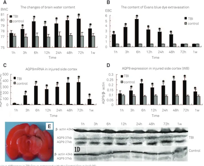
Brain water content was increased obviously at three hours and reached the irst peak at the sixth hour (p<0.05). Following a slight decrease at the 12th hour, it increased
grad-ually and reached the highest point at 72 hours (p<0.05). Until one week, it still remained higher than normal. However, there was no obvious change in the Control Group (Fig 1A).
Brain Evans blue content
he concentration of Evans blue (EB) was rapidly increased after one hour and reached a maximal level at the sixth hour after TBI. hen, it decreased gradually, 72 hours later it had no obvious diference to the Control Group (Fig 1B).
Extent of cerebral ischemia
TTC stain showed that no obvious ischemic focus was noticed in one to six hours. he ischemic tissue surrounding the injured core was increased gradually after 24 hours and reached the peak at 72 hours after TBI (Fig 1E).
The expression of AQP9 mRNA
he expression of AQP9 mRNA in the Control Group was at a low level and no obvious change was found. In the TBI Group, it increased at the third hour and reached the irst peak in six hours, then it slightly decreased after 12 hours, but it was still higher than normal, gradually it increased and reached the highest point at 48 to 72 hours until one week it was still higher than normal (Fig 1C).
The abundance of AQP9 protein
Western blot showed that in the TBI Group AQP9 expres-sion was not obviously changed in one hour, however it was markedly increased at three hours and reached the irst peak at the sixth hour, following a slight decrease that was still higher than normal at the 12th hour. It gradually increased
and reached the highest point at 48 to 72 hours until one week it was still higher than normal. However, the obvious change of AQP9 expression was not observed in the Control Group (Fig 1D).
The expression of AQP9
Immunohistochemistry and immunoluorescence showed AQP9 expressed in astrocytes at very low level in normal rats. AQP9 expression was also at a low level in one hour after TBI in rat brains and obviously increased after three hours in astrocytes of hippocampus, dentate gyrus, and brain tis-sues surrounding ventricle and subarachnoid space, also in blood endothelium near the optic chiasma and subfor-nical organ gliocytes (Fig 2). It reached the first peak at the sixth hour. Following a slightly decrease at 12 hours, the expression of AQP9 increased gradually with time and reached the highest point at 48 to 72 hours after TBI (Fig 2). Until one week, it was still higher than normal. After three hours, AQP9 immunoreactivity was also observed in neurons in hypothalamus, increasing with time and reach-ing the peak at 48 to 72 hours (Fig 3). In hippocampus, the slight expression of AQP9 in neurons was also observed after six hours, even at 72 hours, it was not very strong. A significant change of AQP9 expression was not found in the Control Group.
Table 2. Antibodies used in immunohistochemistry and immunofluorescence reactions.
Primary antibody Secondary antibody
Anti-AQP9, Rabbit, 1:300, Alpha Diagnostic International FITC-labeled goat anti- rabbit IgG, 1:100, Santa Cruz Anti-GFAP, Goat, 1:400, Santa Cruz Cy5-labeled donkey anti-Goat IgG, 1:200, millipore Anti-NeuN, Mouse, 1:400, Chemicon TRITC-labeled rabbit anti-mouse IgG, 1:200, Santa Cruz
DISCUSSION
AQP9 is a member of the aquaporins family, known as water channel proteins, which are small, hydrophobic, inte-gral membrane proteins and expressed widely in animals and plants. AQP9 has the speciic feature of broad range perme-ability, such as water, urea, glycerol, mannitol, sorbitol, pu-rines (adenine), pyrimidines (uracil and chemotherapeutic agent 5-luorouracil), monocarboxylates (lactate and beta-hydroxybutyrate), and ammonia8. In the rodent brain, AQP9
is distributed in three cell types: glial (tanycytes and astro-cytes), catecholaminergic neurons and endothelial cells of sub-pial blood vessels4-6,8. It is concerned with water
homeo-stasis, osmotic regulation, and energy metabolism in brain8.
TBI is a very common disease with metabolic disorders of water, electrolyte, and energy. According to the diferences of pathophysiological processes, TBI can be divided into pri-mary and secondary traumatic brain injury. he former, usu-ally in the early stage of TBI, is the result of mechanical forces
producing tissue deformation at the moment of injury. he latter, usually in the later stage of TBI, occurs as a complica-tion of the primary brain injury and includes ischemic, hy-poxic damage, and cerebral welling1.
In the early stage of TBI (1 to 12 hours), due to the dam-aged brain tissue and badly impaired BBB, the mass in blood, like blood cells, plasma protein, plasma, electrolytes and so on, lowed into intercellular space and caused the in-crease of intercellular luid, change of osmotic pressure, and disorder of electrolyte balance. Our study found that AQP9 protein and mRNA expression increased at three hours and reached the irst peak at six hours, then they decreased slightly at the 12th hour. his change was in accordance with
the one of the brain edema showed by brain water content (BWC). Although in this stage BBB was severely destructed, the brain edema was not the worst compared with the ede-ma in the later stage of TBI. Interestingly, the distribution of AQP9 was expanded, which was observed in cerebrospi-nal luid contacting interface, such as surrounding ventricle
Fig 1. (A) The changes of brain water content; (B) The content of Evans blue dye extravasation; (C) Aquaporin-9 (AQP9)
mRNA expression in injured side cortex showed by real time polymerase chain reaction ; (D) AQP9 expression in injured side cortex showed by western-blot; (E) The badly brain ischemic at 72 hours after traumatic brain injury (TBI) showed by 2,3,5-triphenyltetrazolium chloride staining.
The changes of brain water content
TBI control TBI control TBI control TBI control Time Time Time Time BWC 81 80 79 78 77 75
1h 3h 6h 12h 24h 48h 72h 1w
1h 3h 6h 12h 24h 48h 72h 1w
1h 3h 6h 12h 24h 48h 72h 1w
1h 3h 6h 12h 24h 48h 72h 1w
1h 3h 6h 12h 24h 48h 72h 1w
EBC 6 5 4 3 2 1 0
The content of Evans blue dye extravasation
AQP9mRNA in injured side cortex AQP9 expression in injured side cortex (WB)
500 400 300 200 100 0 AQP 9 (r eal -time PCR) AQP 9/ G - ac tin 0.3 0.25 0.15 0.05 0.1 0.2 0 TBI Control
G- actin 43kb
G- actin 43kb AQP9 37kb AQP9 37kb AQP9 27kb
E
A
C
D
B
Fig 3. The increased aquaporin-9 (AQP9) immunoreactivity (green), Anti-NeuN-positive neurons and glial fibrillary acid protein (GFAP)-positive astrocytes in hypothalamus at 48 hours after traumatic brain injury (TBI) are respectively shown in 3A, 3B and 3C; 3AB (×400) is the combined image of 3A and 3B; 3ABC (×400) is the combined image of 3A, 3B and 3C. AQP9 positive neurons in hypothalamus at 24 hours after TBI is shown in 3D(×400). ↑: AQP9 positive neurons, : The third ventricle.
and subpial tissue near the optic chiasma in pavimentum cerebri in this stage.
hese results suggested that AQP9 in these areas might contribute to the clearance of the excess water in intercellular space at this stage, similar to the roles of AQP49. Additionally,
the obvious increase of AQP9 immunoreactivity was also detected in astrocytes of hippocampus and dentate gyrus after TBI. his inding was in accordance with Hwang’s re-search, which showed that AQP9 expression was induced in hippocampus 6 and 12 hours after global ischemia in region of the gerbil10. Schurr’s presumed that the AQP9 expression
could favor lactate and glycerol clearance from the extracel-lular space during ischemia11. Ischemia was one of the main
pathologic processes of TBI in the early stage. herefore, in this stage the increased expression of AQP9 might indicate its involvement in the clearance of lactate and glycerol caused by ischemia after TBI in extracellular space.
In the later stage of TBI (after 24 hours), brain damage was related to many factors, such as inlammatory response, edema formation, iniltration of peripheral blood cells, nitric oxide and nitric-oxide synthas, oxygen radicals, excess ions, and so on. hese factors were thought to exacerbate the in-jury following TBI and could induce brain hypoxia and isch-emia, then leading to energy supply deiciency. Our experi-ment found that the brain ischemia got worse with time and
the worst one was observed at 72 hours after TBI, which was showed by TTC staining. In this stage, aerobic metabolism could be inhibited and anaerobic glycolysis was active, pro-ducing abundant lactic acid.
Magistretti et al.12 assumed a “lactate shuttle” model,
where glucose was transformed by the astrocytes into lactate and difused from astrocytes to neurons using the monocar-boxylate transporters (MCTs). Tsukaguchi et al.13 found that
AQP9 permeability to lactate increased four-fold when the pH decreased to 5.5. Recent data suggested AQP9 could fa-cilitate the reuptake of lactate and glycerol, which could then be used as energetic substrates and was involved in the lac-tate movement between astrocytes and neurons that could use lactate as energetic substrates14. Lactate seemed to
fa-cilitate the recovery of neurons after ischemic insults11. Our
by a rapid water low involving AQP914, thus a badly
cellu-lar brain edema was observed in our study at this stage. Moreover, AQP9 expression was changed accordingly to the development of the brain edema in this stage. herefore, AQP9 might also contribute to the formation of cellular brain edema in the later stage of TBI.
AQP9 was expressed in neurons of hypothalamus and in-creased slowly and gradually by time throughout nearly all the stages after TBI (at three and after three hours). A very weak expression of AQP9 in neurons in hippocampus was also ob-served by immunoluorescence after three hours. It was simi-lar with that after a metabolic stress, AQP9 expression was induced in pyramidal neurons, which did not express this channel in physiological conditions10. Up to date, indings
of many experiments under many pathological situations in brain about AQP9-positive neurons suggested AQP9 plays an
active role in energy balance as a glycerol-lactate-channel in neuron8,15,16. Our outcomes also suggested AQP9 might be
in-volved in the energy metabolism of neurons after TBI. In conclusion, this study indicated that AQP9 expression increased in diferent stages after TBI. he increased AQP9 might contribute to clearance of excess water, lactate and glycerol in extracellular space in early stage of TBI. he wide-ly-spread AQP9-positive astrocytes in later stage of TBI might help the lactate move from astrocytes to neurons and protect neurons. In addition, AQP9 could also contribute to the for-mation of cellular edema in brain after TBI. As it was seen, AQP9-positive neurons were observed after three hours, TBI suggested AQP9 might play a role in energy balance as a glyc-erol-lactate-channel in neuron. he regulation mechanisms of the changed AQP9 expression and the exact roles of AQP9 in brain after TBI still need further studies.
References
1. Finnie JW, Blumbergs PC. Traumatic brain injury. Vet Pathol 2002;39:679-689.
2. Verweij BH, Amelink GJ, Muizelaar JP. Current concepts of cerebral oxygen transport and energy metabolism after severe traumatic brain injury. Prog Brain Res 2007;161:111-124.
3. Marmarou A. A review of progress in understanding the pathophysiology and treatment of brain edema. Neurosug Focus 2007;22:1-10.
4. Unterberg AW, Stover J, Kress B, Kiening KL. Edema and brain trauma. Neuroscience 2004;129:1021-1029.
5. Badaut J, Petit JM, Brunet JF, Magistretti PJ, Charriaut-Marlangue C, Regli L. Distribution of Aquaporin 9 in the adult rat brain: preferential expression in catecholaminergic neurons and in glial cells. Neuroscience 2004;128:27-38.
6. Ribeiro MC, Hirt L, Bogousslavsky J, Regli L, Badaut J. Time course of aquaporin expression after transient focal cerebral ischemia in mice. J Neurosci Res 2006;83:1231-1240.
7. Feeney DM, Boyeson MG, Linn RT, Murray HM, Dail WG. Responses to cortical injury: I. Methodlogy and local effects of contusions in the rat. Brain Res 1981;211:67-77.
8. Badaut J. Aquaglyceroporin 9 in brain pathologies. Neuroscience 2010;168:1047-1057.
9. Verkman AS. More than just water channels: unexpected cellular roles of aquaporins. J Cell Sci 2005;118:3225-3232.
10. Hwang IK, Yoo KY, Li H, et al. Aquaporin 9 changes in pyramidal cells before and is expressed in astrocytes after delayed neuronal death in the ischemic hippocampal CA1 region of the gerbil. J Neurosci Res 2007;85:2470-2479.
11. Schurr A. Lactate, glucose and energy metabolism in the ischemic brain. Int J Mol Med 2002;10:131-136.
12. Magistretti PJ, Pellerin L. Cellular mechanisms of brain energy metabolism and their relevance to functional brain imaging. Philos Trans R Soc Lond B Biol Sci 1999;354:1155-1163.
13. Tsukaguchi H, Shayakul C, Berger UV, et al. Molecular characterization of a broad selectivity neutral solute channel. J Biol Chem 1998;273:24737-24743.
14. Badaut J, Regli L. Distribution and possible roles of aquaporin 9 in the brain. Neuroscience 2004;129:971-981.
15. Badaut J, Brunet JF, Petit JM, Guérin CF, Magistretti PJ, Regli L. Induction of brain aquaporin 9 (AQP9) in catecholaminergic neurons in diabetic rats. Brain Res 2008;1188:17-24.