https://doi.org/10.1590/0004-282X20170092
ARTICLE
Curcumin decreases astrocytic reaction after
gliotoxic injury in the rat brainstem
Curcumina reduz a reação astrocitária após injúria gliotóxica no tronco encefálico de ratos
Eduardo Bondan1,2, Carolina Cardoso1, Maria de Fátima Martins1,2
1Universidade Paulista, Patologia Ambiental e Experimental, São Paulo SP, Brasil; 2Universidade Cruzeiro do Sul, Medicina Veterinária, São Paulo SP, Brasil.
Correspondence: Eduardo Bondan; Patologia Ambiental e Experimental, Universidade Paulista; Rua Caconde, 125 / 51; 01425-011 São Paulo SP, Brasil; E-mail: [email protected]
Conflict of interest: There is no conflict of interest to declare.
Received 21 December 2016; Received in final form 15 April 2017; Accepted 04 May 2017.
ABSTRACT
Recent studies have demonstrated that curcumin (Cur) has antioxidant, anti-inflammatory and anti-fibrotic effects. Ethidium bromide (EB) injections into the central nervous system (CNS) are known to induce local oligodendroglial and astrocytic loss, resulting in primary demyelination and neuroinflammation. Peripheral astrogliosis is seen around the injury site with increased immunoreactivity to glial fibrillary acidic protein (GFAP). This investigation aimed to evaluate the effect of Cur administration on astrocytic response following gliotoxic injury. Wistar rats were injected with EB into the cisterna pontis and treated, or not, with Cur (100 mg/kg/day, intraperitoneal route) during the experimental period. Brainstem sections were collected at 15, 21 and 31 days after EB injection and processed for GFAP immunohistochemical staining. Astrocytic reactivity was measured in a computerized system for image analysis. In Cur-treated rats, the GFAP-stained area around the lesion was significantly smaller in all periods after EB injection compared to untreated animals, showing that Cur reduces glial scar development following injury.
Keywords: astrocytes; curcumin; ethidium; gliosis; gliotoxin.
RESUMO
Estudos recentes têm demonstrado que a curcumina (Cur) possui efeitos antioxidantes, anti-inflamatórios e antifibróticos. Sabe-se que a injeção de brometo de etídio (EB) no sistema nervoso central induz a perda oligodendroglial e astrocitária, resultando em desmielinização primária e neuroinflamação. Astrogliose periférica é observada ao redor da lesão com aumento da imunorreatividade à proteína glial fibrilar ácida (GFAP). A presente investigação objetivou avaliar o efeito da Cur sobre a resposta astrocitária após injúria gliotóxica. Ratos Wistar foram injetados com EB na cisterna basal e tratados ou não com Cur (100 mg/kg/dia, via intraperitoneal) durante o período experimental. Amostras do tronco encefálico foram coletadas aos 15, 21 e 31 dias pós-injeção de EB e processadas para estudo imuno-histoquímico para a GFAP. A reatividade astrocitária foi medida em um sistema computadorizado para análise de imagem. Nos ratos tratados com Cur, a área marcada para GFAP foi significantemente menor em todos os períodos pós-injeção de EB, indicando que a Cur reduz o desenvolvimento da cicatriz glial após injúria.
Palavras-chave: astrócitos; curcumina; etídio; gliose; gliotoxina.
Ethidium bromide (EB) injections in the white matter of the central nervous system (CNS) are known to act like a glio-toxin, causing local oligodendroglial and astrocytic death, leading to primary demyelination, neuroinlammation, blood-brain barrier disruption and Schwann cell invasion due to the glia limitans breakdown1,2,3,4. Surviving astrocytes
pres-ent a vigorous reaction around the injury site with increased immunoreactivity to the speciic cell marker glial ibrillary acidic protein (GFAP), as well as re-expression of vimentin3.
Curcumin (Cur) [diferuloylmethane or 1,7-bis-(4-hydroxy-3-methoxyphenyl)-1,6-heptadiene-3,5-dione] is the major yellow-orange pigment of turmeric, a common spice and coloring agent derived from the rhizome of the East Indian
plant Curcuma longa, with a long history in Asian traditional cooking and medicine5,6,7. Recently, Cur has been shown to
exhibit proven therapeutic beneits (including antioxidant, anti-inlammatory, anti-cancer and anti-ibrotic efects) in many pathological conditions, such as Alzheimer’s disease, Parkinson’s disease, multiple sclerosis, epilepsy, cerebral injury, cancer, allergy, asthma, bronchitis, colitis, rheumatoid arthri-tis, renal ischemia, psoriasis, scleroderma, diabetes, obesity,
depression, fatigue and acquired immunodeiciency disease6,7,8.
Cur might beneicially afect astrocyte population in the CNS
inlammatory environment by regulating both NF-κβ and
SOX9 signaling pathways14,15.
In this context, the aim of this study was to evaluate whether Cur had the capacity to afect astrocyte response during the process of demyelination and remyelination fol-lowing gliotoxic injury induced by ethidium bromide (EB).
METHODS
he animal procedures were performed in accordance with the guidelines of the Committee on Care and Use of Laboratory Animal Resources and the Brazilian Institutional Ethics Committee, Universidade Paulista (protocol number 235/14, CEUA/ICS/UNIP). Forty-eight adult (4–5-month-old) male Wistar rats were subjected to a local injection of 10 microlitres of 0.1% EB into the cisterna pontis, an enlarged subarachnoid space below the ventral surface of the pons. All rats were anesthetized with 2.5% thiopental (50 mg/ml) by intraperitoneal (IP) route and a burr-hole was made on the right side of the skull, 8 mm behind the fronto-parietal suture. Injections were given freehand, using a Hamilton syringe itted with a 35o angled polished 26-gauge needle, into the cisterna
pontis. Rats were then distributed into two groups - control rats (group I, n = 24) and rats treated with 100 mg/kg/day of Cur (Sigma Aldrich, St. Louis, MO, USA, C1386; 100 mg of Cur was dissolved in 1.0 mL dimethyl sulfoxide and 0.5 mL 0.9% saline solution) by IP route (group II, n = 24). Group I received an equal volume of dimethyl sulfoxide by IP route. he irst injection was done immediately after surgery and then injec-tions were performed once every 24 hours for the experimen-tal period. he animals were kept under controlled light condi-tions (12 hours light-dark cycle) and water and food were given ad libitum during the experimental period.
For the immunohistochemical study of the expression of the astrocytic marker GFAP, six rats per group were anes-thetized and subjected to intracardiac perfusion with buf-ered 10% formaldehyde solution at each of the following periods — 15, 21 and 31 days post-injection. heir brains were then removed and kept for three days in the same ixative. After a 72 hour period, the brainstem was removed through two coronal cuts, beginning at the cerebral peduncles of the mesencephalon and ending in the posterior part of the pons. A rostrocaudal sequence of coronal sections from the brain-stem was done and 5 μm sections were mounted on silanized slides and subjected to GFAP immunostaining using the avidin-biotin peroxidase complex method. Briely, the sec-tions were dewaxed in xylene and rehydrated in a crescent graded series of ethanol solutions. Antigen retrieval was done by transferring the slides to a 10 mM sodium citrate bufer (pH 6.0) at 95°C for 20 minutes. Endogenous peroxidase was
blocked by 3% hydrogen peroxide for 10 minutes at room temperature. Two washes with Tris/HCl bufer pH 6.0 (Wash
bufer 10x, S3006, Dako, Glostrup, Danmark) were done between incubations. Polyclonal rabbit anti-GFAP immuno-globulin (Z0334, Dako), at a dilution of 1:1000, was used as the primary antibody for 16 hours, followed by the application
of biotinylated secondary antibody (Dako Universal LSABTM
2 System - HRP, K0690), according to the manufacturer’s instructions. Immunoreactivity was visualized by incubat-ing the sections in a solution containincubat-ing 0.1% diaminobenzi-dine (DAB, K3467, Dako). Sections were then counterstained by Harris’ modiied hematoxylin solution, dehydrated and mounted in Entellan (Merck, Germany).
Ten photomicrographs per section, of each animal, were taken from randomly-chosen microscopic ields of the periphery of the lesion site using a Nikon E200 microscope (Kanagawa, Japan; 10x objective) equipped with a Nikon Coolpix digital camera linked to a liquid crystal display mon-itor. Astrocytic evaluation was done in the brainstem of ani-mals from both groups using a computerized image analysis system (Image-Pro-Plus 4.5, Media Cybernetics, Silver Spring, USA), measuring by colorimetry the area stained brown (data are expressed in pixels). Negative controls for immunostain-ing (sections lackimmunostain-ing primary antibody application) were per-formed. Data were analyzed by two-way ANOVA followed by Bonferroni’s test, and statistical signiicance was set at p < 0.05.
RESULTS
he EB-induced lesions found in this study were similar to those previously described in other investigations using this gliotoxin in the rat brainstem1,2,3,16. hese lesions exhibited
extensive demyelination in the ventral surface of the brain-stem and showed phagocytic cells, myelin debris and naked axons in their core. At peripheral locations, oligodendrocytes and Schwann cells were noted, the latter occurring in areas of enlarged extracellular spaces devoid of astrocytic processes. Astrocyte extensions were observed near the incipient oli-godendroglial remyelination at the periphery, and Schwann cells also appeared to contribute to myelin repair. Some lym-phocytes and iniltrating pial cells were occasionally seen, the irst contacting phagocytic cells and myelin debris.
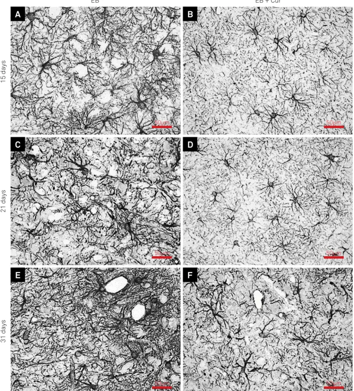
Figure 1 shows the injection site for the gliotoxin. It was observed that the EB-induced lesions from group II (Cur-treated rats) showed a decreased GFAP expression close to the edges of the injury site. Astrocytes presented fewer and thinner GFAP-stained processes at the periphery of the injury site in all periods (Figure 2) and no astrocytes were observed in the cen-tral areas of the lesions, even at 31 days after the EB injection.
test indicated a decreased GFAP expression in the Cur-treated group in relation to the untreated one in all days of observa-tions. At 15 days, the mean brown-stained area was signiicantly smaller in rats treated with Cur (group II - 40,976 ± 2,454 pixels) compared to untreated rats (group I - 68,479 ± 5,487 pixels). Similar indings were seen at 21 days (46,049 ± 5,463 pixels in group II versus 69,689 ± 5,212 pixels in group I) and 31 days (mean areas of 50,338 ± 2,625 pixels and 76,920 ± 4,79 pixels, respectively, in groups II and I).
DISCUSSION
Astrocytes are among the most structurally complex cells in the CNS, and their activation appears in a wide spectrum of CNS injuries and diseases17. Several genes are implicated
in morphological alterations of astrocytes. Glial ibrillary acidic protein, an intermediate ilament-III protein highly expressed in white matter astrocytes and a subset of gray matter astrocytes, is thought to modulate astrocyte motil-ity and shape, providing structural stabilmotil-ity to processes, maintaining their mechanical strength and supporting
neighboring neurons, myelinating oligodendrocytes and the blood-brain barrier17,18,19. Astroglial cells respond to CNS
injury and other neuro-disturbing conditions by undergoing “reactive astrogliosis”, a process whereby astrocytes undergo cellular hypertrophy and proliferation19,20,21,22,23,24. Increased
GFAP is a hallmark of reactive astrocytes and this cytoskel-etal protein contributes to the barrier efect produced by the glial scar that mitigates axonal extension and CNS repair9.
Astrocyte precursors and immature astrocytes present principally nestin and vimentin and, during development as astrocytes mature, nestin expression disappears, GFAP becomes increasingly expressed and vimentin decreases to undetectable levels21. During astrogliosis, astrocytes
re-express vimentin and nestin21. In the EB
demyelinat-ing model, re-expression of vimentin and strong astrocytic immunoreactivity to GFAP were described in the rat brain-stem from the 3rd to the 31st day following gliotoxic injection3.
his increased GFAP expression around the EB-induced lesions was conirmed in the present study.
Activated astrocytes release a variety of factors that participate in neuroinlammation, possibly aggravating
initial injury. he NF-κβ signaling pathway is very
impor-tant for the efects of pro-inlammatory cytokines TNF-α
and IL-1β15, which represent important factors in the initial
activation of astrocytes and are produced in great numbers in brainstem lesions induced by EB25. Under the inluence
of many relevant factors, astrocytes will lead to dense glial scar formation and will produce large amounts of extracel-lular matrix components, such as chondroitin sulfate pro-teoglycan, changing the axonal growth environment and severely inhibiting nerve regeneration15.
Yuan et al.15 observed that Cur suppressed the NF-κβ
signaling pathway, down-regulating the expression of che-mokines MCP-1, RANTES and CXCL10 released by astro-cytes and decreasing macrophage and T-cell iniltration, thus reducing inlammation in the glial scar environment in an experimental model of spinal cord injury. Additionally, by silencing the transcription factor SOX9, Cur reduced the deposition of extracellular matrix chondroitin sulfate pro-teoglycan, contributing to recovery of neurological function. herefore, these authors stated that Cur could both inhibit the formation of intracellular (i.e., GFAP) and extracellular (i.e., chondroitin sulfate proteoglycan) glial scar components and promote neurological recovery after injury.
Any injury to the CNS (caused by trauma, hypoxia, toxin or an infectious agent) represents a complex system of inter-acting cell types that react in a stereotyped way, forming a mature lesion with two distinct components – the periph-ery with hypertrophic astrocytes, whereas the lesion core is composed of NG2 glia/oligodendrocyte precursor cells, men-ingeal- and/or vascular-derived ibroblasts, pericytes, epen-dymal cells and phagocytic macrophages24.
Both oligodendrocyte and astrocyte losses are key events within the EB-induced lesion, while axons remain unafected.
GFAP: glial fibrillary acidic protein
he mechanism of selective glial death supposedly occurs through EB’s action as a minor-groove DNA intercalator. Other evidences suggest that although EB does intercalate both chromosomal and mitochondrial DNA, it only afects
mtDNA transcription26. So, an injection of EB into the white
matter is likely to afect mtDNA in all cells of the lesion site,
although neurons and endothelial cells appear to be less sen-sitive than glia in rodent models4.
Astrocyte disappearance due to the gliotoxic efect and direct mechanical damage due to intracisternal injection are identiied as factors capable of disturbing the blood-brain barrier, thus allowing monocyte and lymphocyte iniltration.
A
B
C
D
E
F
50µm 50µm
50µm 50µm
50µm 50µm
EB + Cur
15 days
21 days
31 days
EB
Lymphocytes are invariably found in the EB demyelinating lesions, sometimes contacting myelin debris in the extracel-lular space and activated macrophages containing phago-cytosed myelin, in a relationship suggestive of antigenic rec-ognition. It is possible that macrophage and lymphocyte products during the inlammatory response triggered by EB injection may represent a more deleterious inluence on nervous tissue than the previous gliotoxin injection itself. herefore, the already described anti-inlammatory and
immunomodulatory efects performed by Cur7,8,12 may
possi-bly be beneicial to tissue repair.
Many distinct signaling molecules released by microg-lia, astrocytes, neurons, oligodendrocyte lineage cells, peri-cytes, endothelia and invasive inlammatory/immune cells, are able to trigger and regulate astrogliosis22,23. hese
molecu-lar signals include: (a) molecu-large polypeptide growth factors and cytokines, such as IL-1, IL-6, TNF-α, IFN-γ, TGF-β, LIF, CNTF, FGF2, among others; (b) mediators of innate immunity such
as LPS and other Toll-like receptor ligands; (c) neurotrans-mitters such as glutamate and noradrenaline; (d) purines (e.g., ATP); (e) reactive oxygen species; ( f) products associ-ated with systemic metabolic activity (e.g., NH4+) and (g)
reg-ulators of cell proliferation, such as endothelin 122,23.
Cur has been shown to have bifunctional antioxidant properties, scavenging reactive oxygen species as well as simultaneously inducing an antioxidant response. Many other beneicial efects were reported for Cur, including the induction of cytoprotective enzymes, such as heme oxygenase-1, glutathione-S-transferase and γ-glutamyl cyste-ine ligase, the inhibition of caspase 1-dependent inlamma-tion, the reversion of mitochondrial dysfunctions in astro-cytes and the inhibition of mitochondria-dependent and -independent apoptosis caused by oxidative damage13.
As Cur is chemically quite unstable at pH > 7 and very poorly absorbed from the gastrointestinal tract, we chose the IP route for this study. According to uptake and bioavail-ability studies, the high proportion of fecal excretion after IP administration indicates good absorption from the
perito-neal cavity and eicient elimination in the bile5. By
compari-son with an IP injected dose, the oral bioavailability of Cur is estimated at about only 1%5.
Lim et al.27 investigated whether Cur could afect
Alzheimer’s disease-like pathology in transgenic mice and noted that with low-dose Cur, but not with high-dose Cur, GFAP expression was decreased, and insoluble Abeta, sol-uble Abeta, and plaque burden were signiicantly reduced (by 43–50%).
In our study, Cur treatment decreased the expected reac-tion of increased GFAP expression in astrocytes around EB-induced lesions at 15, 21 and 31 days compared to the untreated group. Inhibition of GFAP expression following CNS injury was also observed in several other investigations using Cur9,13,15. Cur was able to reduce the expression of both GFAP
mRNA and GFAP protein, as well as to induce autophagy and Table. Areas in pixels with GFAP staining in rats injected with EB, treated (group II) or not (group I) with Cur.
Animal Group I – EB injection Group II – EB injection + Cur
15 days (µm2) 21 days (µm2) 31 days (µm2) 15 days (µm2) 21 days (µm2) 31 days (µm2)
1 65,934 62,477 77,902 40,763 48,372 47,883
2 67,273 67,376 74,873 39,478 46,803 50,631
3 65,206 65,802 69,81 37,392 39,983 51,705
4 63,882 80,163 79,653 43,89 50,382 47,332
5 61,371 71,885 84,926 41,238 55,223 49,77
6 74,982 70,727 73,714 40,943 38,674 55,368
7 73,282 68,526 73,863 39,207 45,87 51,646
8 75,903 70,557 80,623 44,893 43,087 48,372
Mean 68,479 69,689 76,92 40,976 46,049 50,338
SD 5,487 5,212 4,79 2,454 5,463 2,625
GFAP: glial fibrillary acidic protein; EB: ethidium bromide; Cur: curcumin
Total count of pixels (x10
3)
0 10 20
15 days 21 days 31 days
30 40 50 60 70 80 90
EB EB + Cur
GFAP: glial fibrillary acidic protein; EB: ethidium bromide; Cur: curcumin
to rescue the ilamentous organization of the GFAP mutant protein in an in vitro model of Alexander disease, in which het-erozygous mutations of the GFAP gene are responsible for the intracytoplasmic accumulation of ibrous eosinophilic
depos-its known as Rosenthal ibers in dystrophic astrocytes28.
Morphometric analysis in the present investigation unequivocally demonstrated that Cur decreased astrocytic activation until the 31st day after gliotoxic lesion, probably
by suppressing the release of proinlammatory molecules, such as the previously-mentioned TNF-α and IL-1β, which may trigger and promote astrogliosis following CNS injury. hus, our results clearly indicate that this substance may be
used in preventing or reducing glial scar development fol-lowing injury.
Cur has been shown to regulate numerous transcrip-tion factors, cytokines, protein kinases, adhesion molecules,
redox status and enzymes linked to inlammation6,7,8. As the
inlammatory process plays a major role in most chronic ill-nesses, including neurodegenerative, cardiovascular, pulmo-nary, metabolic, autoimmune and neoplastic diseases, Cur undoubtedly presents a potential role in the prevention and
treatment of various proinlammatory diseases8. hese
fea-tures, combined with the pharmacological safety, and negli-gible cost, render Cur an attractive agent to explore further.
References
1. Bondan EF, Lallo MA, Sinhorini IL, Pereira LA, Graça DL. The effect of cyclophosphamide on brainstem remyelination following local ethidium bromide injection in Wistar rats. J Submicrosc Cytol Pathol. 2000;32(4):603-12.
2. Graça DL, Bondan EF, Pereira LA, Fernandes CG,
Maiorka PC. Behaviour of oligodendrocytes and Schwann cells in an experimental model of toxic demyelination of the central nervous system. Arq. Neuropsiquiatr. 2001;59(2B):358-61. https://doi.org/10.1590/S0004-282X2001000300009
3. Bondan EF, Lallo MA, Dagli MLZ, Sanchez M, Graça DL. [Investigation into the astrocytic immunoreactivity to GFAP and vimentin in the brainstem of Wistar rats submitted to the ethidium bromide gliotoxic model]. Arq Neuropsiquiatr. 2003;61(3A):642-9.Portuguese. https://doi.org/10.1590/S0004-282X2003000400022
4. Kuypers NJ, James KT, Enzmann GU, Magnuson DSK, Whittemore SR. Functional consequences of ethidium bromide demyelination of the mouse ventral spinal cord. Exp Neurol. 2013;247:615-22. https://doi.org/10.1016/j.expneurol.2013.02.014
5. Metzler M, Pfeiffer E, Schulz SI, Dempe JS, Curcumin uptake and metabolism. Biofactors. 2013;39(1):14-20. https://doi.org/10.1002/biof.1042
6. Hsu CH, Cheng AL. Clinical studies with curcumin. Adv Exp Med Biol. 2007;595:471-80. https://doi.org/10.1007/978-0-387-46401-5_21 7. Srivastava RM, Singh S, Dubey SK, Misra K, Khar A. Immunomodulatory
and therapeutic activity of curcumin. Int. Immunopharmacol. 2011;11(3):331-41. https://doi.org/10.1016/j.intimp.2010.08.014 8. Aggarwal BB, Harikumar KB. Potential therapeutic effects of
curcumin, the anti-inflammatory agent, against neurodegenerative, cardiovascular, pulmonary, metabolic, autoimmune and
neoplastic diseases. Int J Biochem Cell Biol. 2009;41(1\0:40-59. https://doi.org/10.1016/j.biocel.2008.06.010
9. Lin MS, Lee YH, Chiu WT, Hung KS. Curcumin provides neuroprotection after spinal cord injury. J Surg Res. 2011;166(2):280-9. https://doi.org/10.1016/j.jss.2009.07.001 10. Jin W, Wang J, Zhu T, Yuan B, Ni H, Jiang J et al.
Anti-inflammatory effects of curcumin in experimental spinal cord injury in rats. Inflamm Res. 2014;63(5):381-7. https://doi.org/10.1007/s00011-014-0710-z
11. Wang YF, Zu JN, Li J, Chen C, Xi CY, Yan JL. Curcumin promotes the spinal cord repair via inhibition of glial scar formation and inflammation. Neurosci Lett. 2014;560:51-6. https://doi.org/10.1016/j.neulet.2013.11.050
12. Seyedzadeh MH, Safari Z, Zare A, Gholizadeh Navashenaq J, Razavi SA, Kardar GA et al. Study of curcumin immunomodulatory effects on reactive astrocyte function. Int Immunopharmacol. 2014;22(1):230-5. https://doi.org/10.1016/j.intimp.2014.06.035
13. Daverey A, Agrawal SK. Curcumin alleviates oxidative stress and mitochondrial dysfunction in astrocytes. Neuroscience. 2016;333:92-103. https://doi.org/10.1016/j.neuroscience.2016.07.012 14. Ni H, Jin W, Zhu T, Wang J, Yuan B, Jiang J et al. Curcumin modulates TLR4/NF-κB inflammatory signaling pathway following traumatic spinal cord injury in rats. J Spinal Cord Med. 2015;38(2):199-206. https://doi.org/10.1179/2045772313Y.0000000179
15. Yuan J, Liu W, Zhu H, Chen Y, Zhang X, Li L et al. Curcumin inhibits glial scar formation by suppressing astrocyte-induced inflammation and fibrosis in vitro and in vivo. Brain Res. 2017;1655:90-103. https://doi.org/10.1016/j.brainres.2016.11.002
16. Pereira LA, Dertkigil MS, Graça DL, Cruz- Höfling MA. Dynamics of remyelination in the brain of adult rats after exposure to ethidium bromide. J. Submicrosc. Cytol Pathol. 1998;30(3):341-8.
17. Lee KM, MacLean AG. New advances on glial activation in health and disease. World J Virol. 2015;4(2):42-55. https://doi.org/10.5501/wjv.v4.i2.42
18. Brenner M. Role of GFAP in CNS injuries. Neurosci Lett. 2014;565:7-13. https://doi.org/10.1016/j.neulet.2014.01.055 19. Yang Z, Wang, KW. Glial fibrillary acidic protein: From intermediate
filament assembly and gliosis to neurobiomarker. Trends Neurosc. 2015;38(6):364-74. https://doi.org/10.1016/j.tins.2015.04.003 20. Fitch MT, Silver J. Astrocytes are dynamic participants in central nervous
system development and injury responses. In Jessen KR, Richardson WD. Glial cell development. Oxford: Oxford University Press; 2001. p. 263-77. 21. Pekny M, Pekna M. Astrocyte intermediate filaments in CNS
pathologies and regeneration. J Pathol. 2004;204(4):428-37. https://doi.org/10.1002/path.1645
22. Sofroniew MV. Molecular dissection of reactive astrogliosis and glial scar formation. Trends Neurosci. 2009;32(12):638-47. https://doi.org/10.1016/j.tins.2009.08.002
23. Sofroniew MV, Vinters HV. Astrocytes: biology and pathology. Acta Neuropathol. 2010;119(1):7-35. https://doi.org/10.1007/s00401-009-0619-8
24. Cregg JM, DePaul MA, Filous AR, Lang BT, Tran A, Silver J. Functional regeneration beyond glial scar. Exp Neurol. 2014;253:197-207. https://doi.org/10.1016/j.expneurol.2013.12.024
25. Bondan EF. Propentofylline decreases the production of TNF-alpha and IL-1beta in the rat brainstem after a gliotoxic injury induced by ethidium bromide. J. Neuroimmunol. 2014;275(1-2):139. http://dx.doi.org/10.1016/j.jneuroim.2014.08.373
27. Lim GP, Chu T, Yang F, Beech W, Frautschy SA, Cole GM. The curry spice curcumin reduces oxidative damage and amyloid pathology in an Alzheimer transgenic mouse. J Neurosci. 2001;21(21):8370-7.