Imagem
Mailing address: Marcelo Luiz Campos Vieira •
Rua Cardoso de Melo, 463, ap. 21 - Vila Olímpia - 04548-002, São Paulo, SP - Brazil
E-mail: [email protected], [email protected]
Manuscript received February 10, 2008; revised manuscript received March 03, 2008; accepted March 03, 2008.
Key words
Echocardiography, three-dimensional; mitral valve prolapse.
Three-dimensional Echocardiography in Patient with Mitral Valve
Prolapse
Marcelo Luiz Campos Vieira e Wilson Mathias Jr.
Instituto do Coração (InCor), Faculdade de Medicina da Universidade de São Paulo (FMUSP), São Paulo; SP - Brazil
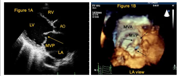
We report a case of a 42-year-old woman with mitral
valve prolapse (Figure 1A,1B). The 3-D view allowed the
visualization of a mitral valve prolapse as seen from the left
atrium, plane in which the surgical correction is performed.
Figure 1 -2-D (Figure 1A) and 3-D ECHO (Figure 1B) of a patient with mitral valve prolapse (MVP); Single arrow indicating the lack of lealet coaptation; double arrow
(Figure 1B) showing MVP, as seen from the left atrium; MVA - Mitral valve annulus.