66 66
Lopes et al
Transposition of the great arteries associated with anomalous pulmonary venous connection
Arq Bras Cardiol 2001; 77: 66-8.
Real e Benemérita Sociedade Portuguesa de Beneficência
Mailing address: Lilian M. Lopes Rua Batista Caetano, 79/111 04108130 -São Paulo, SP, Brazil - e-mail: [email protected]
English version by Stela Maris C. e Gandour
Lilian Maria Lopes, Gláucia Maria Penha Tavares, Fred Luiz Mailho, Vicente de Paulo Cavalcante de Almeida, José Armando Mangione
São Paulo, SP - Brazil
Echocardiographic Diagnosis of Transposition of the Great Arteries
Associated with Anomalous Pulmonary Venous Connection
Brief report
We report 2 cases of transposition of the great arteries associated with anomalous pulmonary venous connection emphasizing the clinical findings, the diagnosis, and the evo-lution of the association. One of the patients had the ano-malous pulmonary venous connection in its total infradia-phragmatic form, in the portal system, and the other patient had a partial form, in which an anomalous connection of the left superior lobar vein with the innominate vein existed. At the time of hospital admission, the patients had cyanosis and respiratory distress with clinical findings suggesting transposition of the great arteries. The diagnosis in 1 of the cases, in which the anomalous connection was partial, was established only with echocardiography, without invasive procedures that would represent risk for the patient; in the other case, in which the anomalous connection was total, the malformation was only evidenced with catheterization. The patients underwent surgery for anatomical correction of the heart disease. Only 1 patient had a good outcome.
The association of transposition of the great arteries and anomalous pulmonary venous connection is very rare 1,2. With
few exceptions, the cases reported in the international literature result from postmortem studies 3 and, many times,
are part of very complex syndromes, usually with death oc-curring a few hours or days after birth.
Therefore, we considered the report of these cases of clinical interest, due to the rarity of the association and the contribution of the sensitivity of the echocardiogram as a preoperative method of diagnosis.
Case Reports
Case1 – The patient was a 22-day-old male infant with a history of mild cyanosis since his birth. On physical exa-mination at the time of admission, the infant was moderately tachydyspneic, and on heart auscultation, an increased in-tensity of the 2nd heart sound predominated.
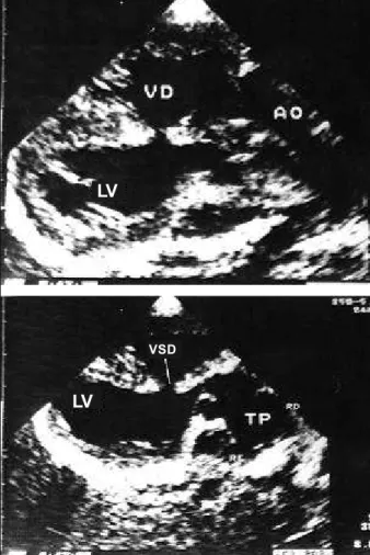
The echocardiogram showed transposition of the great arteries, ventricular septal defect of 3mm in diameter, atrial septal defect of the oval foramen type with a diameter of 4 mm, and pulmonary flow with a pattern of pulmonary hypertension. The great dilation of the right chambers was noteworthy (fig. 1A), but an association with anomalous connection was not suspected. The echocardiography also showed a single coronary ostium, which was confirmed on cardiac catheterization.
The patient underwent cardiac catheterization for as-sessment of the coronary arteries and performance of atrio-septostomy. During the procedure, the echocardiographic-findings were confirmed, and an anomalous infradiaphrag-matic connection of the pulmonary veins of the nonobs-tructive type was shown (fig. 2).
The surgical procedure chosen was the anatomical correction of the transposition of the great arteries, implan-tation of the anomalous pulmonary vein in the left atrium, ventriculoseptoplasty, and resection of a small muscular band from the right ventricular outflow tract that caused a mild subaortic stenosis (intraoperative finding). The patient evolved satisfactorily, and was discharged from the hospi-tal after 2 weeks.
Case 2 – The patient was a 5-month-old male child weighing 4.029kg, who, on physical examination, was mode-rately cyanotic, malnourished, and tachydyspneic. On car-diac auscultation, a systolic murmur (3+/4) predominated on the medium-to-low portion of the left sternal margin, and increased intensity of the 2nd cardiac sound in the
pulmona-ry area was observed.
Echocardiography showed transposition of the great
Arq Bras Cardiol 2001; 77: 66-8.
Lopes et al Transposition of the great arteries associated with anomalous pulmonary venous connection
67 67 arteries (fig. 3), multiple ventricular septal defects, signs of
pulmonary hypertension, mild dilation of the right cham-bers (fig. 1B), and also the presence of partial anomalous pulmonary venous connection, in which, the pulmonary vein of the left superior lobe drained into a vertical vein that followed the innominate vein and superior vena cava (fig. 4).
The child underwent surgery with no complementary hemodynamic investigation, and, during surgery, the echocardiographic diagnosis was confirmed. Anatomic correction of the great arteries and correction of the other defects was performed. The postoperative evolution was unsatisfactory, and the child evolved to death on the 3rd
postoperative day due to renal failure, which was followed by multisystem organ failure.
Discussion
Transposition of the great arteries associated with anomalous pulmonary venous connection is rare 1-5.
Howe-ver, the association of these 2 defects may attenuate the
intensity of the cyanosis and the hemodynamic abnorma-lities caused by the transposition of the great arteries 1,2,4. In
this circumstance, the anomalous pulmonary venous connection in the right atrium avoids an increase in the pul-Fig. 1 – Four-chamber view. A) Observe the severe dilation of the right chambers in
case 1, which was associated with the total form of anomalous pulmonary venous connection; B) Observe the subtle predominance of the right atrium in this echocardiographic view in case 2, which was associated with the partial form of anomalous pulmonary venous connection.
Fig. 2 - Catheterization, case 1. Angiographic diagnosis of the infradiaphragmatic anomalous pulmonary venous connection. Contrasted pulmonary venous return and anomalous vein heading down to the infradiaphragmatic region (arrow).
Fig. 4 – Echocardiogram, case 2. Echocardiographic diagnosis of partial anomalous pulmonary venous connection. Observe the blood flow in red in the vertical vein heading toward the innominate vein on the color-flow mapping (arrows). To the left, the confirmation of the venous flow pattern.
Fig. 3 – Longitudinal view, case 2. Transposition of the great arteries on echocardio-graphy. Observe in the upper part, the aorta emerging from the right ventricle (RV), and, in the lower part, the pulmonary artery emerging from the left ventricle (LV). A ventricular septal defect may also be observed (VSD). RB – right branch of the pulmonary artery; LB – left branch of the pulmonary artery.
LV
LV
68 68
Lopes et al
Transposition of the great arteries associated with anomalous pulmonary venous connection
Arq Bras Cardiol 2001; 77: 66-8.
monary flow and enhances the saturation of the blood that reaches the aorta and the systemic circulation.
In the literature, a few authors 1,2,4,5 document the
sur-gical approach to this association reporting the successful repair in the atrial plane with the modified techniques by Senning or Mustard. One of the studies4 reports that the
anatomical repair in the arterial plane, which is the first choice for the transposition of the great arteries isolated, is contraindicated when an association with total anomalous pulmonary venous connection exists, because the left ven-tricle is not prepared to receive the aorta surgically, due to its decreased flow situation since fetal life.
Analyzing the postoperative evolution of the 2 pa-tients here reported, who underwent anatomical surgical repair, this theory seems not to be applicable, because un-like that which was expected, a good evolution existed in patient 1, whose association was with the total form of anomalous pulmonary venous connection, and no sign of lack of left ventricular preparation or dysfunction occurred in the postoperative evolution. On the other hand, in patient 2, in which a better result should occur according to this theory, because the left ventricle would be better adapted due to the partial form of the anomalous connection, death occurred on the 3rd postoperative day.
Knowing that 4% of the newborn babies with transpo-sition of the great arteries die rapidly before the anatomical surgical repair, Maeno et al 6 tried to establish, through fetal
echocardiography, a group of high-risk fetuses with this congenital heart disease. The intrauterine search for echocardiographic signs of restriction of the oval foramen or ductus arteriosus would be much more important, as a prognostic factor, than the preparation of the left ventricle, which would lead to early neonatal death independent from the aggressive maneuvers of reanimation or early surgical intervention. Therefore, we think that the anatomical repair
for transposition of the great arteries should be considered even when this malformation is associated with anomalous pulmonary venous connection.
In case 1, in which echocardiography failed to diagnose the association with the anomalous pulmonary venous con-nection, a new echocardiographic examination was perfor-med after the cardiac catheterization, to check whether the diagnosis of the anomalous vein was possible or not with the aid of this noninvasive method. We observed that the anomalous vein was perfectly visible and that the diagnostic failure was probably caused by not considering the possibi-lity of this association; this resulted in an inadequate analysis of the subcostal region, which was then restricted to the diag-nosis of the situs. The significant dilation of the right cham-bers was also not considered in case 1, which had much more exuberant findings than did case 2, and could not be explained by only transposition of the great arteries (fig. 1).
Knowing how unexpected the morphological diagno-sis of an examination in a patient with congenital heart di-sease may be, it is of fundamental importance to observe the sequence of the segmentary analysis, which should al-ways be performed in pediatric echocardiography through logical systematics.
In conclusion, the echocardiographic diagnosis of transposition of the great arteries associated with anomalo-us pulmonary venoanomalo-us connection is possible. The severe dilation of the right chambers in a mildly cyanotic child with transposition of the great arteries seems to be the diagnos-tic clue to be remembered for this rare association.
Acknowledgments
We thank Drs. José Pedro da Silva and Gláucio Furla-netto, whose teams assisted clinically and surgically the patients reported.
1. Amadeo A, Corno A, Marino B, Carta MG, Marcelletti C. Combined repair of transposed great arteries and total anomalous pulmonary venous connection. Ann Thorac Surg 1990; 50: 820-1.
2. Barbero-Marcial M, Verginelli G, Vila J, Zerbini EJ. Transposition of the great arteries associated with total anomalous pulmonary venous connection: a surgical approach. Ann Thorac Surg 1984; 37: 92-4.
3. Delisle G, Ando M, Galder AL, et al. Total Anomalous pulmonary venous connection: report of 93 autopsied cases with emphasis on diagnostic and surgical considerations. Am Heart J 1976; 91: 99.
References
4. Gontijo B, Fantini F, Barbosa M, Gomes VM, Gutierrez C, Vrandecic M. Surgical repair of transposition of great arteries and total anomalous pulmonary venous return of the coronary sinus. Eur J Cardio-Thorac Surg 1994; 8: 391-2. 5. Ueda Y, Miki S, Okita Y, et al Transposition of the great arteries associated
with total anomalous pulmonary venous return. Ann Thorac Surg 1994; 57: 470-2.