INTRODUCTION
According to the World Health Organization, age-related macular degeneration (AMD) is the third leading cause of visual impairment worldwide resulting in blindness with a prevalence of 8.7%. AMD is also the leading cause of visual impairment among elderly in in-dus trialized countries. It is likely that AMD prevalence increases in proportion to the increase of the elderly population. Epidemiological
data from recent years indicate that the AMD incidence increased from 5% to 27%(1).
In neovascular AMD, there is a subretinal neovascular membrane and exudation results in intraretinal edema. After treatment with rani-bizumab, retinal edema decreases, leading to retinal thinning. Recent studies have suggested that other assessment methods should be used in neovascular AMD, including optical coherence tomography
Optical coherence tomography and multifocal electroretinography of patients with
advanced neovascular age-related macular degeneration before, during, and after
treatment with ranibizumab
Tomograia de coerência óptica e eletrorretinograia multifocal de pacientes com degeneração macular
relacionada à idade, neovascular avançada, antes, durante e após o tratamento com ranibizumabe
Izabela Negrão Frotade almeIda1, lucIaNa Negrão Frotade almeIda2, edmuNdo Frotade almeIda SobrINho3,4, bruNo duarte gomeS2,
gIvagoda SIlva Souza2,5, alexaNdre aNtoNIo marqueS roSa3,4, luIz carloS l. SIlveIra2,5,6
Submitted for publication: November 5, 2014 Accepted for publication: February 6, 2015
1 Complexo Hospitalar Padre Bento de Guarulhos, Guarulhos, São Paulo, SP, Brazil. 2 Instituto de Ciências Biológicas, Federal University of Pará, Belém, PA, Brazil.
3 Instituto de Ciências da Saúde, Federal University of Pará, Belém, PA, Brazil.
4 Hospital Universitário Bettina Ferro de Souza, Federal University of Pará, Belém, PA, Brazil.
5 Núcleo de Medicina Tropical, Federal University of Pará, Belém, PA, Brazil. 6 Ceuma University, São Luís, MA, Brazil.
Funding: This study was supported by FINEP IBN Net; CNPq-PRONEX/FAPESPA #2268 and #316799/2009; CNPq #620037/2008-3, #476744/2009-1, and #475860/2010-1; and CAPES-PROCAD #182/2007.
Disclosure of potential conflicts of interest: None of the authors have any potential conflict of interest to disclose.
Corresponding author: Izabela Negrão Frota de Almeida. Rua Dr. Sérgio Meira, 230, 94/2 - São Paulo, SP - 01153-010 - Brazil - E-mail: [email protected]
ABSTRACT
Purpose: To evaluate retinal morphology and function of patients with advanced
neovascular age-related macular degeneration (AMD) before, during, and after treatment with ranibizumab.
Methods: Twenty-one eyes diagnosed with advanced AMD were studied with
optical coherence tomography (OCT ) and multifocal electroretinography (mfERG). Three intravitreal injections of ranibizumab were administered at 1-month intervals. Evaluations were performed before the first injection (D0) and at 30 (D30), 60 (D60), and 90 days (D90) after the first injection and compared to an age-matched control group (n=21 eyes).
Results: The thickness of macular retinal layers increased before treatment due to
the presence of intraretinal fluid. A thick retinal pigment epithelium-choriocapillaris complex (RPE-CC) suggested the presence of choroidal neovascular membrane. Intraretinal edema decreased after treatment (P<0.01), but persisting RPE-CC thick-ness resulted in a subretinal scar. Three different annular retinal areas were studied with mfERG (from center to periphery: rings R1, R2, and R3). The amplitude of the first negative component (N1) decreased in R1, R2, and R3 at D30, D60, and D90 when compared with that in controls (P<0.05); the N1 implicit time was delayed in R3 at D30 (P<0.05). The amplitude of the first positive component (P1) was reduced in R1 and R2 at D30, D60, and D90 when compared with that in controls (P<0.01); the P1 implicit time was delayed in R1 at D0 and D60 (P<0.05), in R2 at D0, D30, and D90 (P<0.01), and in R3 at D30 and D60 (P<0.05).
Conclusion: Ranibizumab reduces intraretinal edema, even in advanced cases.
Central macular activity appeared to increase after the initiation of treatment, improving over time.
Keywords: Macular degeneration/drug therapy; Antibodies; monoclonal, huma
ni-zed/therapeutic use; Tomography; optical coherence; Electroretinography; In travitreal injections
RESUMO
Objetivo: Avaliar a morfologia e função da retina em pacientes com doença ma-cular relacionada à idade (DMRI), neovasma-cular avançada, antes, durante e após o tratamento com ranibizumabe.
Métodos: Vinte e um olhos com diagnóstico de DMRI avançada foram avaliados pela tomografia de coerência óptica (OCT ) e eletrorretinografia multifocal (mfERG). Três injeções intravítreas de ranibizumabe foram administradas em intervalos de 1 mês. As avaliações foram realizadas antes da primeira injeção (D0) e aos 30 (D30), 60 (D60), e 90 dias (D90) após a primeira injeção e comparados com um grupo controle (n=21 olhos).
Resultados: A espessura macular estava aumentada antes do tratamento devi-do à presença de fluidevi-do intrarretiniano, e o aumento da espessura devi-do complexo EPR-CC foi compatível com a presença de membrana neovascular coroidal. O edema intrarretiniano diminuiu após o tratamento (P<0,01). Três diferentes áreas retinianas anulares (do centro para a periferia: anéis R1, R2 e R3) foram consideradas no mfERG. A amplitude do componente N1 diminuiu nos anéis R1, R2 e R3 em D30, D60 e D90 comparados com o grupo controle (P<0,05); e o tempo implícito de N1 aumentou no anel R3 em D30 (P<0,05). A amplitude do componente P1 diminuiu em R1 e R2 nos dias D30, D60 e D90 comparados com os controles (P<0,01); o tempo implícito de N1 aumentou no anel R1 em D0 e D60 (P<0,05), no anel R2 em D0, D30 e D90 (P<0,01) e no anel R3 em D30 e D60 (P<0,05).
Conclusão: O ranibizumabe reduziu o edema intrarretiniano, mesmo em casos avan-çados. A atividade central macular parece aumentar após o início do tratamento e melhorar ao longo do tempo.
(OCT) for qualitative and quantitative image analysis and accuracy improvement by characterizing the retinal morphology impairment. The major OCT limitation is the difficulty to perform the tomography of the very same retinal region before and after treatment in order to make an appropriate comparison(2).
Multifocal electroretinography (mfERG) provides a topographic map of retinal function using parameters that ensure a proper evalua-tion of the photopic response. The objective measurements provided by mfERG enable quantification of responses to light from photo-receptors, second order neurons, and inner retinal neurons. mfERG enables simultaneous measurements of responses originating from localized areas in the central and peripheral retina. In this way, mfERG differs from full-field electroretinography (ffERG), which records the global response of the whole retina. Information provided by mfERG for specific retinal areas may assist in explaining functional losses observed by psychophysical methods and complement the structu-ral evaluation provided by ophthalmological observations and OCT imaging. Furthermore, mfERG may reveal the occurrence of retinal dysfunction even when ophthalmological observation, OCT imaging, and psychophysical evaluation suggest little or no impairment of the visual system(3).
One of the few studies using mfERG to evaluate neuroretinal function after the treatment of AMD with ranibizumab described sta-bilization or improvement of visual acuity and reduction of macular thickness after treatment, but also a reduction in mfERG amplitude in patients compared with that in controls(4). The authors suggested that edema reduction could lead to visual acuity improvement, but the drug may also cause tissue hypoxia leading to deterioration of re tinal function(4).
Most studies on the use of ranibizumab for the treatment of neo-vascular AMD evaluated patients with relatively good vision (20/30-20/150)(5-8). The objective of the present study was to use mfERG to study patients with advanced neovascular AMD (visual acuity 20/100 or worse) in order to determine if retinal function improves after ranibizumab treatment.
A short communication of the results of this work was presented in the Annual Meeting of the Association for Research in Vision and Ophthalmology (ARVO)(9).
METHODS
This interventional case series was performed in the Núcleo de Me dicina Tropical and Hospital Universitário Bettina Ferro de Souza, both in Universidade Federal do Pará. We evaluated 17 patients (23 eyes) diagnosed with advanced neovascular AMD; two eyes were excluded because they had a visual acuity better than 20/100. There-fore, a total of 21 eyes with a visual acuity ≤20/100 (advanced neo-vascular AMD) were evaluated. We excluded patients with cataract opacity because this condition prevents reliable OCT imaging and mfERG recording.
Three intravitreal injections of anti-angiogenic ranibizumab were administered at 1-month intervals. OCT and mfERG (103 hexagons) were performed before the first injection (D0) and at 30, 60, and 90 days after injection (D30, D60, and D90, respectively). Macular OCT (fast macular thickness map, 6 mm) was performed at 10-µm axial resolution with Stratus OCT 3.0 (Carl Zeiss Meditec, Jena, Thuringia, Germany)(10). Total macular volume measurements were made using the default software provided by the device manufacturer.
The mfERG was performed with the Veris System 6.010 (Elec-tro-Diagnostic Imaging, EDI, Redwood City, CA, U.S.A.), which was used for stimulation, recording, and data extraction. We used a high spatial resolution (1280 × 1024 pixels) and temporal resolution (60 Hz) FMS III microdisplay (EDI) to present a black and white stimulus for the patient’s eye. The stimulus was composed by an array of 103 hexagons covering 45° squared visual angle. The hexagons were sca-led considering the changes in retinal cone density with eccentricity
in order to elicit a uniform mfERG across retinal areas at different re tinal eccentricities. The luminance modulation of each hexagon was driven by a complete cycle of an m-sequence of 214-1 elements. The same m-sequence was used to control luminance modulation of all hexagons, but the starting point of the m-sequence reading for each hexagon differed. One m-state controlled hexagon lumi-nance at 200 cd/m2 (flash period), while another m-state switched off hexagon luminance to a residual screen luminance of 0.01 cd/m2 (non-flash period). The base period of the m-sequence reading was 16.6 ms. A red cross (1° of visual angle) was used in the center of the hexagon array as a fixation mark. An infrared camera (EDI) was used for eye movements monitoring. The recording duration lasted for approximately 4 min and 33 sec. The retinal activity was recorded by a corneal Dawson-Trick-Litzkow (DTL) electrode (active electrode) and surface electrodes that were used as reference (placed at the temporal canthus of the tested eye) and ground (placed at the center of the forehead) electrodes. The analog signal was amplified × 50,000, sampled at 960 Hz, and on-line filtered between 10 Hz and 300 Hz with a differential amplifier Model 15LT (Grass, Quincy, MA, U.S.A.). The analog signal was digitized using an acquisition board (PCIE series, National Instruments, Austin, TX, U.S.A.). The peak-to-baseline amplitude and implicit time of the first negative component (N1) and first positive component (P1) components were measured. The analysis was made in three different annular retinal areas: R1, which was composed by hexagons corresponding to the central 8° of the retina; R2, which was composed by hexagons located between 8° and 24° of eccentricity; and R3, which was composed by hexagons located between 24° and 45° of eccentricity. The average of the data from each region was obtained. All electrophysiological procedures were performed according to the guidelines of the International Society for Clinical Electrophysiology of Vision (ISCEV)(11).
A sex- and age-matched control group of 21 eyes was also eva-luated using OCT and mfERG. We compared the results from OCT and mfERG patients with the control group using one-way ANOVA followed by Tukey’s post-hoc test (α=0.05).
All patients were studied according to the tenets of Declaration of Helsinki. Institutional approval was granted by the Committee for Research Ethics of the Núcleo de Medicina Tropical (Protocol #027/2010). Informed consent was obtained from all patients.
RESULTS
Patients included 12 women and 5 men, 70.76 ± 2.24 years old. Their visual acuities (logMAR ± standard error) at D0 and D90 were 1.75 ± 0.13 and 1.91 ± 0.14, respectively (P=0.28).
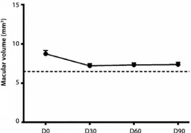
OCT performed prior to the treatment revealed an increased macular thickness probably due to intraretinal edema. We speculated that the increased thickness of the retinal pigment epitheliumcho -riocapillaris complex (RPE-CC) was compatible with the choroidal neovascular membrane. The temporal evolution of the retinal thickness was as follows: D0=8.44 ± 0.35 mm, D30=7.13 ± 0.22 mm, D60=7.16 ± 0.24 mm, and D90=7.23 ± 0.28 mm. Subjects without AMD had 6.52 ± 0.09 mm. Follow-up OCT examination showed a sig-nificant reduction in macular volume when D0 was compared with follow-up examination (P<0.01 one-way ANOVA, Tukey’s post-hoc test), but persistence of the increased retinal thickness compared with controls (Figures 1-2).
The results were grouped in three annular retinal areas for mfERG analysis: R1 (central area, 0°-8°), R2 (parafoveal area, 8°-24°), and R3 (peripheral area, 24°-45°). In each of these rings, we analyzed N1 and P1.
with that in controls over the entire testing period (P<0.01, one-way ANOVA, Tukey’s post-hoc test; Figure 3 A). In R3, the N1 amplitude was significantly reduced at D30, D60, and D90 compared with that in controls (P<0.01, one-way ANOVA, Tukey’s post-hoc test; Figure 3A).
In R1 and R2, N1 implicit time was not significantly different in patients and controls (Figure 3 B). In R3, N1 amplitude was delayed at D30 compared with that in controls (P<0.05, one-way ANOVA, Tukey’s post-hoc test; Figure 3 B).
Prior to treatment (D0), P1 amplitudes in R1 and R2 did not differ between patients and controls, but the amplitude was significantly reduced in patients at D30, D60, and D90 compared with that in con-trols (P<0.01, one-way ANOVA, Tukey’s post-hoc test; Figure 4 A). P1 amplitude in R3 did not differ between the two groups (Figure 4 A).
In R1, P1 implicit time at D0 and D60 was significantly delayed in patients compared with that in the control group (P<0.05, one-way ANOVA, Tukey’s post-hoc test; Figure 4 B). In R2, P1 implicit time was delayed at D30, D60, and D90 in patients compared with that in controls (P<0.01, one-way ANOVA, Tukey’s post-hoc test; Figure 4 B). In R3, P1 implicit time at D30 and D60 was significantly delayed in patients compared with that in controls (P<0.05, one-way ANOVA, Tukey’s post-hoc test; Figure 4 B).
Tables 1 and 2 show the mean values of the amplitude of mfERG components along the therapy.
DISCUSSION
In a multicenter study (the MARINA Study), Boyer and colleagues found improved visual acuity in the ranibizumab-treated group(12), a result corroborated by another multicenter study(13). Most multi-center studies evaluated only patients with good visual acuity, and a few studies evaluated the response of anti-angiogenic therapy in patients with advanced AMD. Despite subjective reports of improved perception in the central scotoma, visual acuity remained altered, with no significant difference before and after treatment. However, a significant reduction in retinal thickness was observed by measuring total macular volume before and after treatment. The results of this work showed a stabilization of visual acuity in patients before and after treatment, but the patients’ visual acuity was different from that observed in previous studies.
There was a significant reduction in retinal thickness after the first month of treatment compared with other days of examination, which is in agreement with other studies(14,15). Keane et al.used OCT to study 95 patients with neovascular AMD after intravitreal injections of ranibizumab(14). The total subretinal fluid (SRF) volume reached its lowest level by 1 month after the injection (P<0.001). Karagiannis et al. evaluated 34 patients treated with monthly injections of intravitreal bevacizumab for six months followed by monthly injections of rani-bizumab for 12 months(15). In these patients, OCT showed a decrease in retinal thickness (P=0.033) after bevacizumab, and this reduction improved after changing to ranibizumab (P=0.09).
Some authors suggested that patient history and age should be considered before analyzing mfERG since these factors may alter the retinal response. Importantly, mfERG responses became smaller and delayed with advancing age(16). The responses mediated by re-tinal cones occurring before rere-tinal damage became observable by ophthalmoscopy(17). Some studies suggested that the implicit time of the photopic mfERG responses was more sensitive for detecting areas with retinal dysfunction than response density(18).
We estimated the amplitude and implicit time mean values for N1 and P1 mfERG components as a function of the time after rani-bizumab injections. We focused on the temporal evolution of these parameters along the therapy. In this study, constant factors that could change these parameters, such as the hexagon areas, were not considered when estimating amplitude density. Previous studies revealed a delay in N1, P1, and the second negative component (N2)
Figure 1. Optical coherence tomography (OCT) of macular volume of controls (dashed
line) and patients (D0, D30, D60, and D90).
A
C
B
D
Figure 2. Optical coherence tomography (OCT) of patients at (A) D0, (B) D30, (C) D60, and (D) D90. An increased thickness
Table 1. mfERG amplitude and implicit time of the N1 component for three retinal areas at diferent days of therapy. Averaged values for controls are shown for comparison
R1 amplitude (µv)
R2 amplitude (µv)
R3 amplitude (µv)
GT-D0 -11.43 ± 0.99** -6.24 ± 0.43* -5.84 ± 0.51*
GT-D30 -8.09 ± 0.61* -4.41 ± 0.37* -4.71 ± 0.50* GT-D60 0-8.54 ± 0.79** -4.41 ± 0.37* -4.49 ± 0.43*
GT-D90 0-9.88 ± 0.99** -5.18 ± 0.48* -4.67 ± 0.46* GC -13.87 ± 0.95 0 -8.46 ± 0.58* -7.26 ± 0.50*
R1 implicit time (ms)
R2 implicit time (ms)
R3 implicit time (ms) GT-D0 17.79 ± 0.69 16.80 ± 0.54 15.47 ± 0.21*
GT-D30 15.23 ± 1.19 17.76 ± 0.91 16.46 ± 0.34*
GT-D60 14.02 ± 0.92 16.18 ± 0.93 16.04 ± 0.41* GT-D90 17.06 ± 0.46 16.81 ± 0.61 15.83 ± 0.23*
GC 15.74 ± 0.48 14.94 ± 0.23 15.16 ± 0.21*
GT= group of patients receiving therapy; GC= group of control subjects; D0= day of the
first injection; D30= 30th day after therapy initiation; D60= 60th day after therapy initiation;
D90= 90th day after therapy initiation; *= P<0.01; **= P<0.05.
Table 2. mfERG amplitude and implicit time of the P1 component for three retinal areas at diferent days of therapy. Averaged values for controls were also shown for comparison
R1 amplitude (µv)
R2 amplitude (µv)
R3 amplitude (µv)
GT-D0 14.24±1.61* 10.26±1.06* 11.10±1.27
GT-D30 11.57±1.33* 07.89±0.93* 08.29±1.08
GT-D60 11.21±0.95* 07.64±0.79* 08.50±0.90
GT-D90 12.39±1.10* 08.39±0.90* 09.14±1.15
GC 18.57±1.25* 13.26±0.89* 12.79±0.94
R1 implicit time (ms)
R2 implicit time (ms)
R3 implicit time (ms)
GT-D0 35.36±0.76** 34.73±0.71* 33.38±0.51*
GT-D30 32.94±0.98** 35.65±0.97* 34.19±0.79*
GT-D60 35.67±1.14** 34.21±0.54* 32.62±0.33*
GT-D90 34.31±1.07** 34.91±0.68* 33.55±0.63*
GC 31.44±0.32** 31.61±0.22* 31.20±0.15*
GT= group of patients receiving therapy; GC= group of control subjects; D0= day of the
first injection; D30= 30th day after therapy initiation; D60= 60th day after therapy initiation;
D90= 90th day after therapy initiation; *= P<0.01; **= P<0.05.
Figure 3. (A) Amplitude and (B) implicit time of the N1 component
of the multifocal electroretinogram (mfERG).
A
B
A
B
Figure 4. (A) Amplitude and (B) implicit time of the P1 component
of the multifocal electroretinogram (mfERG).
the third treatment, N1 and P1 amplitudes in R1 and R2 significantly decreased compared with those in controls. The implicit time of N1 and P1 in R1 and R2 did not change after treatment, but it was sig-nificantly delayed in two out of three patients when compared with those in controls. The authors argued that the mfERG represented AMD neovascularization (mfERG evaluated retinal function in up to 25° of the visual angle and thus represents the area affected by the neovascularization membrane), whereas visual acuity reflected the function of the foveal area, a region <1° of the visual angle. They sugges ted that the adverse effects of anti-VEGF in affected retinal areas were larger than the area clinically affected by AMD(4).
The role of VEGF in choroidal neovascularization is well establi-shed(22). Inflammation and focal retinal ischemia induce VEGF pro-duction, leading to the proliferation of neovascular membranes from the choriocapillaris(23). Ranibizumab was demonstrated as acting as anti-ocular VEGF; it neutralizes VEGF but whether it has an adverse effect on the healthy peripheral retina is not clear(24). Some studies suggest that VEGF inhibition may cause retinal ischemia(25).
The present work suggests a stabilization of visual acuity and reduction of edema after treatment, which is in agreement with other studies(26). Costa and colleagues suggested that the edema reduction alone could result in an improvement and/or stabilization of visual acuity(27). However, conflicting mfERG data were reported in other studies and suggested that AMD interrupted delicate intraretinal mechanisms. The progression of the disease associated or not with the adverse effects of anti-VEGF could interfere with neuroretinal functions(28).
We observed a reduction in the electrical activity of the central macular region (R1) after the initiation of treatment in patients under-going anti-angiogenic therapy with ranibizumab. As AMD affected mainly the central region, changes were expected in R1, which had the largest concentration of retinal neurons affected by the disease and was the place where the drug’s anti-angiogenic effect was expec-tedly higher(4). Some experimental studies suggested a possible toxic effect of high-dose intravitreal VEGF inhibitors on retinal neurons(29,30). The lower retinal electrical activity observed immediately after treat-ment raised the possibility of a deleterious effect of the medication on macular function, which fortunately recovered over time. Perhaps the preferential occurrence of macular lesions in advanced AMD faci-litated a very large increase in the levels of anti-angiogenic factors in the central retina, temporarily causing this deleterious effect.
The present study showed a reduction of retinal thickness due to a reduction of edema and tissue fibrosis. The mfERG findings showed that the amplitude of mfERG components decreased and that the implicit time of these components increased, suggesting a worse-ning of retinal activity. The non-significant reduction in visual acuity indicated that retinal activity impairment did not affect it. Along the therapy, we found an improvement of the mfERG response. More controlled studies will be necessary for more conclusive results of the effects of ranibizumab on AMD.
REFERENCES
1. Oguido AP, Casella AM, Matsuo T, Ramos Filho EH, Berbel R, Silva RM. Prevalence of age-related macular degeneration in Japanese immigrants and their descendants living in Londrina (PR) - Brazil. Arq Bras Oftalmol. 2008;71(3):375-80.
2. Kashani AH, Keane PA, Dustin L, Walsh AC, Sadda SR. Quantitative subanalysis of cys-toid spaces and outer nuclear layer using optical coherence tomography in age-related macular degeneration. Invest Ophthalmol Vis Sci. 2009;50(7):3366-73.
3. Greenstein VC, Chen H, Hood DC, Holopigian K, Seiple W, Carr RE. Retinal function in diabetic macular edema after focal laser photocoagulation. Invest Ophthalmol Vis Sci. 2000;41(11):3655-64.
4. Feigl B, Greaves A, Brown B. Functional outcomes after multiple treatments with ra ni-bizumab in neovascular age-related macular degeneration beyond visual acuity. Clin Ophthalmol. 2007;1(2):167-75.
5. Muether PS, Hoerster R, Hermann MM, Kirchhof B, Fauser S. Long-term effects of ra nibizumab treatment delay in neovascular age-related macular degeneration. Graefes Arch Clin Exp Ophthalmol. 2013;251(2):453-8.
6. Chakravarthy U, Harding SP, Rogers CA, Downes SM, Lotery AJ, Wordsworth S, et al. Ranibizumab versus Bevacizumab to Treat Neovascular Age-related Macular Dege-neration: One-Year Findings from the IVAN Randomized Trial. Ophthalmology. 2012; 119(7):1399-411.
7. Subramanian ML, Ness S, Abedi G, Ahmed E, Daly M, Feinberg E, et al. Bevacizumab vs ranibizumab for age-related macular degeneration: early results of a prospective double-masked, randomized clinical trial. Am J Ophthalmol. 2009;148(6):875-82.e1. 8. Cohen SY, Dubois L, Tadayoni R, Fajnkuchen F, Nghiem-Buffet S, Delahaye-Mazza C,
et al. Results of one-year’s treatment with ranibizumab for exudative age-related macular degeneration in a clinical setting. Am J Ophthalmol. 2009;148(3):409-13. 9. Frota de Almeida IN, Frota de Almeida LN, Frota de Almeida Sobrinho E, Souza GS,
Gomes BD, Rosa AM, et al. Electroretinography and optical coherence tomography in patients with age related macular degeneration before, during, and after treatment with ranibizumab. ARVO Annual Meeting. Inv Ophthalmol Vis Sci. 2011;52(5): E-Abstract 6071/A330.
10. Schuman JS, Puliafito CA, Fujimoto JG. Optical coherence tomography of ocular
disea-ses. 2a ed. NJ: Slack Inc.; 2004.
11. Hood DC, Bach M, Brigell M, Keating D, Kondo M, Lyons JS, et al. ISCEV standard for clinical multifocal electroretinography (mfERG) (2011 edition). Doc Ophthalmol. 2012; 124(1):1-13.
12. Boyer DS, Antoszyk AN, Awh CC, Bhisitkul RB, Shapiro H, Acharya NR. Subgroup analysis of the MARINA study of ranibizumab in neovascular age-related macular degeneration. Ophthalmology. 2007;114(2):246-52.
13. Rosenfeld PJ, Brown DM, Heier JS, Boyer DS, Kaiser PK, Chung CY, et al. Ranibizumab for neovascular age-related macular degeneration. N Engl J Med. 2006;355(14):1419-31. 14. Keane PA, Liakopoulos S, Ongchin SC, Heussen FM, Msutta S, Chang KT, et al.
Quan-titative subanalysis of optical coherence tomography after treatment with rani bizumab for neovascular age-related macular degeneration. Invest Ophthalmol Vis Sci. 2008; 49(7):3115-20.
15. Karagiannis DA, Ladas ID, Parikakis E, Georgalas I, Kotsolis A, Amariotakis G, et al. Chan ging from bevacizumab to ranibizumab in age-related macular degeneration. Is it safe? Clin Interv Aging. 2009;4:457-61.
16. Fortune B, Johnson CA. Decline of photopic multifocal electroretinogram responses with age is due primarily to preretinal optical factors. J Opt Soc Am A Opt Image Sci Vis. 2002;19(1):173-84.
17. Chen C, Wu L, Wu D, Huang S, Wen F, Luo G, et al. The local cone and rod system function in early age-related macular degeneration. Doc Ophthalmol. 2004;109(1):1-8. 18. Gerth C, Hauser D, Delahunt PB, Morse LS, Werner JS. Assessment of multifocal
electro-retinogram abnormalities and their relation to morphologic characteristics in pa tients with large drusen. Arch Ophthalmol. 2003;121(10):1404-14.
19. Gerth C. The role of the ERG in the diagnosis and treatment of Age-Related Macular Degeneration. Doc Ophthalmol. 2009;118(1):63-8.
20. Neveu MM, Tufail A, Dowler JG, Holder GE. A comparison of pattern and multifocal electroretinography in the evaluation of age-related macular degeneration and its treatment with photodynamic therapy. Doc Ophthalmol. 2006;113(2):71-81. 21. Maturi RK, Bleau LA, Wilson DL. Electrophysiologic findings after intravitreal
bevaci-zumab (Avastin) treatment. Retina. 2006;26(3):270-4.
22. Marneros AG, Fan J, Yokoyama Y, Gerber HP, Ferrara N, Crouch RK, et al. Vascular en-dothelial growth factor expression in the retinal pigment epithelium is essential for choriocapillaris development and visual function. Am J Pathol. 2005;167(5):1451-9. 23. Buschini E, Piras A, Nuzzi R, Vercelli A. Age related macular degeneration and drusen:
neuroinflammation in the retina. Progress in neurobiology. 2011;95(1):14-25. 24. Meyer CH, Holz FG. Preclinical aspects of anti-VEGF agents for the treatment of wet
AMD: ranibizumab and bevacizumab. Eye (Lond). 2011;25(6):661-72.
25. Robinson GS, Ju M, Shih SC, Xu X, McMahon G, Caldwell RB, et al. Nonvascular role for VEGF: VEGFR-1, 2 activity is critical for neural retinal development. FASEB J. 2001; 15(7):1215-7.
26. Witmer AN, Vrensen GF, Van Noorden CJ, Schlingemann RO. Vascular endothelial growth factors and angiogenesis in eye disease. Prog Retin Eye Res. 2003;22(1):1-29. 27. Costa RA, Jorge R, Calucci D, Cardillo JA, Melo LA, Jr., Scott IU. Intravitreal bevacizumab for choroidal neovascularization caused by AMD (IBeNA Study): results of a phase 1 dose-escalation study. Invest Ophthalmol Vis Sci. 2006;47(10):4569-78.
28. Jurklies B, Weismann M, Husing J, Sutter EE, Bornfeld N. Monitoring retinal function in neovascular maculopathy using multifocal electroretinography - early and long-term correlation with clinical findings. Graefes Arch Clin Exp Ophthalmol. 2002;240(4):244-64. 29. Myers AC, Lovestam Adrian M, Bruun A, Ghosh F, Andreasson S, Ponjavic V. Retinal
function and morphology in rabbit after intravitreal injection of VEGF inhibitors. Current eye research. 2012;37(5):399-407.