Original Article
3 1 0 Arq Bras Oftalmol. 2015;78(5):310-2 http://dx.doi.org/10.5935/0004-2749.20150081
INTRODUCTION
Eyes are the third most frequently affected organ by trauma, after hands and feet(1). The incidence of traumatic globe dehiscence after penetrating keratoplasty (PK) has been reported to be 0.6%-5.8%(2). Any globe is susceptible to rupture and will do so at its weakest point if subjected to enough force(3-5). Keratoplasty exposes patients to a higher risk of globe rupture because the surgical wound may never regain the strength and stability of an intact cornea.
In this study, the authors present their experience with 11 cases of traumatic graft dehiscence after PK or deep anterior lamellar kerato-plasty (DALK) as seen at the Brasilia Ophthalmologic Hospital (HOB), Brasilia, Brazil, from January 2004 to December 2012.
METHODS
This study is a retrospective, noncomparative, and interventional case series. All cases of traumatic wound dehiscence (WD) treated in the HOB were identified from the records of the only surgeon perfor-ming regular PK and DALK in this hospital from 2005 through 2013. PK was performed in 5 eyes (45.5%), and DALK was performed using the
big-bubble technique in 6 eyes (54.5%). All eyes were phakic before the trauma. No intraoperative microperforation had occurred previously in the DALK patients. Cases of WD caused by infection or suture mani-pulation, such as adjustment or removal of sutures, were excluded. All patients received immediate surgical intervention within several hours after admission. Subsequent secondary surgeries, if any, were noted.
The study was performed according to established ethical stan-dards for clinical research and was approved by the Institutional Review Board (IRB) of the Brasilia Ophthalmologic Hospital, Brazil. De mographic and surgical details were recorded, including the indi-cation for keratoplasty, time from keratoplasty to WD, extent of the WD, associated ocular complications, and the visual outcome after surgical repair.
All data analyses were performed using SPSS statistical software (version 17.0, SPSS, Inc, Chicago, Illinois, USA). Quantitative data are pre-sented as the mean ± standard deviation (range). The Mann-Whitney test was employed to compare the preoperative and postoperative visual acuities. Differences were considered statistically significant when the P value was <0.05.
Traumatic wound dehiscence after corneal keratoplasty
Deiscência de sutura pós-trauma ocular em olhos submetidos a transplante de córnea
Patrick Frensel tzelikis1, eduardo Muniz Fenelon1, rodrigo rodrigues YoshiMoto1, gustavo Peixoto rascoP1, rodrigo laFetá Queiroz1, Wilson takashi hida1
Submitted for publication: April 1, 2015 Accepted for publication: August 3, 2015
1 Cornea and External Diseases Department, Hospital Oftalmológico de Brasília, Brasilia, DF, Brazil.
Funding: No specific financial support was available for this study.
Disclosure of potential conflicts of interest: None of the authors have any potential conflict of interest to disclose.
Corresponding author: Patrick Frensel de Moraes Tzelikis. SQN 203, Bl. K, ap 502 - Brasília, DF - 70833-110 - Brazil - E-mail: [email protected]
ABSTRACT
Purpose: To assess patient characteristics, risk factors, outcomes, and the treatment of wound dehiscence (WD) in patients after corneal keratoplasty.
Methods: Retrospective chart reviewof 11 eyes of 11 patients with corneal grafts who underwent repair of WD from January 1, 2004 to December 31, 2012 at Hos-pital Oftalmologico de Brasilia.
Results: Eight (72.7%) patients were men and three were women. Six (54.5%) patients had deep anterior lamellar keratoplasty (DALK) and 5 had penetrating keratoplasty. The mean age at trauma was 31.1 years. The mean time from corneal keratoplasty to WD was 12.82 months (range, 3-33 months). The mean best-corrected visual acuity of patients before trauma was 20/60 (0.48 logMAR) and after final treatment was 20/160 (0.90 logMAR) (P=0.15). In one case, visual acuity decreased to no light perception because of retinal detachment and phthisis bulbi. Accidental blunt trauma and fall were the most common causes of WD.
Conclusion: Patients who undergo corneal keratoplasty have a life-long risk of WD. The full-thickness rupture at the graft-host junction in our study suggests that the junction remains vulnerable, even following DALK, and can rupture with trauma. In our series, depending upon the severity of the trauma, postkeratoplastic WD can be associated with a good visual prognosis.
Keywords: Surgical wound dehiscence; Corneal transplantation; Risk factors
RESUMO
Objetivo: Avaliar as características, os fatores de risco, resultados, e tratamento de pa-cientes que apresentaram deiscência traumática de sutura após transplante de córnea. Métodos: Estudo retrospectivo em que foram avaliados 11 olhos de 11 pacientes sub metidos a transplante de córnea e que desenvolveram deiscência de sutura entre janeiro de 2004 e dezembro de 2012 no Hospital Oftalmológico de Brasília. Resultados: Oito (72,7%) pacientes eram homens e três mulheres. Seis (54,5%) pacientes foram submetidos a ceratoplastia lamelar anterior profunda (DALK) e 5 pacientes a ceratoplastia penetrante (PK). A média de idade dos pacientes no momento do trauma era de 31,1 anos. O tempo médio entre o transplante de córnea e a deiscência de sutura foi de 12,82 meses (variando de 3 a 33 meses). A melhor acuidade visual corrigida (AVCC) doa pacientes antes do trauma era de 20/60 (0,48 logMAR), e após o trauma era de 20/160 (0,90 logMAR) (P=0,15). Em um caso, a acuidade visual reduziu para sem percepção luminosa devido a descolamento de retina e posterior atrofia bulbar. O trauma ocular acidental e a queda da própria altura foram as principais causas de deiscência de sutura nos olhos transplantados.
Conclusão: Pacientes previamente submetidos a transplante de córnea apresen-tam um risco prolongado de deiscência de sutura. A ruptura completa na junção doador-receptor no nosso estudo sugere que mesmo após um DALK a junção per-manece vulnerável e pode romper com o trauma. Na nossa série, dependendo da severidade do trauma, a deiscência pós-ceratoplastia pode estar associada a um bom prognóstico visual.
Tzelikis PF, et al.
3 1 1 Arq Bras Oftalmol. 2015;78(5):310-2 RESULTS
Our hospital surgeons performed 471 keratoplasties during the 8-year study period, and eleven eyes (2.33%) in this group subse-quently presented with globe rupture. Table 1 shows the demogra-phic data for the 11 patients with globe rupture. The study included 11 eyes of 11 patients, of which 8 (72.7%) were men. The mean patient age at the time of rupture was 31.1 ± 8.81 years (range, 17-44 years). The median interval between keratoplasty and globe rupture was 12.82 months (range, 3-33 months). The mean follow-up time after the repair surgery was 4.36 ± 2.5 years (range, 1-8 years). The most common indication for the original graft was keratoconus.
In all except 1 eye, globe rupture involved the graft-host junction. Three patients (27.3%) had dehiscence involving 1 quadrant, 6 pa-tients (54.5%) had dehiscence involving 2 quadrants, and 2 papa-tients (18.2) had dehiscence involving 3 or more quadrants. Descemet membrane perforation after DALK was observed in 4 eyes (66.6%).
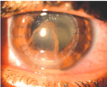
None of the patients were wearing protective eyewear at the time of trauma. Seven patients (63.6%) were using topical steroids at the time of WD. In all except 2 eyes, some or all of the sutures were present at the time of the trauma. In 4 eyes (36.4%), 3 of which un-derwent PK and 1 unun-derwent DALK, the crystalline lens was expelled during trauma. Two patients (18.2%) experienced retinal detachment after WD. Three eyes (27.3%) were noted to have elevated intraocu-lar pressure (defined as intraocuintraocu-lar pressure >22 mmHg), requiring medical therapy. Only 1 patient developed endophthalmitis after WD (Figure 1).
The best-corrected visual acuity (BCVA) at the last visit before trauma ranged between 0.00 logMAR to 1.18 logMAR (mean, 0.48 logMAR). At the final visit after the surgery, the mean BCVA was 0.90 logMAR (range, 0.40 logMAR to no light perception). The difference between the mean BCVA at the last visit before trauma and the last visit after the repair surgery was not found to be statistically
signifi-cant (P=0.15). At the last visit, 8 grafts (72.7%) were clear, 4 underwent secondary intraocular lens implantation for aphakia after the first surgical intervention, 2 (18.2%) experienced graft failure and the eye underwent repeat PK, and 1 eye (9.1%) developed phthisis bulbi.
DISCUSSION
WD is an unusual but serious complication of corneal kerato-plasty. The wound healing occurs mainly at the endothelial and epithelial junctures. At the level of the stroma, the lamellar cut ends reconnect through a tangle of new collagen fibers, rather than through anasto mosis(4). Various laboratory simulations and clinical and his-topathological reports have shown that the cornea never regains its preoperative strength(5,6). The finding of a 2.33% incidence of WD after keratoplasty is approximately the same as that reported in other studies, in which the incidence has been reported to be 1.3-2.6%(6,7). Generally, WD after keratoplasty occurs early in the postoperative course(2,6-8). Our study supports this finding, with an average time between keratoplasty and ocular trauma reported as 12.8 months.
Most of the patients in our study were relatively young males with the mean age of 31 years, which was lower than that of patients in earlier investigations. The mean ages in studies by Williams et al.(7), Das et al.(9) and Renucci et al.(8) were 65, 55, and 69.5 years, respectively. Most of the young patients were males, similarly to what has been reported(7). Tseng et al.(10) also found a predominance of men in their series of traumatic WD cases and attributed their findings to the more physically active lifestyle of men.
Keratoconus was the most common indication for keratoplasty among the patients that experienced traumatic WD in this study. Other graft indications included corneal scar and herpes simplex keratitis. This is consistent with the most common indications for corneal transplantation performed in Brazil, in which keratoconus is the most
Table 1. Demographic and clinical data for eleven patients with traumatic wound dehiscence after corneal keratoplasty
Case Age (years) Gender
Indication for keratoplasty
Type of keratoplasty
Interval between keratoplasty
and wound
dehiscence Nature of trauma
Extent of wound disruption (quadrants) and
other damage
BCVA before trauma
BCVA after rehabilitation
Follow-up (years)
01 26 M KCN DALK 06 m Deliberate blunt
trauma (punched)
2, lens and vitreous loss
20/60 20/60 2
02 41 M KCN DALK 07 m Hit with tennis ball 2 20/80 20/100 3
03 33 M KCN PK 06 m Finger poked into eye 2 20/70 20/60 2
04 33 F KCN PK 23 m Fall 2 20/20 20/200 4
lens and vitreous loss
05 44 M Corneal scar PK 17 m Deliberate blunt
trauma
3 20/25 20/2000 8
lens and vitreous loss and RD
2
06 39 F Corneal scar PK 10 m Fall lens and vitreous loss 20/50 20/50 6
07 28 F KCN PK 05 m Accidental blunt
trauma
3 20/100 NPL 1
endophthalmitis, vitreous hemorrhage,
and RD
08 37 M HSK DALK 19 m Accidental blunt
trauma
2 20/50 20/80 7
09 20 M KCN DALK 33 m Fall 1 20/30 20/50 8
10 24 M KCN DALK 12 m Accidental blunt
trauma
1 20/80 20/60 4
11 17 M KCN DALK 03 m Accidental blunt
trauma
1 20/300 20/50 3
Traumatic wound dehiscence after corneal keratoplasty
312 Arq Bras Oftalmol. 2015;78(5):310-2
common indication, followed by pseudophakic bullous keratopathy and corneal scar(11). In a study by Nagra et al.(12), the most common indication for PK among patients who developed WD was also kerato-conus, followed by Fuch’s endothelial dystrophy and pseudophakic bullous keratopathy. This is in contrast to the most common indica-tion for corneal transplantaindica-tion performed in the United States and United Kingdom, which is graft failure(13,14). Other indications include pseudophakic or aphakic bullous keratopathy, Fuch’s endothelial dys-trophy, keratoconus, and viral keratitis(13,14). In another retrospecti ve case series of 19 traumatic dehiscence with corneal grafts that underwent repair In Australia, the most common indication for the original graft was keratoconus(9). The indication for the original keratoplasty is not considered to influence the risk of subsequent dehiscence. However, grafts for keratoconus are less likely to vascularize, a process that would probably strengthen the wound.
The most prevalent cause of trauma in our series was accidental blunt trauma (4 eyes, 36.4%). This reflects the younger ages of our patients who engaged in more social activities and were, therefo re, more prone to ocular injury. Three patients sustained a fall from their own height, two received a deliberate blunt trauma (they were pun-ched), and one was hit with a tennis ball. The force of the trauma was great enough to expel the lens through the WD in four eyes (36.4%), given that three had undergone PK and only one had undergone DALK.
In each of these cases the patient received primary resuturing of his corneal graft within several hours after the trauma. With follow-up varying from 1 to 8 years, the grafts remained clear in 8 eyes (72.7%).
Other reports have found that WD after keratoplasty can be associa-ted with a good visual outcome, depending upon the nature and severity of the wound disruption and the type of keratoplasty per-formed. Our series supports this finding. The difference between the mean BCVA at the last visit before trauma and the last visit after the repair surgery was not found to be statistically significant (P=0.15). Among patients with worsening of vision, the major cause of vision loss was posterior segment damage at the time of trauma. In 5 of 11 cases with posterior segment involvement, only one had undergone DALK, whereas the other 4 had undergone PK.
Although DALK offers advantages over PK, including less damage to the endothelium and fewer immunologic reactions, it has compli-cations similar to that of PK. It is reasonable to assume that the intact Descemet’s membrane provides reinforcement against dehiscing traumas after DALK surgery. However, a weakness at the graft-host junction seems to persist, and a severe deforming force can result in WD with lens and vitreous loss.
In summary, this study found that the risk of traumatic corneal graft rupture after keratoplasty is significant, even years after the surgery, and may cause a bad visual outcome depending upon the severity of WD. This should be clearly emphasized during preopera-tive counseling, particularly for young male patients. To minimize the risk of WD after keratoplasty, a thorough education of transplant candidates encouraging the use of protective eyewear and a low-risk lifestyle is warranted.
REFERENCES
1. Négrel AD, Thylefors B. The global impact of eye injuries. Ophthalmic Epidemiol. 1998; 5(3):143-69.
2. Lam FC, Rahman MQ, Ramaesh K. Traumatic wound dehiscence after penetrating keratoplasty-a cause for concern. Eye (Lond). 2007;21(9):1146-50.
3. Maurice DM. The biology of wound healing in the corneal stroma. Castroviejo lecture. Cornea. 1987;6(3):162-8.
4. Calkins JL, Hochheimer BF, Stark WJ. Corneal wound healing: holographic stress-test analysis. Invest Ophthalmol Vis Sci. 1981;21(2):322-34.
5. Simonsen AH, Andreassen TT, Bendix K. The healing strength of corneal wounds in the human eye. Exp Eye Res. 1982;35(3):287-92.
6. Rehany U, Rumelt S. Ocular trauma following penetrating keratoplasty: inci dence, out co me, and postoperative recommendations. Arch Ophthalmol. 1998;116(10):1282-6. 7. Williams MA, Gawley SD, Jackson AJ, Frazer DG. Traumatic graft dehiscence after
penetrating keratoplasty. Ophthalmology. 2008;115(2):276-8.
8. Renucci AM, Marangon FB, Culbertson WW. Wound dehiscence after penetrating ke-ratoplasty: clinical characteristics of 51 cases treated at Bascom Palmer Eye Institute. Cornea. 2006;25(5):524-9.
9. Das S, Whiting M, Taylor HR. Corneal wound dehiscence after penetrating keratoplasty. Cornea. 2007;26(5):526-9.
10. Tseng SH, Lin SC, Chen FK. Traumatic wound dehiscence after penetrating kerato-plasty: clinical features and outcome in 21 cases. Cornea. 1999;18(5):553-8. 11. Calix Netto MJ, Giustina ED, Ramos GZ, Peccini RF, Sobrinho M, de Souza LB. [Major
indications for corneal penetrating keratoplasty at a reference service in São Paulo state]. Arq Bras Oftalmol. 2006;69(5):661-4. Article in Portuguese.
12. Nagra PK, Hammersmith KM, Rapuano CJ, Laibson PR, Cohen EJ. Wound dehiscence after penetrating keratoplasty. Cornea. 2006;25(2):132-5.
13. Kang PC, Klintworth GK, Kim T, et al. Trends in the indications for penetrating kerato-plasty, 1980-2001. Cornea. 2005;24(7):801-3.
14. Al-Yousuf N, Mavrikakis I, Mavrikakis E, Daya SM. Penetrating keratoplasty: indications over a 10 year period. Br J Ophthalmol. 2004;88(8):998-1001.