Vitrectomia pars plana e tamponamento por óleo de silicone para o
tratamento de endoftalmite aguda
Trabalho realizado no Centro de Pesquisa Rubens Si-queira de São José do Rio Preto (SP) - Brasil, no Hos-pital do Olho de Rio Preto - São José do Rio Preto (SP) e Universidade de São Paulo - USP - Ribeirão Preto (SP) - Brasil.
1Responsável pelo Centro de Pesquisa Rubens Siqueira - CPRS - de São José do Rio Preto (SP) - Brasil, Médico colaborador em pesquisa da Universidade de São Paulo - USP - Ribeirão Preto (SP) - Brasil. Responsável pelo Departamento de Oftalmologia da Faculdade de Medi-cina de Catanduva - Catanduva (SP) - Brasil. 2Médico assistente do CPRS de São José do Rio Preto
(SP) - Brasil.
3Fellow de retina do CPRS de José do Rio Preto (SP) -Brasil.
4Residente em Oftalmologia do Hospital do Olho de São José do Rio Preto (SP) - Brasil.
5Responsável pelo Departamento de Retina e Vítreo da USP - Ribeirão Preto (SP) - Brasil.
Corresponding author: Rubens Camargo Siqueira. Rua Saldanha Marinho, 2.815 - Conj. 42 - São José do Rio Preto (SP) Code 15010-100
E-mail: [email protected] Recebido para publicação em 07.06.2007 Última versão recebida em 09.12.2008 Aprovação em 11.12.2008
Rubens Camargo Siqueira1 Aline Degasperi Cote Gil2 Fabio Canamary3
Mirian Minari4 Rodrigo Jorge5
acute endophthalmitis treatment
Keywords: Vitrectomy; Silicone oils; Endophthalmitis
Purpose: To evaluate the outcomes of pars plana vitrectomy and silicone oil injection for the treatment of infectious endophthalmitis. Methods:35 cases of endophthalmitis secondary to phacoemulsification (20 patients), trabeculectomy (8 patients), perforating trauma (2 patients), trauma (2 patients), corneal transplantation (1 patient), vitrectomy (1 patient) and corneal ulceration (1 patient) were retrospectively studied. Patients were separated into two groups: Group 1 (n=24): intravitreal antibiotic injection, associated with topical and oral antibiotics; Group 2 (n=11): vitrectomy with intravitreal antibiotic injection and silicone oil injection. The follow-up ranged from 1 to 48 months (mean of 16 months). Results: From 24 patients in group 1, 11 patients (45.83%), had infection controlled with intravitreal antibiotic injection only; 13 patients (54.15%) regressed to uncontrolled endophthalmitis, in which two patients (8.33%) were submitted to evisceration and one patient (4.16%) had corneal melting. The remaining 10 patients (41.66%) with uncontrolled endophthalmitis were submitted to pars plana vitrectomy and silicone oil injection. Six patients (25%) from Group I had retinal detachment during the first month of follow-up and also required pars plana vitrectomy and silicone oil injection. In Group 2 patients (n=11), all of them had controlled infection at the first procedure. In one case (9.09%), a severe proliferatative vitreo-retinopathy induced loss of vision. Conclusion:These results suggest that silicone oil tamponade might be beneficial in the treatment strategy of infectious endophthalmitis.
ABSTRACT
INTRODUCTION
Endophthalmitis is most commonly reported after cataract surgery but may occur after any intraocular procedure, after trauma, or from endoge-nous sources. The incidence of endophthalmitis after cataract surgery varies according to the surgical technique, but in recent large series, it has ranged from 0.04% to 0.13%(1-4). Endophthalmitis after filtering surgery is
probably the second most commonly encountered etiologic category. Because of its typically delayed onset, the exact incidence is more dif-ficult to ascertain. With longer follow-up, bleb-associated endophthal-mitis may occur in at least 1% of patients and has been described in up to 1.7% of eyes per year when antimetabolites are used(2,5-6). This incidence
multiple routes of antibiotic administration, including intra-vitreal, systemic, topical, and subconjunctival(7). With the
advent of pars plana vitrectomy techniques, vitrectomy com-bined with injection of intravitreal antibiotics became the standard treatment for virtually all forms of endophthalmi-tis(8-9). Favorable sensitivity profiles and acceptably low
to-xicity rates have resulted in vancomycin and ceftazidime (more recently supplanting aminoglycosides) becoming a widely utilized regimen of intravitreal antibiotics(10-12). Topical and
subconjunctival antibioticsare also commonly used. The role of pars plana vitrectomy (PPV) in the treatment of endophthalmitis is controversial. The results of the Endoph-thalmitis Vitrectomy Study (EVS) suggested that patients, with a visual acuity of light perception, have better prognosis if treated with immediate PPV(13). Silicone oil has been used
in-creasingly in vitreoretinal surgery as an internal tamponade, with a significant improvement of the anatomical and visual outcome in many bad prognosis cases(14). The dynamics of
sili-cone involve surface tension and viscosity, but also limit the free movements of aqueous humor in the eye. No microorga-nisms seem to be able to penetrate and develop within this inert material(15). Moreover, antibacterial properties of silicone oil
have been recently reported in vitro(16). Since the early nineties,
vitreoretinal surgeons have used silicone oil in some cases of endophthalmitis with extensive retinal damage as intraocular tamponade in order to prevent retinal detachment(16-21).
The purpose of this retrospective study was to compare the results of treating endophthalmitis using 3 options: 1) in-travitreal antibiotic injection (IVAB) 2) vitrectomy with injection of silicone oil and antibiotics as a first treatment 3) vitrectomy with injection of silicone oil and antibiotics as a second treatment after the failure of IVAB.
METHODS
Thirty-five charts from thirty-five patients with endoph-thalmitis treated at HORP-Hospital do Olho de Rio Preto-São Paulo-Brazil, between January 2001 and 2006 were reviewed. The clinical diagnosis was based on the standard sign and symptoms of endophthalmitis: pain, loss of vision, lid edema, conjunctival chemosis and injection, corneal ede-ma, anterior chamber flare, hypopyon, and vitreous opacifi-cation. Culture studies of vitreous samples were performed in all cases.
The patients were separated into two groups:
Group 1 (24 patients): Intravitreal antibiotic injection (IVAB), vancomycin 1 mg/0.1 ml, ceftazidime 2.25 mg/0.1 ml, dexamethasone 0.4 mg/0.1 ml). Periocular (subconjunctival) vancomycin 25 mg, ceftazidime 100 mg, dexamethasone 12 to 24 mg. Topical (started on first postoperative day) van-comycin 50 mg/ml q 1 hour, ceftazidime 50 mg/ml q 1 hour. Topical steroids and cycloplegics. Systemic: vancomycin 1 g IV q 12 hours, ceftazidime 1 g IV q 12 hours or ciprofloxacin 750 mg p.o. q 12 hours for susceptible organisms.
Group 2 (11 patients): Pars plana vitrectomy (PPV) with injection of silicone oil (SOI) and antibiotics as a first treat-ment. The patients were submitted to standard three-port pars plana vitrectomy using ACCURS vitrectomy machine (Alcon, Fort Worth, Texas, USA). Initially, the central vi-treous was removed (core vitrectomy), with later peeling of dense epiretinal membranes. Next, the remaining traction points were identified; including those of hard removal, i.e., star folds with intraretinal fibrosis. At those sites, instead of removing membranes with the use of forceps, a puncture retinotomy was performed using endodiathermy. After rela-xing the traction areas, subretinal fluid was aspirated with a long extensible silicone-tipped cannula, taking advantage of the retinotomy. After this procedure, we checked whether the entire retina was attached and, if this was not the case, additional retinotomies were made to release the traction, and a new aspiration of subretinal fluid was performed. After the retina was entirely flattened against the retinal pigment epithelium RPE, photocoagulation was applied surrounding all retinal breaks and along the entire edge of the puncture retinotomies. At the end of the procedure silicone oil was injected into the eye with IVAB.
The follow-up ranged from 1 to 48 months (mean=16 months). Anatomical status and visual outcome were recor-ded in order to compare the prognosis of different approa-ches.
The criteria for control of infection were: a) absorption of hypopyon, b) decrease or absence of cells and flare in the anterior chamber and vitreous, c) improvement of symptoms such as pain, d) improvement in congestion and conjunctival hyperemia and eyelids.
RESULTS
Clinical characteristics of all patients are shown in tables 1 and 2.
The average age of the patients was 66.82 years (range, 12-85 years). A vitreous positive culture was found in 12 cases (34.28%). Of these 12 cases, 6 (50%) were due to Sta-phylococcus epidermidis: 2 (16.66%) cases were due to S trep-tococcus sp; 1 case each was due to Staphylococcus sapro-fiticus, Staphylococcus aureus and Morganella morganii.
In Group 1, the average visual acuity ranged from 20/30 to light perception (before treatment) and 20/60 to light perception (after treatment). Nine patients (37.5%) had wor-sening of visual acuity, 10 patients (41.6%) improved and 5 patients (20.83%) did not change.
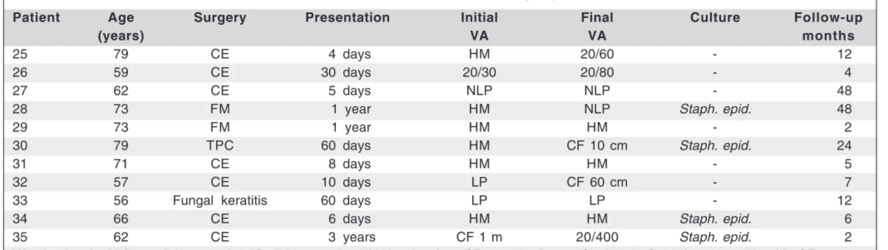
In Group 2, the average visual acuity ranged from 20/40 to light perception (before treatment) and 20/30 to light percep-tion (after treatment). One patient (9.09%) showed worsening of visual acuity, 5 patients (45.45%) improved and 5 patients (45.45%) did not change.
Table 1. Clinical characteristics of group 1
Patient Age Surgery Time Infection Second Infection Compli- Initial Final Culture
Follow-(y) presen- control treatment control cation VA VA up
tation (1) (2) months
01 63 CE 30 d no PPV+sil oil yes - CF 1m CF 1m - 5
02 75 CE 4 d no PPV+sil oil yes RD HM HM Strep. sp 14
03 78 CE 5 d no PPV+sil oil yes - HM LP Strep.viridans 4
04 66 CE 3 d no PPV+sil oil yes RD HM HM - 6
05 60 CE - no AC wash yes Melting LP LP - 24
06 69 CE 30 d no PPV+sil oil yes RD HM LP - 2
07 67 CE 5 d yes - - - LP 20/80 - 8
08 12 Trauma 3 d no Evisceration - - LP NLP Bacillus G + 24
09 30 Trauma 3 d no PPV+sil oil yes - HM HM Staph. 30
saprofiticus
10 79 CE 5 d no PPV+sil oil yes - HM 20/30 Staph. aureus. 24
11 85 FM 4 y yes - - - LP HM - 12
12 77 CE 10 d no PPV+sil oil yes - HM 20/200 - 8
13 53 FM 2 y no - - RD HM NLP - 48
14 73 CE 15 d no PPV+sil oil yes - 20/40 20/30 Morganella 32
morganii
15 81 CE 10 d no PPV +sil oil yes - HM CF 60 cm Staph. epid. 7
16 81 FM 2 y no PPV+sil oil yes - 20/100 HM - 2
17 69 PCIOL 6 d yes - - - CF 60 cm 20/30 - 36
18 65 CE 15 d yes - - - CF 20 cm 20/60 - 6
19 78 FM 2 y yes - - - 20/60 20/100 - 17
20 64 FM 5 y no PPV+sil oil yes - LP HM - 1
21 66 CE 4 d no Evisceration - - NLP NLP Staph. epid. 13
22 77 CE 120 d no PPV+sil oil yes - HM CF 30 cm - 32
23 51 CE 1 d no PPV+sil oil yes - HM 20/200 - 20
24 83 FM 1 y no PPV+sil oil yes RD HM LP - 7
VA= visual acuity; NLP= non light perception; LP= light perception; HM= hand motions; CF= counting fingers; AC= anterior chamber; Staph.= Staphylococcus; epid.=
epidermidis; Strep.= Streptococcus; CE= cataract extraction; PCIOL= posterior chamber intraocular lens; FM= filtering surgery; d= days; y= years; PPV= pars plana vitrectromy; sil oil= silicone oil; RD= retinal detachment
Table 2. Clinical characteristics of group 2
Patient Age Surgery Presentation Initial Final Culture Follow-up
(years) VA VA months
25 79 CE 4 days HM 20/60 - 12
26 59 CE 30 days 20/30 20/80 - 4
27 62 CE 5 days NLP NLP - 48
28 73 FM 1 year HM NLP Staph. epid. 48
29 73 FM 1 year HM HM - 2
30 79 TPC 60 days HM CF 10 cm Staph. epid. 24
31 71 CE 8 days HM HM - 5
32 57 CE 10 days LP CF 60 cm - 7
33 56 Fungal keratitis 60 days LP LP - 12
34 66 CE 6 days HM HM Staph. epid. 6
35 62 CE 3 years CF 1 m 20/400 Staph. epid. 2
VA= visual acuity; NLP= non light perception; LP= light perception; HM= hand motions; CF= counting fingers; Staph. epi.= Staphylococcus epidermidis; CE= cataract
extraction; PCIOL= posterior chamber intraocular lens; FM= filtering surgery; TPC
uncontrolled endophthalmitis, in which two patients (8.33%) were submitted to evisceration and one patient (4.16%) had corneal melting. The remaining 10 patients (41.66%) with uncontrolled endophthalmitis were submitted to PPV and SOI. Six patients (25%) from Group I had retinal detachment
DISCUSSION
The relationship between the use of silicone oil and endoph-thalmitis has been little discussed. Endophendoph-thalmitis is considered one of the most devastating complications of ophthalmic surgery. The incidence of endophthalmitis after cataract surgery ranges from 0.04 to 0.13%(22-25). And after vitrectomy surgery
from 0.046 to 0.07%(12,26). The incidence of endophthalmitis after
vitrectomy where silicone oil was used is more rare with only 2 cases reported in the literature with positive culture(3,20). Other
authors reported 2 cases of endophthalmitis in patients under-going surgery with silicone oil, but the culture was negative and because the inflammatory reaction was probably consequent to the components of silicone oil(7-10).
The likely antimicrobial potential of silicone oil has re-cently been reported. The authors demonstrated in vitro that the silicone oil decreases the proliferation of bacteria respon-sible for endophthalmitis(18). In the eyes of humans, the
ac-tion of silicone oil against the microorganisms could be explained by the following mechanisms: 1) the silicone oil can be toxic to the bacteria, as suggested in in vitro experi-ments, however this hypothesis still needs to be confirmed by more experiments; 2) the silicone oil is highly hydrophobic with a high interfacial tension and thus presents a low per-meability to the passage of cells and bacteria(8). Therefore,
this limits the space for the movement of infectious agents and keeps them in touch with the ciliary body and vessels of the retina, which can improve the effectiveness of the defense mechanisms, by a high concentration of biochemical media-tors, antibodies and inflammatory cells within an aqueous compartment of the vitreous cavity.
In the literature, endophthalmitis is suspected to increase the risk of retinal detachment. EVS reported 20 cases of retinal detachment in a series of 420 patients, 6 in the vitrectomy group (2.8% of 218 patients) and 14 in the non vitrectomy group (6.9% of 202 patients). Other authors(17) described
reti-nal detachment to occur in 21% (7/34) of endophthalmitis patients treated with vitrectomy and intraocular antibiotics.
In the other study a similar complication rate was descri-bed with 26% retinal detachments (9/34): 8 patients (23%) in the non silicone groups (1 and 2), 1 patient (7%) in the silicone groups (3 and 4). The authors postulated that retinal detach-ment could be either the result of surgical complications or be provoked by the changes in retina and vitreous as a reaction to severe ocular inflammation(21).
Our series showed that in Group 1, 11 patients (45.83%), had infection control with intravitreal injections only. Howe-ver, 16 (66.66%) patients required pars plana vitrectomy and silicone oil, 10 patients (41.66%) had uncontrolled endoph-thalmitis and 6 (25%) had retinal detachment. Two patients (8.33%) were submitted to evisceration and one patient (4.16%) had corneal melting.
In Group 2, all patients had controlled infection at the first procedure and did not need further surgery, except for removal of silicone oil three months later. In one case
(9.09%), a severe PVR induced loss of vision (NLP) but the cosmetic results were acceptable.
Many authors also reported that concurrent endophthalmitis and retinal detachment have a poor visual and anatomical out-come, especially when retinal detachment is an intraoperative complication(2,11,15,19-20). The use of silicone oil can therefore help
in controlling the infectious process and reduce the risk of retinal detachment, contributing to a better outcome in the treatment of endophthalmitis. After control of the infectious and inflammatory process the silicone oil can be removed, which in our study was conducted on average 3 months after the procedure.
CONCLUSION
This retrospective study suggests that silicone oil might be beneficial in the treatment strategy of severe endophthalmitis. Patients treated with silicone oil have a better control of infec-tion, better anatomical stabilization and better final visual acuity. Additionally, silicone oil seems to reduce the risk of retinal detachment and the need for additional procedures.
The role of silicone oil in the treatment of endophthal-mitis should be better assessed by a prospective, controlled study and with the highest number of cases to confirm the evidence of this study.
RESUMO
Objetivo: Avaliar os resultados da vitrectomia pars plana com tamponamento com óleo de silicone no tratamento de endof-talmite aguda. Métodos: Trinta e cinco pacientes com endoftal-mite, sendo 20 secundário à facoemulsificação, 8 por trabecu-lectomia, 2 por trauma perfurante, 2 por trauma, 1 por trans-plante de córnea, 1 por vitrectomia, e 1 por úlcera de córnea, foram estudados retrospectivamente. Os pacientes foram separa-dos em dois grupos. Grupo 1 (n=24): injeção de antibiótico intravítreo (AIV), associado com antibióticos oral e sistêmico; Grupo 2 (n=11): vitrectomia com AIV e óleo de silicone. O seguimento variou de 1 a 48 meses (média de 16 meses). Resul-tados: Dos 24 pacientes no Grupo 1, 11 (45,83%) tiveram con-trole da infecção apenas com injeção AIV, 13 (54,15%) não controlaram a endoftalmite, sendo que, dois destes (8,33%) fo-ram submetidos à evisceração e um (4,16%) evoluiu para “mel-ting” corneano. Os outros 10 (41,66%) pacientes foram submeti-dos à vitrectomia pars plana e óleo de silicone. Seis pacientes (25%) do Grupo 1 tiveram descolamento de retina e também necessitaram de vitrectomia pars plana e óleo de silicone. No Grupo 2 (n=11), todos tiveram controle da infecção no primeiro procedimento e não necessitaram de mais intervenções, exceto pela remoção do óleo de silicone três meses depois. Conclusão:
Os resultados sugerem que o tamponamento por óleo de silico-ne parece ser benéfico na estratégia de tratamento da endof-talmite infecciosa aguda.
REFERENCES
1. Affeldt JC, Flynn HW Jr, Forster RK, Mandelbaum S, Clarkson JG, Jarus D. Microbial endophthalmitis resulting from ocular trauma. Ophthalmology. 1987;94(4):407-13.
2. Brinton GS, Topping TM, Hyndiuk RA, Aaberg TM, Reeser FH, Abrams GW. Posttraumatic endophthalmitis. Arch Ophthalmol. 1984;102(4):547-50. 3. Chong LP, De Juan E Jr, McCuen BW 2nd, Landers MB 3rd. Endophthalmitis
in a silicone oil-filled eye. Am J Ophthalmol.1986;102(5):660-1.
4. Cohen SM, Flynn HW Jr, Murray TG, Smiddy WE. Endophthalmitis after pars plana vitrectomy. The Postvitrectomy Endophalmitis Study Group. Oph-thalmology. 1995;102(5):705-12.
5. Results of the Endophthalmitis Vitrectomy Study: A randomized trial of immediate vitrectomy and of intravenous antibiotics for the treatment of postoperative bacterial endophthalmitis. Endophthalmitis vitrectomy Study Group. Arch Ophthalmol. 1995;113(12):1479-96. Comment in: Arch Oph-thalmol. 1995;113(12):1555-7; Arch OphOph-thalmol. 1996;114(8):1025-6; author reply 1026-7; Arch Ophthalmol. 1996;114(8):1025; author reply 1026-7; Arch Ophthalmol. 1996;114(8):1027-8; author reply 1028-9; Arch Ophthalmol. 1996;114(8):1029-30; author reply 1028-9; Arch Ophthalmol. 1996;114(8): 1029; author reply 1028-9, 1030; Arch Ophthalmol. 2002;120(2):230-1; Arch Ophthalmol. 2002;120(2):231-3.
6. Foster RE, Rubsamen PE, Joondeph BC, Flynn HW Jr, Smiddy WS. Concurrent endophthalmitis and retinal detachment. Ophthalmology. 1994;101(3):490-8. 7. Gabel VP, Kampik A, Burkhardt J. Analysis of intraocularly applied silicone
oils of various origins. Graefes Arch Clin Exp Ophthalmol. 1987;225(3):160-2. 8. Giordano GG, Refojo MF. Silicone oils as vitreous substitutes. Prog Polym
Sci. 1998;23:509-32.
9. Johnson RN, Flynn HW Jr, Parel JM, Portugal LM. Transient hypopyon with marked anterior chamber fibrin following pars plana vitrectomy and silicone oil injection. Arch Ophthalmol. 1989;107(5):683-6. Comment in: Arch Opthalmology. 1989;107(11):1566.
10. Kampik A. Silicone oil interaction with ocular tissue. Int Ophthalmol Clin. 1987;10:85.
11. Kattan HM, Flynn HW Jr, Pflugfelder SC, Robertson C, Forster RK. Noso-comial endophthalmitis survey. Current incidence of infection after intraocular surgery. Ophthalmology. 1991;98(2):227-38. Comment in: Ophthalmology. 1991;98(8):1147-8.
12. Kresloff MS, Castellarin AA, Zarbin MA. Endophthalmitis. Surv Ophthal-mol. 1998;43(3):193-224.
13. Landers JH, Chappell CW. Bilateral metastatic endophthalmitis. Retina. 1981;1(3):175-8.
14. Lucke K, Laqua H. Silicone oil in the treatment of complicated retinal de-tachments. Berliln: Springer-Verlag. 1990.
15. Mao LK, Flynn HW Jr, Miller D, Pflugfelder SC. Endophthalmitis caused by streptococcal species. Arch Ophthalmol. 1992;110(6):798-801.
16. Mieler FW, Glazer LC, Bennett SR, HaN DP. Favourable outcome of trau-matic endophthalmitis with associated retinal breaks or detachment. Can J Ophthalmol. 1992;27(7):348-52.
17. Nelsen PT, Marcus DA, Bovino JA. Retinal detachment following endoph-thalmitis. Ophthalmology. 1985;92(8):1112-7.
18. Ozdamar A, Aras C, Ozturk R, Akin E, Karacorlu M, Ercikan C. In vitro antimicrobial activity of silicone oil against endophthalmitis causing agents. Retina. 1999;19(2):122-6. Comment in: Retina. 2001;21(1):92-3.
19. Vahey JB, Flynn HW Jr. Results in the managementof bacillus endophthal-mitis. Ophthalmic Surg. 1991;22(11):681-6. Comment in: Ophthalmic Surg. 1992;23(5):368.
20. Zimmer-Galler IE, Santos A, Haller JA, Campochiaro PA. Management of endophthalmitis in a silicone filled eye. Retina. 1997;17(6):507-9. 21. Bali E, Huyghe PH, Caspers L, Libert J. Vitrectomy and silicone oil in the
treatment of acute endophthalmitis. Preliminary results. Bull. Soc Belge Ophtalmol. 2003;(288):9-14.
22. Benz MS, Scott IU, Flynn HW Jr, Unonius N, Miller D. Endophthalmitis isolates and antibiotic sensitivities: A 6-year review of culture-proven cases. Am J Ophthalmol. 2004;137(1):38-42. Comment in: Am J Ophthalmol. 2004; 137(6):1167-8; author reply 1168; Am J Ophthalmol. 2004;137(6):1169; author reply 1169-70.
23. Schiedler V, Scott IU, Flynn HW Jr, Davis JL, Benz MS, Miller D. Culture-proven endogenous endophthalmitis: clinical features and visual acuity outcomes. Am J Ophthalmol. 2004;137(4):725-31.
24. Miller JJ, Scott IU, Flynn HW Jr., Smiddy WE, Newton J, Miller D. Acute-onset endophthalmitis after cataract surgery (2000-2004): Incidence, clinical settings, and visual acuity outcomes after treatment. Am J Ophthalmol. 2005; 139(6):983-7. Comment in: Am J Ophthalmol. 2005;139(6):1097-8. 25. Miller JJ, Scott IU, Flynn HW Jr, Smiddy WE, Corey RP, Miller D.
Endoph-thalmitis caused by streptococcus pneumoniae. Am J Ophthalmol. 2004; 138(2):231-6. Comment in: Am J Ophthalmol. 2005;139(6):1147; author reply 1147-8.