Toxicidade intravítrea da rapamicina em olhos de coelhos
Trabalho realizado na Tulane University - New Orleans - USA.
1MD, Department of Ophthalmology, Universidade de São Paulo - USP - São Paulo (SP) - Brazil. MD, Depart-ment of Ophthalmology, Santa Casa de Misericórdia de São Paulo - São Paulo (SP) - Brazil.
2MD,Department of Ophthalmology, University of Arizona - Tucson - EUA.
3MD, Department of Ophthalmology, Tulane University, New Orleans - EUA.
4MD, Department of Ophthalmology, Tulane University, New Orleans. MD,Department of Ophthalmology, University of Arizona - Tucson - EUA.
5MD,Department of Pathology, The Methodist Hospital - Houston - EUA.
6MD, Department of Ophthalmology, Universidade de São Paulo - USP - São Paulo (SP) - Brazil.
Corresponding author: Roberta P. A. Manzano. Rua Aramanaí, 307 - São Paulo (SP) CEP 05450-030 E-mail: [email protected]
None of the authors has any proprietary interest in any technique or product described herein.
Recebido para publicação em 12.03.2008 Aprovação em 25.11.2008
Roberta Pereira de Almeida Manzano1 Gholam A. Peyman2
Palwasha Khan3 Muhamet Kivilcim4 Patricia Chevez-Barrios5 Walter Takahashi6
in rabbit eyes
Keywords: Choroidal neovascularization; Sirolimus/toxicity; Uveitis; Vascular endothelial growth factor receptor-1; Rabbits
Purpose: To evaluate retinal toxicity of varying doses of rapamycin when injected intravitreally in rabbits. Rapamycin is a potent immunosup-pressive agent with significant antitumor and antiangiogenic properties, clinically approved for prevention of organ transplant rejection. Methods:
Twelve New Zealand albino rabbits were divided into four groups. Four different doses of rapamycin were prepared in 0.1 ml: 20 µg, 50 µg, 200 µg, and 1000 µg. Each concentration was injected in one eye of three rabbits,
and 0.1 ml volume of sterile BSS was injected into the contralateral eye of the three rabbits. Slit-lamp and fundoscopic examinations were perfor-med and the animals were observed for 2 weeks for signs of infection, inflammation, and toxicity. A baseline ERG was performed before drug treatment and at day 14, after which the rabbits were euthanized. Histology of the enucleated eyes was studied to look for retinal toxicity. Results:
ERG results showed some decrease in scotopic response; however this was not dose related. ERG results were normal at 20 µg. Histological
results showed no retinal toxicity in all groups. Conclusion: Although ERG changes were identified at dosages between 50-1000 µg, the histology
of all groups up to 1000 µg did not show any discernable abnormalities.
ABSTRACT
INTRODUCTION
Treatment of severe ocular inflammation may require long-term use of immunosuppressive drugs. Long-term use of these drugs is problematic because of their adverse side effects. To decrease these adverse side effects, intravitreal administration is a possible treatment option.
Rapamycin (sirolimus; A.G. Scientific, San Diego, CA) is a cyclic ma-crolide antibiotic isolated from the fungus Streptomyces hygroscopicus(1).
It is a potent immunosuppressive agent, clinically approved for prevention of organ transplant rejection(2).It has been used in sirolimus eluting stents
in patients with coronary artery disease(3), as it also has antiproliferative
effects on smooth muscle cells and on arterial intimal thickening(4). Similar
Rapamycin has significant antitumor and antiangiogenic activities. Several rapamycin-related compounds are in phase I, II and III clinical trials in patients with renal cancer and other malignancies(6). In an in vivo experiment with rats, rapamycin
dramatically inhibited tumor growth, with a marked decrease in tumor vascularization(7). Guba et al. demonstrated the
anti-VEGF activity of sirolimus in a murine tumor model(8). Its
antiangiogenic properties were also demonstrated in a 1995 study by Olsen et al. who showed that rats receiving sirolimus following orthotopic allogenic corneal grafts had reduced amounts of corneal neovascularization(9). Recently, it was
demonstrated that rapamycin significantly reduced the extent of neovascularization in both experimental choroidal neovas-cularization (CNV) and retinopathy of prematurity (ROP) in mice(10).
Rapamycin has been shown to reduce inflammation in animal models of experimental autoimmune uveitis, in combi-nation with tacrolimus and cyclosporine(11-12). Recent
inves-tigation has revealed that rapamycin is an effective and potent immunosuppressive treatment for patients with non-infec-tious uveitis and can reduce the need for long-term supple-mentary corticosteroid therapy(13).
One possible option of treatment for chronic non-infec-tious uveitis, choroidal, or retinal neovascularization could be an intravitreal injection of rapamycin based on its immuno-suppressive and antiangiogenic properties. However, retinal toxicity is a primary concern when using intravitreal injec-tions. The purpose of our study was to analyze the retinal toxicity of rapamycin at various doses.
METHODS
Animals
Twelve New Zealand albino rabbits weighing between 2 and 3 kg were used for this study. The rabbits were treated according to guidelines of the Association for Research in Vision and Ophthalmology (ARVO). Slit-lamp and indirect fundoscopic examinations were performed on all eyes prior to the study and on days 1, 7, and 14 after intravitreal injection. Any rabbit that demonstrated corneal or lens opacity or retinal damage prior to the study was excluded. Rabbits were anes-thetized before all procedures using approximately 1 ml of a mixture of ketamine hydrochloride (50 mg/kg) and xylazine hydrochloride (5 mg/kg). The eyes were dilated by topical application of phenylephrine (2.5%) and tropicamide (0.5%). Topical anesthesia was applied using proparacaine (0.5%).
Intravitreal injections
All procedures were performed under sterile conditions using an operating microscope for visualization. Anterior cham-ber tap was performed with a 25 G needle withdrawing 0.1 cc of aqueous fluid to reduce intraocular pressure and to minimize drug reflux following injection. Intravitreal injection was per-formed using a 30 G needle attached to a tuberculin syringe
inserted bevel up approximately 2 mm posterior to the limbus. Four different doses of rapamycin were prepared in 0.1 ml: 20 µg,
50 µg, 200 µg, and 1000 µg. Each dose was injected in one eye
of three rabbits, and 0.1 ml volume of sterile BSS was injected into the contralateral eye of the three rabbits. Slit-lamp and fundoscopic examinations were performed and the animals were observed for 2 weeks for signs of infection, inflammation, and toxicity.
Electrophysiological tests
ERG using the UTAS-2000 system (LKC Technology, Gaithersburg, MD) was performed prior to and 14 days after intravitreal injection. Rabbits were dark-adapted for at least 30 minutes after pupillary dilation. Unipolar contact lenses (ERG jet electrodes) were put on both corneas with Goniosol (IOLab Corporation, Claremont, CA); the negative electrode was placed in the subcutaneous space of the forehead, and the ground electrode was clipped to the earlobe with some electric gel. The dark-adapted scotopic response (step 1, Rod response), scotopic flash response (step 2, Maximal response, cone+ rod) and (after waiting 3 minutes) light-adapted pho-topic response (step 4, Cone response) were recorded. The average of five sweeps was determined for each step. The difference between a and b waves was calculated for each step. The baseline was compared to the response 2 weeks after injection.
Histological examination
Following the final ERG session, all rabbits were eutha-nized with an intravenous injection of sodium pentobarbital. The eyes were enucleated and fixed in Karnovsky fixative for 48 hours and then processed, sectioned, and stained with hematoxylin and eosin for light microscopy.
RESULTS
Clinical examination
No corneal opacity, cataract, vitreous hemorrhage, retinal detachment, or optic atrophy was seen in any of the rabbit eyes by fundoscopic examination.
Electrophysiological tests
Follow-up ERGs showed some decrease in 2 of 3 eyes va-rying in the amplitude of the following groups: 50 µg, 200 µg,
and 1000 µg. However, this decrease was not dose related and was never extinguished (Figures 1, 2).
Histological examination
Figure 1 - ERG step 1, 2, and 4 of one rabbit eye before (left) and 14 days after (right) intravitreal injection of 200 µµµµµg rapamycin. There is a decrease in the amplitude of step 1.
Figure 2 - ERG step 1, 2, and 4 of one rabbit eye before (left) and 14 days after (right) intravitreal injection of 1000 µµµµµg rapamycin. There is a non-significant decrease in step 1 and 2, and an increase in amplitude in step 4.
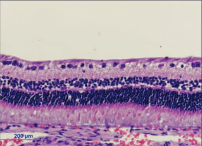
Figure 3 - Normal retinal histology in rabbit eye after intravitreal injection of 200 µµµµµg rapamycin (H&E stain)
DISCUSSION
Rapamycin appeared to be non-toxic to the retina in albino rabbits; however, we observed some decrease in ERG amplitu-de, mainly in the scotopic response in a few eyes. The ERG findings were not uniform in each group nor were they dose related. We think this finding might have been related to reduced body temperature occurring in a cold room in anes-thetized animals during dark adaptation(14-15). Hypothermia
decreases general body metabolism, leading to a reduction in the rate of chemical reactions and the amplitude of the ERG in mice and rabbits(14-15).
Another possibility for this slight decrease in the a-wave amplitude is that rapamycin (a lipophilic compound) might be localized in the region of photoreceptor or a pigment epithe-lium, thus creating some dysfunction of the photoreceptor. Further studies including immunohistochemical assays of drug localization should be done to confirm this possibility.
Maybe the histology is not sensitive enough since doses below 50 µg did not cause any ERG changes. We can have
some functional damage without any change in the histology. Nevertheless, normal ERG does not rule out functional and possible structural changes at the level of ganglion cells or the nerve fiber layer. The lack of changes by light microscopy does not exclude possible alterations at a submicroscopic level. Further studies must be done to evaluate this possi-bility.
An important characteristic of rapamycin is its antiangio-genic property.
Numerous clinical and experimental observations have in-dicated that ischemia or hypoxia triggers retinal neovascula-rization through an excessive production of angiogenic factors. Among multiple factors, many investigators have reported vas-cular endothelial growth factor (VEGF) to be an important(16-17)
and major stimulus responsible for neovascularization in exu-dative age-related macular degeneration (ARMD)(18).
Intravitreal injections of VEGF inhibitors such as pegap-tanib(19-20), ranibizumab(21-22), and bevacizumab(23) have been
commonly used to treat CNV.
Intravitreal injection of rapamycin may have a potential in the treatment of CNV in ARMD. The antiangiogenic proper-ties of rapamycin are associated with a decrease in VEGF production and a reduction in the response of vascular en-dothelial cells to stimulation by VEGF(8). The mechanism by
which rapamycin elicits its effects is not clear.mTOR is an upstream activator of hypoxia inducible factor 1α (HIF-1α), a
transcription factor that mediates the transcriptional regula-tion of VEGF(24-25). In hypoxic cells, rapamycin can interfere
with HIF-1α activation by increasing the rate of its degrada-tion(24).
Other proangiogenic agents such as basic fibroblast growth factor (bFGF), platelet derived growth factor B (PDGF-B), nitric oxide synthase, and angiopoietin are upregulated by hypo-xia(26). Hypoxia can stimulate gene expression by different
me-chanisms including gene transcription, regulation at the trans-lational level, and mRNA stabilization. Rapamycin affects mTOR, thereby influencing all these pathways(5,27). Therefore,
rapamy-cin may also be exerting its effects through additional non-VEGF related pathways.
Our study showed that rapamycin did not cause any histologic changes to the rabbit retina when injected intra-vitreally up to a concentration of 1000 µg. Further studies are
warranted to evaluate the long-term safety of high doses of this drug. Intravitreal rapamycin may offer another option for the treatment of CNV in ARMD, retinal neovascularization, and chronic uveitis.
RESUMO
Objetivo: Avaliar a toxicidade da injeção intravítrea de dife-rentes doses de rapamicina para a retina de coelhos. Rapami-cina é uma potente droga imunossupressora aprovada clinica-mente para a prevenção da rejeição de transplantes de orgãos.
Métodos: Doze coelhos albinos da Nova Zelândia foram usa-dos neste estudo. Foram dividiusa-dos em quatro grupos. Quatro diferentes doses de rapamicina foram preparadas nas seguin-tes concentrações: 20 µg, 50 µg, 200 µg, 1000 µg. Foram
rea-lizadas injeções intravítreas de 0,1 ml de cada concentração em um olho de três coelhos e 0,1 ml de solução salina foi injetada no olho contralateral de cada coelho. Foram realizadas bio-microscopia e fundoscopia e observamos sinais de inflama-ção, infecção ou toxicidade durante duas semanas. Fizemos um ERG antes do tratamento e outro 14 dias depois da injeção intravítrea. Os animais foram sacrificados, fizemos a enuclea-ção dos olhos e preparamos o tecido para a avaliaenuclea-ção histo-lógica. Resultados: Os resultados do ERG e da histologia demonstraram diminuição da resposta escotópica, entretanto essa diminuição não foi dose dependente. A histologia foi normal em todos os grupos. Conclusão: A injeção intravítrea de rapamicina levou a alterações eletrorretinográficas nos gru-pos de 50-1000 µg, entretanto a histologia foi normal em todos os grupos até 1000 µg.
Descritores: Neovascularização coroidal; Sirolimo/toxici-dade; Uveite; Receptor 1 do fator de crescimento do endotélio vascular; Coelhos
REFERENCES
1. Sehgal SN, Baker H, Vézina C. Rapamycin (AY-22,989), a new antifungal antibiotic. II. Fermentation, isolation and characterization. J Antibiot (Tokyo). 1975;28(10):727-32.
2. Dupont P, Warrens AN. The evolving role of sirolimus in renal transplanta-tion. QJM. 2003;96(6):401-9.
4. Gregory CR, Huie P, Billingham ME, Morris LE. Rapamycin inhibits arte-rial intimal thickening caused by both alloimmune and mechanical injury. Its effect on cellular, growth factor, and cytokine response in injured vessels. Transplantation. 1993;55(6):1409-18.
5. Sehgal SN. Rapamune (RAPA, rapamycin, sirolimus): mechanism of action immunosuppressive effect results from blockade of signal transduction and inhibition of cell cycle progression. Clin Biochem. 1998;31(5):335-40. Repu-blished in: Clin Biochem. 2006;39(5):484-9.
6. Bjornsti MA, Houghton PJ. The TOR pathway: a target for cancer therapy. Nat Rev Cancer. 2004;4(5):335-48.
7. Liu M, Howes A, Lesperance J, Stallcup WB, Hauser CA, Kadoya K, et al. Antitumor activity of rapamycin in a transgenic mouse model of ErbB2-dependent human breast cancer. Cancer Res. 2005;65(12):5325-36. 8. Guba M, von Breitenbuch P, Steinbauer M, Koehl G, Flegel S, Hornung M,
et al. Rapamycin inhibits primary and metastatic tumor growth by antiangio-genesis: involvement of vascular endothelial growth factor. Nat Med. 2002; 8(2):128-35. Comment in: Kidney Int. 2003;63(3):1160-1.
9. Olsen TW, Benegas NM, Joplin AC, Evangelista T, Mindrup EA, Holland EJ. Rapamycin inhibits corneal allograft rejection and neovascularization. Arch Ophthalmol. 1994;112(11):1471-5. Erratum in: Arch Ophthalmol 1995; 113(2):144.
10. Dejneka NS, Kuroki AM, Fosnot J, Tang W, Tolentino MJ, Bennett J. Systemic rapamycin inhibits retinal and choroidal neovascularization in mice. Mol Vis. 2004;10:964-72.
11. Ikeda E, Hikita N, Eto K, Moshisuki M. Tacrolimus-rapamycin combination therapy for experimental autoimmune uveoretinitis. Jpn J Ophthalmol. 1997; 41(6):396-402.
12. Martin DF, DeBarge LR, Nussenblatt RB, Chan CC, Roberge FG. Syner-gistic effect of rapamycin and cyclosporin A in the treatment of experimental autoimmune uveoretinitis. J Immunol. 1995;154(2):922-7.
13. Shanmuganathan VA, Casely EM, Raj D, Powell RJ, Joseph A, Amoaku WM, Dua HS. The efficacy of sirolimus in the treatment of patients with refractory uveitis. Br J Ophthalmol. 2005;89(6):666-9.
14. Mizota A, Adachi-Usami E. Effect of body temperature on electroretinogram of mice. Invest Ophthalmol Vis Sci. 2002;43(12):3754-7.
15. Lachapelle P, Benoit J, Guité P. The effect of in vivo retinal cooling on the electroretinogram of the rabbit. Vision Res. 1996;36(3):339-44.
16. Ferrara N. VEGF and the quest for tumour angiogenesis factors. Nat Rev Cancer. 2002;2(10):795-803.
17. Leung DW, Cachianes G, Kuang WJ, Goeddel DV, Ferrara N. Vascular
endothelial growth factor is a secreted angiogenic mitogen. Science. 1989; 246(4935):1306-9.
18. Kvanta A, Algvere PV, Berglin L, Seregard S. Subfoveal fibrovascular mem-branes in age-related macular degeneration express vascular endothelial growth factor. Invest Ophthalmol Vis Sci. 1996;37(9):1929-34.
19. Gragoudas ES, Adamis AP, Cunningham ET Jr, Feinsod M, Guyer DR; VEGF Inhibition Study in Ocular Neovascularization Clinical Trial Group. Pegaptanib for neovascular age-related macular degeneration. N Engl J Med. 2004;351(27):2805-16. Comment in: ACP J Club. 2005;143(1):18. N Engl J Med. 2004;351(27):2863-5. N Engl J Med. 2005;352(16):1720-1; author reply 1720-1.
20. VEGF Inhibition Study in Ocular Neovascularization (V.I.S.I.O.N.) Clinical Trial Group, D’Amico DJ, Masonson HN, Patel M, Adamis AP, Cunnin-gham ET Jr, Guyer DR, Katz B. Pegaptanib sodium for neovascular age-related macular degeneration: two-year safety results of the two prospec-tive, multicenter, controlled clinical trials. Opthalmology. 2006;113(6): 992-1001.e6.
21. Michels S, Rosenfeld PJ. Ranibizumab therapy for neovascular age related macular degeneration. Retinal Physician. 2004;1(1):16-22.
22. Rosenfeld PJ, Heier JS, Hantsbarger G, Shams N. Tolerability and efficacy of multiple escalating doses of ranibizumab (Lucentis) for neovascular age-related macular degeneration. Ophthalmology. 2006;113(4):623.e1.
23. Avery RL, Pieramici DJ, Rabena MD, Castellarin AA, Nasir MA, Giust MJ. Intravitreal bevacizumab (Avastin) for neovascular age-related macular degenera-tion. Ophthalmology. 2006;113(3):363-72.e5.
24. Hudson CC, Liu M, Chiang GG, Otterness DM, Loomis DC, Kaper F, et al. Regulation of hypoxia-inducible factor 1alpha expression and function by the mammalian target of rapamycin. Mol Cell Biol. 2002;22(20):7004-14. 25. Gerber HP, Condorelli F, Park J, Ferrara N. Differential transcriptional
regulation of the two vascular endothelial growth factor receptor genes. Flt-1, but not Flk-1/KDR, is up-regulated by hypoxia. J Biol Chem. 1997;272(38): 23659-67.
26. Humar R, Kiefer FN, Berns H, Resink TJ, Battegay EJ. Hypoxia enhances vascular cell proliferation and angiogenesis in vitro via rapamycin (mTOR)-dependent signaling. FASEB J. 2002;16(8):771-80. Erratum in: FASEB J. 2006;20(9):1573.