Two-phase treatment of patients with crossbite and
tendency toward skeletal Class III malocclusion*
Maria de Lourdes Machado Bayerl1
Angle Class III malocclusion is characterized by an inadequate anteroposterior dental relationship which may or may not be accompanied by skeletal changes. In general, patients are distressed by a significantly compromised facial aspect which, when associated with a deficient middle third, encourages patients to seek treatment. This article reports a two-phase treatment carried out in a female patient aged six years and six months with a tendency towards a Class III skeletal pattern. This case was presented to the Brazilian Board of Orthodontics and Facial Orthopedics (BBO). It is representative of the Discrepancy Index (DI) category, and fulfills part of the require-ments for obtaining BBO Diploma.
Keywords:Angle Class III malocclusion. Palatal expansion technique. Maxilla.
How to cite this article: Bayerl MLM. Two-phase treatment of patients with crossbite and tendency toward skeletal Class III malocclusion. Dental Press J Orthod. 2014 July-Aug;19(4):122-35. DOI: http://dx.doi.org/10.1590/2176-9451.19.4.122-135.bbo.
Submitted: April 3, 2014 - Revised and accepted: April 15, 2014
» Patients displayed in this article previously approved the use of their facial and in-traoral photographs.
Contact address: Maria de Lourdes Machado Bayerl
Av. Nossa Senhora da Penha, 1255 salas 201/202, ed. Omega Center Vitória / ES - Brazil — E-mail: [email protected] » The author reports no commercial, proprietary or financial interest in the
prod-ucts or companies described in this article.
1 Specialist in Orthodontics and Facial Orthopedics, PUC/Minas. Specialist in
Pediatric Dentistry, Federal University of Minas Gerais (UFMG). MBA in Health management, FGV. Certified by the Brazilian Board of Orthodontics and Facial Orthopedics (BBO).
* Clinical case report, DI 18, approved by the Brazilian Board of
Orthodontics (BBO). INTRODUCTION
This report describes the case of a healthy female pa-tient presented for treatment at the age of six years and six months. Her medical and dental history was within the normal range, with no history of illness or trauma. She had good oral hygiene and saw her pedodontist every six months. Her parents’ chief complaint was that her upper
teeth were positioned behind her lower teeth. The pa-tient’s father had Angle Class III malocclusion with a skeletal Class III pattern as well. Given that his daughter’s dental arch resembled his own, he wanted her treatment to start as early as possible since he had been informed that in so doing she might avoid future surgery, or at least minimize it in case it was needed in adulthood.
DOI: http://dx.doi.org/10.1590/2176-9451.19.4.122-135.bbo
A má oclusão de Classe III de Angle é caracterizada por uma relação dentária anteroposterior inadequada, que pode ou não estar acompanhada de alterações esqueléticas. Em geral, o aspecto facial fica bastante comprometido, principal-mente quando associado à deficiência no terço médio, sendo esse, na maioria das vezes, o principal fator que motiva o paciente a procurar pelo tratamento ortodôntico. Este artigo relata o tratamento realizado em duas fases, de uma paciente do sexo feminino, com seis anos e seis meses de idade, com tendência a um padrão esquelético de Classe III. Este caso foi apresentado à diretoria do Board Brasileiro de Ortodontia e Ortopedia Facial (BBO), representando a categoria com índice de grau de complexidade (IGC) igual ou acima de dez pontos, como parte dos requisitos para obtenção do título de Diplomada pelo BBO.
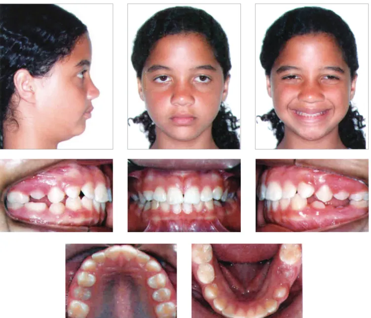
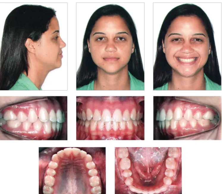
Figure 1 - Initial facial and intraoral photographs.
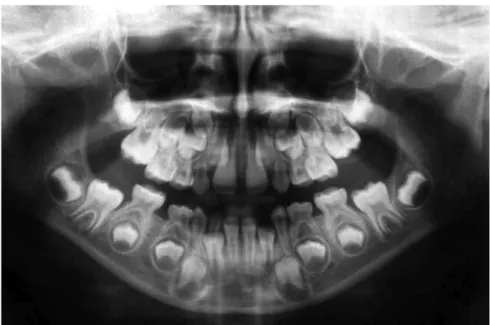
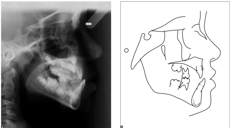
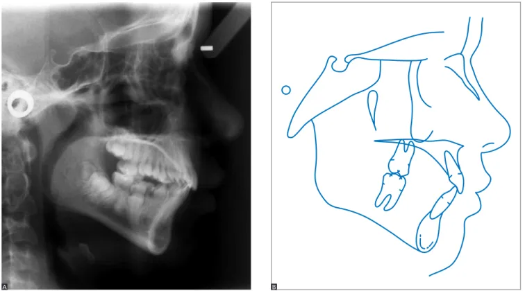
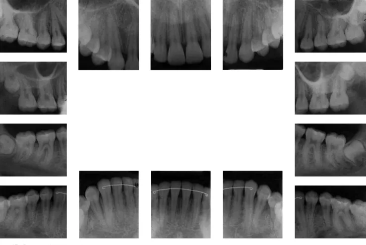
In analyzing the panoramic radiograph (Fig 3), it was observed that all permanent teeth were present, indict-ing that bone and tooth structures were within normal limits. Cephalometrically (Fig 4 and Tab 1), although the value of the ANB angle was within normal range (ANB = 3.5°), it could be veriied through Wits analy-sis (Wits = -3 mm) that the patient had a skeletal pat-tern tending towards Class III. This fact, coupled with her unfavorable family history — given that the patient’s father had a dental and skeletal Class III pattern — rein-forced the need for early intervention. A growth pattern with vertical tendency (SN-GoGn = 36°), severe axial in-clination of upper incisors towards palatal (1-NA = 0°), and well positioned lower incisors (NB-1 = 26°) were also observed. It is noteworthy that mandibular manipulation caused no change from centric relation to maximum and habitual intercuspation (MHI). In calculating the Dis-crepancy Index (DI) a total of 18 points (Fig 5) was found.
DIAGNOSIS
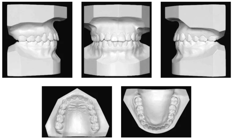
Figure 2 - Initial casts.
Figure 4 - Initial profile cephalometric radiograph (A) and cephalometric tracing (B).
TREATMENT PLAN (PHASE 1)
Planning involved a two-phase treatment. Initially, special attention would be given to the skeletal dis-harmony between maxilla and mandible which, if let uncorrected, would contribute signiicantly to the worsening of this condition during the whole pro-cess of growth and development of the bony bases. Thus, the anterior and posterior crossbites would be corrected, enlarging the maxilla in the transverse di-rection, thereby gaining space in the anterior region for proper eruption of teeth #12 and #22.
The patient and her parents would then be informed of the importance of compliance in wearing the appli-ances and the need to perform treatment in two phases. In this irst phase, the patient was six years and six months old. The posterior crossbite would be corrected using a modiied Haas palate expansion appliance supported on the second primary molars with acrylic covering the in-cisor surface of primary canines and occlusal surfaces of primary molars . Additionally, hooks would be soldered to the buccal surface of orthodontic bands itted on second primary molars, combined with inger springs to procline
teeth #11 and #21. Figure 5 - Calculation of the Discrepancy Index (DI) adapted by BBO.
1- Overjet 7- Lingual posterior crossbite
8- Buccal posterior crossbite
9- Cephalometric measurements
10- Other conditions 2- Overbite
3- Anterior open bite
4- Lateral open bite
5- Crowding
6- Occlusal relationship (Angle classification) Total Total Total Total Total Total Total Total Total Total DI Total
≥ 0 to < 1 mm (edge-to edge) buccal cusp > 0 mm to lingual 1 point per maxillary tooth
2 points per maxillary tooth palatal cusp > 0 mm to buccal
SN-MP ≥ 38º
SN-MP ≤ 26º
1 to MP ≥ 99º
Skeletal asymmetry (non-surgic.) Additional treatment complexities Tooth transposition Diastema Mx. central incisor ≥ 2 mm
Note: Shaded cells must be preserved to evaluators’ notes.
Spacing (≥ 0.5 mm on ≥ 4 teeth) Missing teeth (except 3rd molars)
» Non-congenital » Congenital Midline discrepancy ≥ 3 mm Impaction (except for 3rd molars)
Anomalous morphology Ankylosis of permanent teeth Supernumerary
For each degree > 99º For each degree < 26º For each degree > 38º For each degree > 6º or < -2º ANB ≥ 6º or ≤ -2º > 0 to ≤ 3 mm
1.1 - 3 mm 0 - 1 mm > 3 to ≤ 5 mm
3.1 - 5 mm > 5 to ≤ 7 mm
5.1 - 7 mm > 100% overbite
> 7 mm
1 point
4 points
+ 1 point
2 points/tooth 2 points/tooth 2 points/tooth 1 point/tooth 2 points/tooth 2 points/arch 2 points/tooth
2 points for each occurrence 2 points 2 points 3 points 1- 2- 3- 4- 5-1 point/tooth + 1 point
1 point 1 point 2 points + 1 point
+ 2 point
1 point 0 point 0 point 12 2 0 0 0 0 0 0 0 0 0 0 0 0 0 0 0 0 0 0 0 0 0 2 18 2 0 0 0 2 = = = = = = = = = = = = = = = = = = = = = = = = = = = = = = = = = = = = = = = = = = = = = = = = = = = = = = = ≥ 1 to ≤ 3 mm 0 point
0 point > 3 to ≤ 5 mm 2 points
2 points
+ 1 addl. point / mm / side 4 points per side 2 points per side
2 points > 5 to ≤ 7 mm 3 points
3 points > 7 to ≤ 9 mm 4 points
4 points 5 points
1 point / tooth 1 point / addl. mm / tooth
2 points / mm / tooth > 9 mm 5 points
1 pt / mm / tooth
7 points Negative
Open bite (> 0 mm) 0 mm (edge-to-edge)
≥ 0.5 mm (maxillary posterior tooth)
Beyond Class II or III Full cusp Class II or III End to end Class II or III Class I up to end to end
Figure 6 - Intermediate facial and intraoral photographs.
Maxillary protraction would be performed (a) to correct the anteroposterior discrepancy, and (b) to pro-cline the incisors with inger springs. This procedure would make use of Hickham’s chin cup. Ater this stage, the patient would be monitored every six months until the ideal moment came to implement the second treatment phase.
At this stage, no orthodontic appliance was placed in the patient’s lower dental arch.
TREATMENT PROGRESS
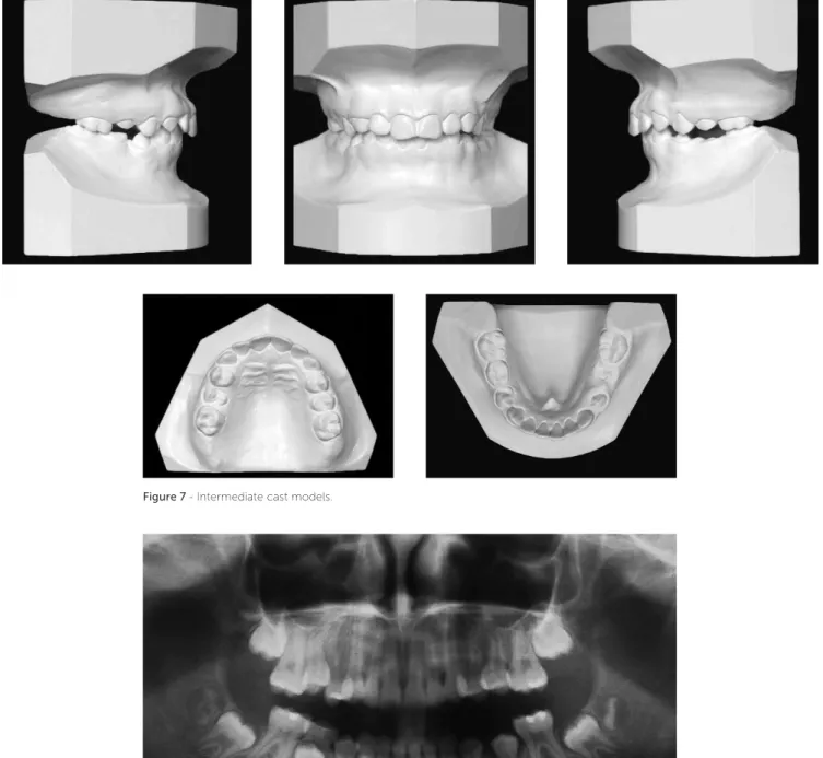
Figure 7 - Intermediate cast models.
Figure 8 - Intermediate panoramic radiograph.
RESULTS
After analysis of the examinations carried out in planning the second treatment phase, it was observed that the objectives proposed for the first phase had been fully accomplished (Figs 6 to 10 and Tab 1). From a dental perspective, anterior and posterior
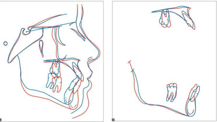
Figure 10 - Total (A) and partial (B) superimposition of initial (black) and intermediate (blue) cephalometric tracings.
Figure 9 - Intermediate profile cephalometric radiograph (A) and cephalometric tracing (B).
A B
Cephalometrically, there was an increase in the ANB angle to 5° (SNA = 82° and SNB = 77°) and a signiicant improvement in Wits value (Wits = 0 mm). This change was especially due to displacement of the maxilla forward and downward, although there was a slight counterclock-wise rotation of the mandible (SN-GoGn increased from 36° to 33° and FMA went from 21° to 17°).
TREATMENT PLAN (PHASE 2)
Ater four years monitoring patient’s development with appointments every six months, the second treat-ment phase started during the period of pubertal growth spurt, when the patient was 11 years and 10 months old. The goals in this phase were to align and level the teeth, achieving a perfect molar relationship and ulti-mately inishing the treatment. Ater inishing, extrac-tion of third molars would be indicated. To this end, a preadjusted ixed orthodontic appliance, Roth prescrip-tion, 0.022 x 0.028-in slot would be bonded to upper and lower dental arches. In the inishing stage, indi-vidualized bends would be placed as required in stain-less steel, 0.018 x 0.025-in archwires, and intermaxillary elastics would be prescribed. Ater inishing, the reten-tion phase would begin with the use of a wraparound re-movable appliance placed in the upper arch and a lingual canine-to-canine retainer in the lower arch.
TREATMENT PROGRESS
The upper arch received orthodontic bands with convertible triple tubes (0.051-in) welded to them, placed on teeth #16 and #26. Thereater, preadjusted metal brackets (Roth prescription, 0.022 x 0.028-in slot) were bonded to upper incisors for alignment and leveling with the aid of Twistlex 0.018-in wire and stainless steel 0.016-in and 0.018-in wire, with ex-pansion loop. Ater slight laring of incisors, brackets were bonded to upper canines and premolars.
As of his moment, straight archwires made with 0.016-in, 0.018-in and 0.020-in were used. Eighteen months into treatment, teeth #17 and #27 erupted and were included in alignment and leveling of teeth. Upon inishing, as planned, rectangular 0.018 x 0.025-in stainless steel archwires were formed, torqued and in-dividually coordinated as needed.
Orthodontic bands with convertible rectangu-lar double tubes were itted to teeth #36 and #46 in the lower arch, and 20 months into treatment, brack-ets were bonded to teeth #37 and #47. Alignment and leveling was performed using 0.018-in twist-lex archwires and round 0.016-in, 0.018-in and 0.020-in stainless steel archwires. The case was inished with 0.018 x 0.025-in rectangular stainless steel arch-wires with form, torques and coordination performed as needed on an individual basis. Class II intermaxil-lary elastics in Class II direction were also employed to decrease lower anchorage and thus achieve a per-fect molar relationship. Ater performing this step, positions of maximum habitual intercuspation (MHI), centric relation and functional guidances (right and let laterality, and protrusion) were veriied.
RESULTS
At the end of treatment, additional exams were re-quired (Figs 11 to 14). They revealed that results were very favorable as all objectives were clearly achieved. Concerning the facial features, as shown in Figure 11, the smile exhibits an adequate exposure of upper inci-sors, and the lower lip is parallel to the upper teeth, dis-playing an excellent smile arc. Balanced facial symmetry and lip competence can also be observed. In analyzing the proile, it appears that despite an overall harmony, the mandible still shows slight protrusion.
In examining the teeth (Figs 11 and 12), it was ob-served that proper alignment and leveling were achieved,
as well as proper occlusion with a perfect relationship be-tween upper and lower molars, and bebe-tween the upper and lower canines, on both sides. Overbite, overjet, pos-terior crossbite and lower midline were corrected. The spaces in the lower dental arch were closed and proper intercuspation obtained. There was an increase in in-tercanine and intermolar widths in both dental arches. In the upper arch, the intercanine width was 25 mm to 36 mm, and intermolar width 41 mm to 51 mm. In the lower arch, the intercanine width was 20 mm to 29 mm, and intermolar width 39 mm to 44 mm. Func-tional harmony was attained in protrusive position, as well as right and let laterality and the centric relation
coincided with the intercuspid position. It is notewor-thy that all these results were achieved with only mild apical remodeling. No sign of signiicant root resorption was observed at the end of treatment (Fig 13).
As expected, several skeletal changes were observed. Mandibular growth occurred in the anterior direction, which caused a decrease in the ANB angle from 3.5° to 1° while the SNA angle remained unchanged and the SNB angle rose from 77.5° to 80°. The vertical dimen-sion was controlled, with a signiicant decrease in the mandibular plane angle (SN-GoGn changed from 36° to 29° and FMA from 21° to 17°). These data can be viewed in Figure 14 and Table 1.
Cephalometric total superimpositions revealed that the maxilla and mandible moved forward and downward. Partial maxillary superimpositions dur-ing the first phase showed vertical development of the alveolar process in the molars, and a substantial change in the inclination of upper incisors. In the second phase, molar mesialization and extrusion were observed along with mild flaring and extru-sion of incisors. Partial mandibular superimposi-tions during the first phase showed mesialization and extrusion of molars, with flaring and extrusion of incisors. In the second phase, molar extrusion and incisor uprighting were noted.
Figure 13 - Final periapical radiographs.
Figure 14 - Final profile cephalometric radiograph (A) and cephalometric tracing (B).
Figure 15 - Total (A) and partial (B) superimpositions of intermediate (blue) and final (red) cephalometric tracings.
Figure 16 - Total (A) and partial (B) superimpositions of initial (black) and final (red) cephalometric tracings.
A A
FINAL CONSIDERATIONS
Diagnosis and treatment planning of Class III mal-occlusion should be performed judiciously. Moreover, patients and their families should be made aware of the entire process as results can be unpredictable. Even if goals and a success rate have been established, the prognosis is unclear, since it depends largely on case development and patient compliance. Even so, it is likely that once patient’s growth has ended the need
for retreatment by dental compensation1 or surgical
in-tervention may still exist.2
No consensus has yet been reached in the litera-ture regarding the ideal time to start treatment since some authors posit that growth and development of the craniofacial complex is genetically determined, and therefore unalterable. For these authors, correc-tion of most Class III cases necessarily involves sur-gery. Furthermore, orthodontic treatment should be started immediately after the growth spurt period has ceased. Other authors however — although agreeing with the role of heredity in the etiology of Class III — believe they can change growth pattern and direc-tion by means of a non-surgical approach capable
of minimizing the malocclusion or even treating it successfully.² When the patient is in the phase that precedes the pubertal growth spurt, early treatment is indicated given that Class III malocclusions tend to become increasingly severe over time; the reason being that mandibular growth remains active for a
longer period than maxillary growth.²,³ Treatment of
the case reported here was conducted in two phases. It was initiated when the patient was six years and six months old. Younger individuals tend to achieve more favorable results between four and ten years of age, although patients whose ages range from 10 to 14
years old also present positive results.3,4
In the irst phase, rapid maxillary expansion was
combined with maxillary protraction using Hickham’s5
chin cup. This device is easy to fabricate and individual-ize. It has met with wide acceptance by patients, besides
being more stable, especially during sleep.6 Although
there are several types of appliances for orthopedic pro-traction of the maxilla, the literature contains no studies comparing their eiciency. The skill of the professional handling the device and patient comfort are important
variables when choosing one of these appliances.2,7,8
Table 1 - Initial (A), intermediate (A1) and final (B) cephalometric values.
Measurements Normal A A1 B A/B dif.
Skeletal pattern
SNA (Steiner) 82° 81° 82° 81° 0
SNB (Steiner) 80° 77.5° 77° 80° 2.5
ANB (Steiner) 2° 3.5° 5° 1° 2.5
Angle of convexity (Downs) 0° 8° 13° 1° 7
Axis Y (Downs) 59° 52° 55° 54.5° 2.5
Facial angle (Downs) 87° 93.5° 92° 95° 1.5
SN-MP (Steiner) 32° 36° 33° 29° 7
FMA (Tweed) 25° 21° 17° 17° 4
Dental pattern
1 to MP (Tweed) 90° 93° 99° 94.5° 1.5
1.NA (degrees) (Steiner) 22° 0° 20° 29° 29
1-NA (mm) (Steiner) 4 mm 0 mm 4 mm 8 mm 8
1.NB (degrees) (Steiner) 25° 26° 30° 25° 1
1-NB (mm) (Steiner) 4 mm 4 mm 7 mm 5 mm 1
1
1 - Interincisal angle (Downs) 130° 151° 125° 126° 25
1-APo (Ricketts) 1 mm 4 mm 4 mm 4 mm 0
Proile Upper lip — S line (Steiner) 0 mm 2 mm 2 mm 0 mm 2
1. Bittencourt MAV. Má oclusão de Classe III de Angle com discrepância Antero-posterior acentuada. Rev Dental Press Ortod Ortop Facial. 2009;14(1):132-42
2. Araújo EA, Araújo CV. Abordagem clínica não-cirúrgica no tratamento da má oclusão de Classe III. Rev Dental Press Ortod Ortop Facial. 2008;13(6):128-57.
3. Baccetti T, McGill JS, Franchi L, McNamara JA, Tollaro I. Skeletal efects of early treatment of Class III malocclusion with maxillary expansion and face-mask therapy. Am J Orthod Dentofacial Orthop. 1998;113(3):333-43. 4. Brunetto AR. Má oclusão de Classe I de Angle, com tendência à Classe
III esquelética, tratada com controle de crescimento. Rev Dental Press Ortod Ortop Facial. 2009;14(5):129-45.
5. Hickham JH. Maxillary protraction therapy: diagnosis and treatment. J Clin Orthod. 1991;25(2):102-13.
REFERENCES
6. McDonald KE, Kapust AJ, Turley PK. Cephalometric changes after the correction of Class III malocclusion with maxillary expansion/facemask therapy. Am J Orthod Dentofacial Orthop. 1999;116(1):13-24. 7. Turley PK. Managing the developing Class III malocclusion with palatal
expansion and facemask therapy. Am J Orthod Dentofacial Orthop. 2002;122(4):349-52.
8. Turley PK. Orthopedic correction of Class III malocclusion with palatal expansion and custom protraction headgear. J Clin Orthod. 1998;22(5):314-25.
9. Andrews LF. The six keys to normal occlusion. Am J Orthod. 1979;62(3):296-309.
The second treatment phase began in late mixed dentition. At this stage, there was large overcorrection, and the patient was in Angle Class II malocclusion. Fur-thermore, the mandible was slightly retruded, which is indicated in this case since she would otherwise
con-tinue to present with an unfavorable growth pattern.8
At this stage, a conventional ixed orthodontic appli-ance was used. Treatment outcome was very favorable,
both functionally and esthetically. Upper arch expan-sion, which had been performed in the irst phase, was stable, indicating that the morphological limits were re-spected. Dental alignment and leveling were achieved in the second stage. Canine and molar occlusion was well established and anterior and posterior crossbites
cor-rected,9 with pleasing facial esthetics, good symmetry