C
ASER
EPORT| R
ELATO DEC
ASO123
Renal hydatid cyst: medical treatment
Cisto hidático do rim: tratamento médico
Authors
Ana Teresa Trindade Soares 1
Catarina Couto 1
Maria João Cabral 1
Luísa Carmona 1
Isabel Vieira 1
1 Hospital Garcia de Orta.
Submitted on: 06/08/2015. Approved on: 07/08/2015.
Correspondence to: Ana Teresa Soares. Hospital Garcia de Orta. Rua Actor José Pinheiro Amaro, nº 16 - 2esq, Carnaxide, Portugal. CEP: 2790-005.
E-mail: anateresasoares@gmail. com
DOI: 10.5935/0101-2800.20160017
Introdução: A infecção por Echinococcus
ou hidatidose constitui-se em uma zoonose causada pelo parasita Echinococcus granu-losus. Apresenta distribuição mundial, sendo Portugal considerado endêmico. A doença hidática do rim é rara, ocorrendo em apenas 2% dos casos. Caso Clínico: Adolescente de
14 anos, do sexo masculino, com doença hidática do rim. O diagnóstico foi efetuado por exames de imagem e por teste sorológico específico. Foi instituída terapêutica com al-bendazol durante quatro ciclos de quatro se-manas cada, tendo-se verificado diminuição significativa do tamanho do cisto e negativi-dade dos anticorpos específicos. Discussão:
Embora a terapêutica recomendada seja a excisão cirúrgica, estudos demonstram que a terapêutica médica pode ser uma alterna-tiva a considerar, permitindo a preservação do órgão.
R
ESUMOIntroduction: Hydatid disease is a zoonosis caused by the parasite Echinococcus granulosus. It has a worldwide distribution, being endemic in Portugal. Hydatid disease of the urinary tract is rare, occurring in less than 2% of all cases. Case report: 14-year-old male with hydatid disease of the kidney. The diagnosis was established by imaging studies and specific serologic testing. Treatment consisted of four 4-week cycles of albendazol and resulted in a progressive reduction of the cyst as well as in a negative specific serology. Discussion: Even though the recommended treatment is surgical excision, studies have demonstrated that medical treatment may be an alternative allowing for organ preservation.
A
BSTRACTI
NTRODUCTIONEchinococcosis or hydatid disease is a zoonotic condition more frequently caused by parasite Echinococcus granulosus.1 Cases of the disease have been reported all over the world, but higher prevalence rates have been described in Australia, New Zealand, Northern Africa, India, the Mediterranean Basin, and in some regions in South America.2 The World Health Organization has categorized Portugal as an endemic country for this disease, with an estimated incidence of 2.2 cases/100,000 population.1,3 Studies at a national level have found that most cases are confined to the Évora district in the Alentejo region. The number of cases of the disease has decreased over the years.3
Palavras-chave: criança; equinococose; terapêutica.
Keywords: child; echinococcosis; kid-ney.
The disease may affect any organ, but liver and lung involvement is found in 90% of the patients. The kidneys are affected in only 2% of the cases.3-5
Kidney involvement is usually observed in the form of a single cyst located at the level of the renal cortex. Symptoms vary depending on the size, extension, and location of the cyst. Patients may be asymptomatic for long periods of time or present with lower back pain, hematuria, or an abdominal lump.1,5,6
Hydatiduria, a pathognomonic pre-sentation of the disease, is observed in only 10-20% of the patients when the cyst ruptures into the collecting system.5,7
J Bras Nefrol 2016;38(1):123-126
Renal hydatid cyst
124
computed tomography (CT), or nuclear magnetic resonance (NMR). Additional workup may include ELISA or indirect hemagglutination assays to titer for specific antibodies, follow patients, and determine cases of recurring disease.5,6,8 However, humoral response sensitivity varies from 35% to 90% depending on a series of factors including the genetics of the host and the parasite, and the location and viability of the hydatid cyst.1,9
The gold standard therapy for the disease is surgical excision of the cyst combined with antiparasitics.1,8,10 Less invasive methods such as echo or CT-guided percutaneous drainage of the cyst with suction and instillation of saline solution or alcohol may be prescribed in select cases.6
C
LINICAL CASEThe patient was a 14-year-old male individual residing in the Setúbal district. He was admitted into the Pediatric Emergency Unit with lower back pain and hematuria after having sustained a back injury. He had no significant previous medical events, but alluded to having regular contact with dogs, sheep, and goats at his grandparents’ property in Alentejo, where he often spent weekends. The patient had no alterations in his objective examination upon admission.
Renal ultrasound (Figure 1) and CT (Figure 2) scans revealed an encapsulated expansive lesion in the middle third of the left kidney measuring 9x9x6.8 cm in its longitudinal, anteroposterior, and cross-sectional axes, respectively. The lesion had millimetric calcium particles close to its walls and was heterogeneous inside, with a dense central component consistent with recent bleeding. Workup findings included a hemoglobin level of 12.8 g/dL, a leukocyte count of 5.8x10⁹/L, neutrophils 52.5%, eosinophils 3.9%, C-reactive protein 0.7 mg/dL, urea 25 mg/ dL, creatinine 0.6 mg/dL, a sedimentation rate of 40 mm/h (reference value < 16 mm/h), and the presence of antibodies specific for Echinococcus granulosus. The patient did not present with hydatiduria.
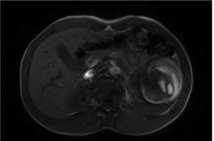
NMR scans (Figure 3) were ordered to better characterize the lesion. The images revealed a pre-dominantly cystic exophytic node suggestive of a hydatid cyst located in the renal sinus without infil-trative or obstructive components. The patient was diagnosed with renal hydatid disease complicated by bleeding secondary to trauma. In order to preserve
Figure 1. Abdominal ultrasound scan showing cyst in the left kidney.
Figure 2. Abdominal CT scan showing an expansive encapsulated lesion in the middle third of the kidney.
J Bras Nefrol 2016;38(1):123-126
Renal hydatid cyst
125
Figure 3. Abdominal NMR scan showing a cystic exophytic node without iniltrative or obstructive components located in the renal sinus consistent with hydatid cyst.
Figure 4. Evidence of decrease in cyst size, now measuring 3.6x3.4x2.3cm in the greater longitudinal, cross-sectional, and anteroposterior.
at the end of the protocol described above showed the cyst had decreased in size to 3.6x3.4x2.3 cm in its longitudinal, cross-sectional, and anteroposterior axes respectively (Figure 4), and blood tests revealed the patient had no disease-specific antibodies. Four years into follow-up the patient has been symptom-free, negative for disease-specific antibodies, and the cyst has not changed size.
D
ISCUSSIONRenal hydatid disease is a rare condition. It may be associated with potentially severe complications such as vascular compression, cyst infection, shock, sepsis, and death.1,5 Dogs are the main hosts and herbivores are intermediate hosts. Human beings may acquire the disease by direct contact with infected animals or be eating foods contaminated with parasite eggs, found
usually in fruit, raw or undercooked vegetables, and untreated water. Larvae first hatch from the eggs to populate the bowels and then invade other organs through the bloodstream.1,5,8 The patient described in this case mentioned he traveled regularly to Alentejo, an endemic region in Portugal, where he was in contact with animals categorized as potential hosts of the parasite.
Surgical removal of the cyst is considered the first line treatment whenever there is involvement of the renal parenchyma. Depending on the size of the cyst a partial or a radical nephrectomy is performed. In specific cases the cyst alone may be excised, although relapse rates range between 10% and 30%.8,9 Kidney-sparing less invasive procedures have been recently described in the literature. One of them is echo or CT-guided percutaneous drainage combined with adjuvant antiparasitics.6 The main disadvantages of this procedure are the risk of recurrence, infection dissemination, and anaphylaxis.6
Drug therapy alone with antiparasitics such as albendazole or mebendazole has been described to treat hydatid disease involving the liver or lungs as an alternative to surgery, with good outcomes in terms of reducing cyst size and volume.4 However, antiparasitics alone are less efficacious and may elicit adverse events such as liver toxicity, leukopenia, allergic reactions, and alopecia.4,6 Nonetheless, according to the latest recommendations published by the WHO-IWGE concerning the treatment of liver hydatid disease, the choice of treatment should be based on the characteristics of the cyst, the experience of the medical staff and the availability of drug therapy/surgery, and the possibility of following the patients in the long term. The recommendations do not define an ideal course of treatment.6
J Bras Nefrol 2016;38(1):123-126
Renal hydatid cyst
126
drug at a dosage of 10-15 mg/kg/day (maximum of 400 mg), orally, before and after surgery.10
A conservative approach was chosen for the patient described in this report to preserve his clinical and hemodynamic stability and due to the fact that the location and size of the cyst meant he would have to be offered a radical nephrectomy with the added risk of the cyst rupturing during the procedure. Therefore, he was started on a protocol similar to the one prescribed to patients with liver hydatid disease, with 400 mg of albendazole divided in two daily doses, in order to spare the affected kidney. The size of the cyst decreased gradually and the patient had no associated adverse events. The cyst’s size and the patient’s serologic tests remained unaltered, while disease-specific antibody levels were at an undetectable level after the end of the drug therapy.
Although only a few studies have described drug therapy as the treatment of choice for individuals with renal hydatid disease, this might be a particularly good alternative for younger patients, as it allows the affected kidney to be spared.
This choice of treatment requires the collaboration of the patient’s caregivers, proximity to specialized emergency care, long-term follow-up, and the means to detect cases of recurring disease early on.
R
EFERENCES1. Reis T, Vilares A, Ferreira I, Martins S, Furtado C, Gargaté MJ. Hidatidose quística humana: análise retrospetiva de casos diag-nosticados e em monitorização entre 2008 e 2013. Porto: Insti-tuto Nacional de Saúde Doutor Ricardo Jorge; 2014. p.30-3 2. Vaz PS, Pereira E, Usurelu S, Monteiro A, Caldeira A, Melo
G, et al. Hepatic hydatid cyst: a non-surgical approach. Rev Soc Bras Med Trop 2012;45:774-6. DOI: http://dx.doi. org/10.1590/S0037-86822012000600025
3. Morais JAD. Hidatidose humana. Estudo clínico-epidemiológi-co no distrito de Évora durante um quarto de século. Acta Med Port 2007;20:1-10.
4. Rinaldi F, De Silvestri A, Tamarozzi F, Cattaneo F, Lissandrin R, Brunetti E. Medical treatment versus "Watch and Wait" in the clinical management of CE3b echinococcal cysts of the liver. BMC Infect Dis 2014;14:492. DOI: http://dx.doi. org/10.1186/1471-2334-14-492
5. Moscatelli G, Moroni S, Freilij H, Salgueiro F, García Bournis-sen F, Altcheh J. A five-year-old child with renal hydatidosis. Am J Trop Med Hyg 2013;89:554-6. PMID: 23897992 DOI: http://dx.doi.org/10.4269/ajtmh.13-0243
6. Brunetti E, Kern P, Vuitton DA; Writing Panel for the WHO--IWGE. Expert consensus for the diagnosis and treatment of cystic and alveolar echinococcosis in humans. Acta Trop 2010;114:1-16. DOI: http://dx.doi.org/10.1016/j.actatropi-ca.2009.11.001
7. Rexiati M, Mutalifu A, Azhati B, Wang W, Yang H, Sheyhedin I, et al. Diagnosis and surgical treatment of renal hydatid disease: a retrospective analysis of 30 cases. PLoS One 2014;9:e96602. DOI: http://dx.doi.org/10.1371/journal.pone.0096602 8. Marcelino J, Dias J, Lopes T, Martins F. Quisto hidático do
rim: dificuldades no diagnóstico diferencial. Acta Urol Port 2000;17:73-5.
9. Moscatelli G, Abraham Z, Morini S, Iriart EM, Rodrí-guez M, Mirón L, et al. Descripción del caso presentado en el número anterior: Hidatidosis pulmonar. Arch Argent Pediatr 2012;110:265-7. DOI: http://dx.doi.org/10.5546/ aap.2012.265