Physiology of Accommodation and Presbyopia
Presbyopia is one of the earliest universal signs of aging and the basic pathophysiology involved in its development has been a matter of controversy for centuries. This article discusses many aspects of presbyopia by reviewing the literature on the multitude of age-related changes that occur in the eye.
Fisiologia da acomodação e presbiopia
From the Department of Ophthalmology, São Geral-do Eye Hospital, Federal University of Minas Ge-rais, Belo Horizonte, Minas GeGe-rais, Brazil. The authors thank the Storm Eye Institute (Director: David J. Apple), Medical University of South Caro-lina, Charleston, USA, for the schematic drawing and photomicrographs.
Address for correspondence: Dr. Fernando Trinda-de - Rua Manaus, 595 - Belo Horizonte (MG) CEP 30.150-350. Email: [email protected]
Leonardo Werner Fernando Trindade Frederico Pereira Liliana Werner
SUMMARY
Keywords: Accommodation; Presbyopia.
The Mechanism of Accommodation
Studying the basic mechanism of accommodation and presbyopia is fundamental to understand the pathophysiology of the eye. The normal, young human eye can easily focus on near and distant objects, i.e., it can change focus or accommodate. The word “accommodation” has a relatively recent origin and was definitely introduced by Burow in 1841 1, 2. Certain
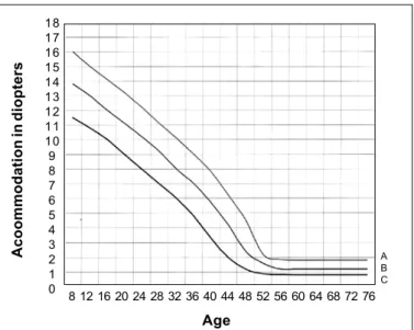
standard textbooks before that time used the term “adaptation”, now accepted as connoting the changes in the sensitivity of the retina to varying intensities of light. Explanations of how accommodation occurs have been speculated upon for centuries. By reviewing the literature on accommodation and presbyopia one finds much that is assumed to be known is still controversial. If the innervation responsible for accommo-dation remains unclear, also the mechanism of the development of presbyopia is no longer theoretical. Scientists have studied the change in the eye’s ability to focus (amplitude of accommodation) in relationship to age. They have found that the amplitude of accommodation declines in a linear fashion with age and that this decline occurs universally and predictably (Fig. 1). If a patient is properly corrected for distance, his/her age can be determined within one and a half years by measuring his/her amplitude of accommodation. Therefore, an adequate theory of the mechanism of accommodation and presbyopia in man must take into account the changes observable in the human eye during the effort of accommodation, and provide a reasonable explanation for the decline in this function with age. In viewing the great number of theories proposed since many years ago, we have found that the mechanism and interrelation of these have not yet been completely clarified and some analysis has been attempted in other to provide a more accurate understanding of accommo-dation and presbyopia.
was first demonstrated by Scheiner (1619). In his experiment, two pinholes are made in a card at a distance apart less than the diameter of the pupil, and the eye, looking through them, is focused on a needle held at right angles to a line joining the two holes: the needle appears single. If, however, the eye is focused on some other object nearer or further away, the needle appears double. If three holes are made, three needles are seen, and so on (Fig. 2). This experiment proves that in the eye there is a mechanism controlling the adjustment of focus. However, the true explanation of this classical experiment was offered by William Porterfield (1759) who suggested that accommodation was effected by a change in the lens. Other possible hypothesis have been put forward to explain the rationale of accommodation. Albrecht von Haller (1763), considered that the contraction of the pupil diminished the blur-circles sufficiently to account for the phenomenon, a mechanism resembling a camera obscura which is present in some animals. Some authors suggested that an elongation of the eyeball caused by contraction of the extra-ocular muscles was responsible for the phenomenon. The original theory of Kepler (1611) that changes in focus were attained by forward
and backward movements of the lens (as occurs in some fishes) received support from other investigators, until it was demonstrated that an impossible excursion would be required in order to obtain the requisite change in focus: it would, indeed, require the lens to move forwards by 10 mm. The remaining possibility, that accommodation was accomplished by a change in the shape of the lens, was suggested at a very early date by Descartes (1677). Later, Helmholtz 3 (1853-1856)
was able to demonstrate that the action of accommodation provided by the ciliary muscle was accompanied by an increase in curvature of both surfaces of the lens and an increase in its thickness. The histology of the anterior segment and the ciliary muscle of the human eye is reviewed in figure 3 (A and B).
8 12 16 20 24 28 32 36 40 44 48 52 56 60 64 68 72 76 18
17 16 15 14 13 12 11 10 9 8 7 6 5 4 3 2 1 0
A B C
A
c
o
o
m
m
o
d
a
ti
o
n
in
d
io
p
te
rs
Age
Fig. 1 - Duane’s 33 standard curve of accommodation in diopters in
relation to age (A: lowest values; B: average values; C: highest values).
Fig. 2 - Schematic drawing showing Scheiner’s experiment (1619). If the card is perforated at E and E, the object, O, is brought to a focus on a screen, R, at I, where one image will appear. If the screen is held
at R’ or R’’, however, two images appear (E’F’ and E’’F’’).
Fig. 3 - Photomicrographs from a human globe obtained postmortem showing the anterior segment. These histological sections are stained with Masson’s trichrome, which stains collagen fibers in blue and smooth muscle fibers in red; A. Cornea (C), sclera (S), iris (I) and ciliary body (CB) can be identified. The ciliary muscle extends from the scleral spur to the choroid, although some of its fibers are confined to the pars plicata. The ciliary process and the entire inner surface of the ciliary body are lined with two layers of epithelium, the innermost of which lacks melanin pigment (Masson’s trichrome; original magnification X 200); B. Same section in higher magnification showing the ciliary body. The ciliary muscle is traditionally divided into three parts. The outermost, lying next to the sclera, is the meridional portion (M). The innermost fibers nearest the ciliary processes constitute the circular portion (C), and the radial portion (R) lies between the meridional and circular portions. The divisions are not sharp and the boundaries are
just approximate.
A
B
S
C B
C
M
R
C
In 1965, the American Committee on Optics and Visual Physiology adopted the slogan: “Put Helmholtz back into Ophthalmology”. Hermann von Helmholtz (1821-1894) trained as a physician, became professor of physiology and physics, returning all his life to study the Physiologic Optics 1. He
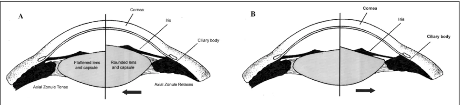
observed in 1855 that the center of the human lens thickened during accommodation. Based on this observation, he theorized that when the eye accommodates, the ciliary muscle contracts, reducing the tension on the zonules that span the circumlental space extending between the ciliary body and the lens equator. This releases the outward-directed equatorial tension on the lens capsule and allows this elastic capsule to contract, causing an increase in the anterior-posterior diame-ter of the lens and resulting in an increase in its optical power. Thus, the act of accommodation should result from a con-traction of the ciliary muscle which reduces the ciliary body diameter and releases the resting zonular tension. This allows young lenses to undergo elastic recovery which causes an increase in the lens curvatures and an increase in lens power to enable near objects to be focused on the retina. When accommodation ceases, the ciliary muscle relaxes and returns to its unaccommodated configuration, the zonular tension is once again increased and the lens is pulled back into a relatively flattened state to increase the focal length 3. The
movement of the equatorial edge of the lens is thus away from the sclera during accommodation (Fig. 4A) and toward the sclera during disaccommodation. Although the influence of the capsule in determining the shape of the lens is undoubted, Helmholtz’s theory cannot stand in its original form, since it does not explain the shape assumed by the anterior surface of the lens. At a later date, Fincham 4 suggested that the peculiar
form taken in this molding might be due to the structure of the capsule. It is much thicker in front than behind and the anterior and posterior portions are thicker laterally, just within the attachment of the zonular fibers, than at the poles. The variations of its thickness in different parts suggest that, on the application of tension, a flattening of the lens would occur preferentially in the periphery, where the capsule is thickest and strongest, and a bulging in the axial region where it is
weakest. At the posterior pole the capsule is very thin, and here the maximal curvature of the lens occurs even in the unaccommodated state 5. It is this difference in the thickness
of the central and lateral parts of the anterior capsule which Fincham believes is responsible for the hyperbolic form of the anterior surface of the lens during accommodation.
During many years, there was a consensus of opinion on the mechanism of accommodation derived from the theory of Helmholtz. However, Schachar et al. 6 have recently proposed
an alternative accommodative mechanism for the primate eye that is similar to a theory originally proposed by Tscherning 7.
Both theories 7-12 state that the equatorial zonules insert into
the anterior ciliary muscle at the root of the iris and the anterior and posterior zonules insert into the posterior ciliary body. Schachar and Anderson 12 allege that during ciliary muscle
contraction, through the action of the radial and longitudinal fibers, the anterior portion of the ciliary muscle curls toward the sclera at the iris root. This movement increases tension on the equatorial zonular fibers while releases tension on the anterior and posterior zonular bundles. Schachar believes that this provides a net outward-directed force at the lens equator through the equatorial zonular fibers. This force, putatively, would pull the lens equator toward the sclera during accom-modation and, together with the concurrent relaxation of the anterior and posterior zonular bundles, would cause a flattening of the peripheral lens surfaces while increasing the central anterior and posterior lens surface curvatures. The movement of the equatorial edge of the lens is thus toward the sclera during accommodation (Fig. 4B) and away from the sclera during disaccommodation. From a theoretical stand-point, pulling on the lens equator could cause an increase in the central lens curvatures, depending on the viscoelastic properties of the lens. Schachar’s theory, however, differs from that of Tscherning because it does not depend on the vitreous to explain the changes in lens shape that occur during accommodation 13. The background of Schachar’s
theory is that the lens equatorial diameter increases with accommodation. However, a group of recent studies showed, using various imaging techniques, that the crystalline lens
Fig. 4 - Schematic drawings showing Helmholtz’s (A) and Schachar’s (B) theories of accommodation. The arrows indicate the movement of the equatorial lens edge away from the sclera (A) and towards the sclera (B) during accommodation; A. The left side of the drawing shows the unaccommodated state. On the right side, the ciliary muscle has contracted during accommodation; the lens is thicker and more steeply curved; B. The left side is as in A. On the right side, note the flattening of the peripheral lens surfaces while the central anterior lens surface
diameter decreases with accommodation, as the classical lite-rature maintains and contrary to Schachar’s contention (Fig. 5 A-B). Wilson 14 also has shown lens equatorial movements
away from the sclera during accommodation using transillu-minated infrared light in a young human subject with ocular albinism. Glasser and Kaufman 13 studied the movements of
the lens equator and the ciliary body using ultrasound biomicroscopy and goniovideography during accommo-dation and disaccommoaccommo-dation. They found that despite the systematic eye movement occurring with electrical stimulation and the nonsystematic eye movements occurring with phar-macologic stimulation, in all instances the ciliary body and the lens equator moved away from the sclera during accommo-dation. Another study conducted by Glasser and Campbell 15
have shown that mechanically stretching the zonule of the human lens increases lens focal length in accordance with classic teachings from Helmholtz. In addition, there has been no independent confirmation of the anatomic arrangements of the ciliary region, the accommodative mechanism, or the causes of presbyopia described by Schachar. The localized posterior or outward movement of the anterior portion of the ciliary muscle toward the sclera, as suggested by this author from histologic analysis, was not visible by imaging tech-niques 13. Also, his description concerning the insertion of the
zonule conflicts with evidences provided by analysis of fresh human tissues and from scanning electron microscopic studies 15,16. They show no insertion of equatorial bundles or
any other zonular fibers at the iris root and anterior ciliary muscle (Fig. 6A and B). The fibers destined for the anterior and equatorial lens capsule have been shown to be strongly adherent to the valleys of the ciliary processes. They part company with the posterior zonules by continuing in an
Fig. 5 - Ultrasound biomicroscope (UBM) images of the human lens equator (arrows) in relation to the scleral spur (asterisks) during unaccommodated state induced by tropicamide (1.0%) and accommodate state induced by pilocarpine (2%). The probe is positioned over the temporal ciliary region. Cornea (C), sclera (S), iris (I) and ciliary body (CB) can be identified in both pictures; A. In the unaccommodated state the distance between the lens equator and the scleral spur is 1.401 mm; B. In the accommodated state the distance
between the lens equator and the scleral spur is 1.695 mm.
A
B
A
B
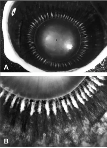
Fig. 6 - Photographs of a human globe obtained postmortem taken from an operating microscope after sectioning the globe at the equator showing the inner surface of the anterior segment from a posterior view; A. The crystalline lens (L) is in the center, surrounded by a series of radial lines, which are the ciliary processes, the pars plicata (PP). This latter is surrounded by the pars plana, which terminates in a scalloped edge, the ora serrata, where the retina (R) begins; B. Same view with higher magnification. Note the zonules (arrows) attaching the
almost straight course to their insertion. Both the anterior and posterior zonules exit from the pars plicata in ribbonlike swaths, lining up parallel to the ciliary processes. The few zonules passing to the equator of the lens arise from the midsides of the processes or from the valleys and usually derive from anterior or posterior zonular bundles 5.
Presbyopia
Presbyopia is the most common refractive disorder of later life, related to decrease of accommodative amplitude. Some 4 million new patients emerge as presbyopes in the U.S. population each year 17. In emmetropes and hyperopes, it is
usually manifested at 40 years of age by the need for reading with glasses or contact lenses. Although normal myopes are benefited at this age because of their shortsighted, their accommodative amplitude also diminishes with age in a more or less regular manner. The symptoms begins with an annoying inertia of focus when gazing from far to near objects and advances to an inability to carry out prolonged near work without stinging, smarting, or tearing, which eventually leads to disinterest in reading. Fine print and small targets can no longer be resolved at the costumary reading distance, and when the object is habitually brought nearer, the blur strangely increases. Soon even ordinary print begins to blur, smudge, smear, run together, and disappear. These symptoms are intensified under inadequate light, amplified by poor contrast, and exaggerated at the end of the day 18.
Despite its ubiquity and high annual costs (National Advisory Eye Council, 1983), the underlying cause(s) of presbyopia remain unclear. Many studies based on Helmhol-tz’s theory have been tried out to explain the loss of accommo-dation of the aging eye. By considering many possibilities, any proposed theory must take into account the known decline in the ability of the eye to alter its focus as age advances. Some suggest a loss of zonules or capsule elas-ticity with aging, thus, when the zonules are relaxed the lens is not able to change its shape 19. There are some conflicting
reports on whether the ciliary muscle atrophies with age 20.
There is also continued deposition of the lens fibers within the lens as it ages, causing the lens to become more compact and stiff. A major factor in the loss of accommodation may be the increased stiffness of the aging lens with inability to respond to accommodative stimuli 21. In general, the multitude
of changes that occur in the eye resulting in presbyopia can be broadly grouped into three categories: lens and capsule-based theories, which consider changes in the elasticity and compliance of the lens and capsule; extralenticular theories which consider changes in the ciliary muscle and choroid; and geometric theories which consider changes in the geometry of the zonular attachments to the lens 15.
Fincham 22 added additional experiment support to the
accommodative theory of Helmholtz and also offered evidence that presbyopia was caused by the inability of the lens capsule to mold the hardened lens substance into the
accommodated form. Fischer 23 and Pau and Kranz 24 also
supported Fincham’s theory of presbyopia by attributing accommodative loss to changes in the elastic properties of the lens. In addition, Fischer 25 found that Young’s modulus of
elasticity of the lens capsule decreases by half between youth and 60 years of age. Based on such evidences, we can assume that reduced capsular elasticity alone cannot explain presbyo-pia, only diminished capacity to change curvature. Further-more, by evaluating capsular molding pressure vs lenticular strain, Fischer 23 concluded that decreased amplitude can be
accounted for reduced elasticity of the capsule, changes in the elasticity of the lens substance, and flattening of the lens. This finding supports Fincham’s theory of presbyopia because a less elastic lens capsule would exert less force on the hardening substance of the aging lens. When the capsule is stripped off (in young monkeys), the lens becomes thinner and flatter 2. These observations suggest that the lens
subs-tance houses restoring forces, which tend to maintain it in the unaccommodated form. These forces are in turn antagonized by capsular elasticity. Weale 26 proposed that in the young
eye, elastic capsular forces are dominant, while restoring forces predominate in presbyopia.
The lens and capsule-based theories accept an indirect evidence that ciliary muscle is capable of providing the same magnitude of force in presbyopic as in pre-presbyopic eyes. Impedance cyclography has been used to measure ciliary muscle contraction and to show that it remains normal up to the age of 60 years, supporting lens or capsule-based theories of presbyopia 27. However, these findings have been criticized
owing to the uncertainties of exactly what impedance cyclography measures. This stems, in part, from the observa-tion that a given accommodative demand does not consis-tently produce the same impedance 28. Also, in the rhesus
monkey, which has an accommodative apparatus similar to that of the human and develops presbyopia on a comparable time scale relative to its lifespan 29, the ability of the ciliary
muscle to alter its configuration in response to topical cholinomimetic drugs or electrical stimulation of the Edinger-Westphal nucleus clearly declines with age 30-32.These
poten-tially confounding results may suggest a possible loss of ciliary muscle function concurrent with the development of presbyopia. Supporting this concept in agreement with extralenticular theories, Fuchs 2 first reported that cycloplegia
is more effective in young eyes than old, and Duane 33 found
the onset of cycloplegia to be more rapid in early presbyopes than nonpresbyopes. Moreover, if ciliary muscle wasting was significant, its vigorous exercise (e.g., hyperopia, prolonged near work) should postpone presbyopia and there is no evidence that this occurs 15,34.
Brown 35 has suggested that presbyopia is associated with
liquefaction of the vitreous, since the two processes occur at about the same point in the human lifespan. However, this theory does not explain why the age-related decline in accommodative amplitude begins so early in life 36. Another
because it arises from the observation that the location of the zonulo-lenticular attachments relative to the lens equator and the ciliary muscle changes with age, as the lens increases in size 37. Such an alteration in the anterior segment geometry
would result in greater retention of zonular force applied to the lens during ciliary muscle contraction, without necessarily re-quiring other changes in the anterior segment properties 38-40.
The observations also provided by Brown 41 of an increase of
the lens in size with age changing its curvatures and by Farnsworth and Shyne 37 of an anterior shift of the zonular
attachments onto the lens led to the suggestion that distinct factors may be interacted to contribute to the failure of the older lens to accommodate, as it becomes retained in an unaccommodated state.
More recently, presbyopia has been described as a geome-tric disorder only attributed to changes in the size and volume of the lens. Schachar 6 proposed that zonular tension is
increa-sed during accommodation in young subjects in contrast with the classic Helmholtz’s theory, as we described above. In support of his hypothesis, Schachar and colleagues 8 believes
that presbyopia results from a decrease in zonular tension caused by the normal growth of the crystalline lens with age. The lens is of ectodermal origin and continues to grow throughout life and the equatorial diameter increases at approximately 0.02 mm/year. However, except for the progres-sive myope, the dimensions of the scleral shell do not change significantly after 13 years of age. The distance between the ciliary muscle and the equator of the lens decreases throu-ghout life. Therefore, the effective force that the ciliary muscle can apply to the lens equator is reduced in a linear fashion with age. The amplitude of accommodation decreases linearly with age resulting in presbyopia and is a consequence of normal lens growth 8-12.According to Schachar 10, surgical
expansion of the sclera surrounding the ciliary body can restore accommodation as a remedy for presbyopia. Scleral
expansion surgery involves the implantation of a plastic ring or arches of plastic in the sclera surrounding the ciliary body to increase the space between the ciliary body and the lens equator. However, the efficacy of scleral expansion surgery in the treatment of presbyopia was not completely determined and there are some evidences accumulating that the impro-vement obtained in some patients may represent a consequen-ce of lenticular aberrations resulting in a multifocal optical system, rather than true accommodation 13, 42.
This concept of age-related loss of accommodation related to decreased zonular tension resulting from continued growth of the lens throughout life, rather than lenticular sclerosis was also described by Weale 26 and Bito and Miranda 43 .
Tscher-ning 7 also postulated that there was increased zonular tension
during accommodation, as described by Schachar and mentioned above. However, he thought the lens equator moved posteriorly during accommodation and he attributed presbyopia to enlargement of the lens nucleous. Such lenticular contribution to presbyopia was suggested because of a change in the ratio of the lens capsular and also lens matrix elasticity. The continuous equatorial growth of the lens claimed by Schachar and other authors is without experimen-tal support and is no longer generally accepted due to the fact that Farnsworth and Shyne 37 showed that the distance from
the ciliary body to the zonular insertion onto the lens does not change with increasing age. Furthermore, Weale and Bito and Miranda provided no experimental evidence to support their claim that lenticular sclerosis does not occur, and their contention is not consistent with subsequent experimental finding 23, 25.
Although many theories on the causes of presbyopia have invoked changes in the make-up of the lens (such as it would be concurrent with a change in the refractive index of the lens), relatively few studies have directly measured the age-related optical changes in the lens. Glasser and Campbell 15 used an in
vitro scanning laser technique to measure the optical properties of crystalline lenses from 27 human eyes that ranged in age from 10 to 87 years. They found that crystalline lenses beyond 58 years of age would not change focal length when increasing and decreasing radial stretching forces were applied through the ciliary body-zonular complex. Schachar’s theory propose that presbyopia is due purely to lens growth and that the lens remains pliable with increasing age. Contrary to Schachar’s theory, the study of Glasser and Campbell strongly supports the classical theories of presbyopia based on the crystalline lens becoming unmalleable with age.
In conclusion, the eye ages in structure and function and, although part of the physiological aging process, presbyopia has until now been considered an irreversible optical failure, an intriguing evolutionary blunder that comes as a psychological shock. However, its diagnosis and treatment with spectacles are probably the most common, if not the simplest, refractive problem. We have seen that none of the experiments that tried out to explain the loss of
accommodation with age is crucially conclusive and certain of the results may be influenced by the training of the subject. By reviewing many theories proposed since early dates, it must be admitted that the dispute is still unresolved, but the mechanism of the lens itself appears to be the most important factor in the determination of presbyopia. Up to date, Helmholtz generated the most widely accepted theory of the physical mechanism of accommodation. However, further experimental work is still necessary to dispel many of the incorrect notions about the development of presbyopia and to identify those age-related changes in the eye that contribute to the loss of accommodative ability with increasing age.
RESUMO
A presbiopia é um dos mais precoces sinais do envelheci mento natural e a fisiopatologia básica envolvida no seu desenvolvimento tem sido um tema de controvérsia durante séculos. Este artigo discute vários aspectos da presbiopia através de uma revisão literária das alterações que ocorrem no olho durante este processo, e que já foram descritas previamente por diversos autores.
Palavras-chave: Acomodação; Presbiopia.
REFERENCES
1. Michaels DD. Accommodation, Vergences, and Heterophorias. In: Michaels DD, eds. Visual Optics and Refraction, 3rd ed. St. Louis: C.V. Mosby, 1985: Chap. XVIII.
2. Elder S. Adjustments to the optical system: accommodation. In: Duke-Elder S, eds. System of Ophthalmology: Ophthalmic Optics and Refraction, St. Louis: C.V. Mosby, 1970; Vol.V, Chap. IV.
3. von Helmholtz H. Physiological Optics. New York: Dover, 1962; Vol.I, 143-172,375-415.
4. Fincham EF. The mechanism of accommodation. Br J Ophthalmol 1937;8(Suppl):5-80.
5. Last RJ. The eyeball. In: Wolff E, eds. The Anatomy of the Eye and Orbit, 6th ed. Philadelphia, Pa: W.B. Saunders, 1968; Chap. II.
6. Schachar RA, Cudmore DP, Black TD. Experimental support for Schachar’s hypothesis of accommodation. Ann Ophthalmol 1993;25:404-9.
7. Tscherning M. Physiologic Optics: Dioptrics of the Eye, Functions of the Retina, Ocular Movements, and Binocular Vision, 2nd ed. Philadelphia: Keystone, 1904;160-89.
8. Schachar RA, Black TD, Kash RL, Cudmore DP, Schanzlin DJ. The mechanism of accommodation and presbyopia in the primate. Ann Ophthalmol 1995;27:58-67.
9. Schachar RA, Cudmore DP, Torti R, Black TD, Huang T. A physical model demonstrating Schachar’s hypothesis of accommodation. Ann Ophthalmol 1994;26:4-9.
10. Schachar RA. Cause and treatment of presbyopia with a method for increasing the amplitude of accommodation. Ann Ophthalmol 1992;24:445-52. 11. Schachar RA, Tello C, Cudmore DP, Liebmann JM, Black TD, Ritch R. In
vivo increase of the human lens equatorial diameter during accommodation. Am J Physiol 1996;271:670-6.
12. Schachar RA, Anderson DA. The mechanism of ciliary muscle function. Ann Ophthalmol 1995;27:126-32.
13. Glasser A, Kaufman PL. The mechanism of accommodation in primates. Ophthalmology 1999;106:863-72.
14. Wilson RS. Does the lens diameter increase or decrease during accommodation? Human accommodation studies: a new technique using infrared retro-illumination video photography and pixel unit measurements. Trans Am Ophthalmol Soc 1997;95:261-70.
15. Glasser A, Campbell MCW. Presbyopia and the optical changes in the human crystalline lens with age. Vision Res 1998;38:209-29.
16. Rohen JW. Scanning electron microscopic studies of the zonular apparatus in human and monkey eyes. Invest Ophthalmol Vis Sci 1979;18:133-44. 17. Milder B, Rubin ML. Progressive power lenses. Surv Ophthalmol
1987;32:189-98.
18. Eichenbaum JW, Simmons DH, Velazquez C. The correction of presbyopia: a prospective study. Ann Ophthalmol 1999;31:81-4
19. Brown N. The change in shape and internal form of the lens of the eye on accommodation. Exp Eye Res 1973;15:441-59.
20. Fischer RF. The force of contraction of the human ciliary muscle during accommodation. J Physiol 1977;270:51-74.
21. Van Heyningen R. What happens to the human lens in cataract? Sci Am 1975;233:70-72,77-81.
22. Fincham EF. The mechanism of accommodation. Br J Ophthalmol 1937;8:5-80.
23. Fisher RF. Elastic constants of the human lens. J Physiol 1971;212:147-80. 24. Pau H, Kranz J. The increasing sclerosis of the human lens with age and its relevance to accommodation and presbyopia. Graefes Arch Clin Exp Ophthalmol 1991;229:294-6.
25. Fisher RF. Presbyopia and the changes with age in the human crystalline lens. J Physiology 1973;228:765-79.
26. Weale RA. Presbyopia. Br J Ophthalmol 1962;46:660-8.
27. Swegmark G. Studies with impedance cyclography on human ocular accommodation at different ages. Acta Ophthalmologica 1969;46:1186-206. 28. Sladin JJ, Stark L. Presbyopia: New evidence from impedance cyclography
supporting the Hess-Gullstrand theory. Vision Research 1975;15:537-41. 29. Bito LZ, DeRousseau, CJ, Kaufman PL, Bito JW. Age-dependent loss of
accommodative amplitude in rhesus monkeys: an animal model for presbyopia. Invest Ophthalmol Vis Sci 1982;23:23-31.
30. Neider MW, Crawford K, True B, Kaufman PL Bito LZ. Functional studies of accommodation and presbyopia in rhesus monkeys. Invest Ophthalmol Vis Sci 1986;27(S):81.
31. Bito LZ, Kaufman PL, Neider M, Miranda OC, Antal P. The dynamics of accommodation (ciliary muscle contraction, zonular relaxation and lenticular deformation) as a function of stimulus strength and age in iridectomized rhesus eyes. Invest Ophthalmol Vis Sci 1987;28(S):318.
32. Lutjen-Drecoll E, Tamm MD, Kaufman PL. Age-related loss of morphologic responses to pilocarpine in rhesus monkey ciliary muscle. Arch Ophthalmol 1988;106:1591-8.
33. Duane A. Normal values of the accommodation at all ages. JAMA 1912;59:1010-3.
34. Koretz JF, Kaufman PL, Neider MW, Goeckner PA. Accommodation and presbyopia in the human eye-aging of the anterior segment. Vision Res 1989;29:1685-92.
35. Brown NP. In the human lens in relation to cataract. CIBA Foundation Symposium 1973;19:65-78.
36. Duane A. Studies in monocular and binocular accommodation with their clinical applications. Am J Ophthalmol 1922;5:867-77.
37. Farnsworth PN, Shyne SE. Anterior zonular shifts with age. Exp Eye Res 1979;28:291-7.
38. Handelman GH, Koretz JF. A mathematical representation of lens accommodation. Vision Res 1982;22:924-7.
39. Koretz JF, Handelman GH. A model of the accommodative mechanism in the human eye. Vision Res 1982;22:917-24.
40. Koretz JF, Handelman GH. A model for accommodation in the young human eye. Vision Res 1983;23:1679-86.
41. Brown NP. The change in lens curvature with age. Exp Eye Res 1974;19:175-83.
42. Mathews S. Scleral expansion surgery does not restore accommodation in human presbyopia. Ophthalmology 1999;106:873-7.