Mário Mateus Sugizaki
, Ana Paula Lima Leopoldo
, Sandro José Conde
, Dijon Salome Campos
, Ricardo Damato
,
André Soares Leopoldo
2, André Ferreira do Nascimento
2, Silvio de Assis Oliveira Júnior
2, Antonio Carlos Cicogna
2 Instituto de Ciências da Saúde - Universidade Federal de Mato Grosso - UFMT, Campus de Sinop1, Cuiabá, MT; Faculdade de Medicina - Universidade Estadual Paulista - UNESP, Campus de Botucatu2, Botucatu, SP - BrazilAbstract
Background: Chronic exercise and food restriction (FR) have directionally opposite changes in transcription of molecular structures of calcium handling and thyroid hormone (TH) status.
Objective: Evaluate the association of chronic exercise and FR on serum thyroid hormones and gene transcription of molecular structures of intracellular calcium transients and thyroid receptors in myocardium of rats.
Methods: Male Wistar Kyoto rats, divided into two groups: control (C, n = 7), FR (R50, n = 7), chronic exercise (EX, n = 7) and chronic exercise + FR (EX50, n = 7). FR was of 50% and exercise was swimming (1 hour/day, 5 days/
week, during 12 weeks). Serum concentrations of T3, T4 and TSH were determined. The mRNA gene expression of the sarcoplasmatic reticulum calcium pump (SERCA2a), phospholamban (PLB), Na+/Ca+2 exchanger (NCX), calcium
channel L-type (L-channel), ryanodine (RYR), calsequestrin (CQS) and HT receptor (TRa1 and TRb1) of the myocardium was performed by PCR real-time.
Results: FR reduced serum levels of T4 and TSH and TRa1 mRNA and increased the expression of PLB, NCX and L-channel.
Exercise increased the TRb1 receptor, L-channel and NCX. The association of exercise and FR reduced plasma T4 and TSH, TRb1 mRNA increase, SERCA2a, NCX and PLB, and there was a significant correlation of TRb1 with CQS and NXC.
Conclusion: Chronic exercise and food restriction increased the mRNA of transient Ca2+ proteins; however, TH – receptor
axis cannot participate in the transcription of mRNA of myocardial calcium transient proteins. (Arq Bras Cardiol. 2011; [online].ahead print, PP.0-0)
Keywords: Exercise; caloric restriction; thyroid hormones; myocardium; carrier proteins; calcium.
Mailing address: Mário Mateus Sugizaki •
Av. Brasília, 1200 - Setor Industrial - 78550-000 - Sinop, MT - Brazil E-mail: [email protected], [email protected]
Manuscript received September 07, 2010; revised manuscript received September 09, 2010; accepted February 02, 2011.
Introduction
Interventions such as physical activity and food restriction (FR) have often been used to maintain health or esthetics. However, depending on the intensity, duration and frequency of FR and physical activity, there may be damage to the organism1-7. Research shows that this association may depress8
or improve cardiac performance9,10.
Molecular structures found in the sarcolemma, slow L-type calcium channel (L-channel) and sodium/potassium exchanger (NCX), also found in the sarcoplasmic reticulum, ryanodine channel, the calsequestrin (CQS), the sarcoplasmic reticulum calcium pump (SERCA2a) and phospholamban, are involved in the intracellular calcium handling and participate in the cardiac muscle contraction and relaxation11. Studies show that
FR decreases the gene expression of ryanodine12, SERCA213,
L-channel14, while physical training (PT) increases the mRNA of
SERCA215-17, ryanodine18,19 and L-channel20. The transcription
of the molecular structures involved in the intracellular calcium handling can be modulated by thyroid hormones (TH), via nuclear receptors of HT21-23. It is known that food restriction
reduces the synthesis and release of TH hormones13 and
exercise can increase the release and/or the sensitivity of these hormones21. However, the relationship between FR, PT and
thyroid hormones has not been established.
There is little literature that examines the relationship between FR, physical training, thyroid hormones and gene transcription of molecular structures of myocardial calcium handling. Katzeff et al13 found decreased plasma levels of
TH hormones and decreased gene expression of SERCA2 in restricted rats. Iemitsu et al22 showed that exercise training
Methods
Animal models and experimental protocol
This study used 28 Wistar-Kyoto (WKY) 60 day-old rats from the Experimental Animal Laboratory, Department of Clinical Medicine, Faculty of Medicine of Botucatu. During the experiment, the animals were kept in individual cages in a temperature-controlled environment (24 ± 2o C), light-dark
cycle (12:12 h) and fed with commercial food and water. The project followed the guidelines established in Guide for the Care and Use of Laboratory Animals and the Ethical Principles in Animal Experiments of the Brazilian College of Animal Experimentation (Cobea), and was approved by the Ethics Committee of the Faculty of Medicine of Botucatu - UNESP, under protocol 575/2006.
The rats were divided into four groups: control with ad libitum food (C); food restricted to 50% (R50); chronic exercise with ad libitum food (EX); chronic exercise with food restriction of 50% (EX50).
Animals in groups C and EX were fed with commercial food Labina (Purina, Paulina, SP, Brazil) consisting of 3.76% fat, 20.96% protein, 52.28% carbohydrate, 9.60% ash and 13.40% humidity. Animals in groups R50 and EX50 received the same commercial diet but with a 50% reduction of the average amount consumed by the control group. The FR period was 90 days. Food intake was monitored daily and the weight of the animals was monitored weekly.
Training was swimming with a load of 5% of body weight. For swimming sessions, we used two tanks of fiber glass with the following dimensions: 100 cm long, 80 cm wide and 80 cm high containing heated water (32 ± 1o C), maintained at
the level of 60 cm. The weight overload was performed using fishing sinkers wrapped in bandages and attached to the thorax of the rats by means of elastic bands. Up to six animals per training session were maintained.
Animals in groups EX and EX50 underwent five weekly sessions of swimming for 12 consecutive weeks. In the first, second and third weeks, the animals swam for 30, 45 and 60 minutes, respectively, without any additional load. From the fourth week, the swimming sessions were 60 minutes and overloaded. The rats of these groups had been previously subjected to a period of adaptation to the water. During one week, they were kept for 15 minutes inside the tank containing heated water, whose initial level of water was 7.0 cm, and gradually increased throughout the week, reaching a height of 60 cm.
T3, T4 and TSH serum level
Prior to sacrifice, animals were fasted for 12 hours and then were killed by decapitation, anesthetized with sodium pentobarbital (0.1 ml/kg) and blood was immediately transferred into test tubes. The doses of triiodothyronine (T3), thyroxine (T4) and thyroid-stimulating hormone (TSH) were determined using a specific commercial kit for rats, thyroid panel (LINCOplex) at the Gênese clinical laboratory, São Paulo - Brazil.
Assessment of general characteristics of animals
After collecting blood, the heart was removed, and the atrium, right ventricle and left ventricle were dissected and
then weighed and frozen for analyses of gene expression. The morphological variables used to characterize each group of animals were: initial body weight (IW) and final body weight (FW), left ventricular weight (LV), ratio of left ventricular weight and final body weight (LV/FW x 103), right ventricular weight
(RV), ratio of right ventricular weight and final body weight (RV/FW x 103).
Evaluation of gene expression by real-time polymerase chain reaction after reverse transcription
RNA extraction
Left ventricular fragments were quickly frozen in liquid nitrogen and stored in a freezer at -80o C. The frozen
sample was homogenized in Polytron apparatus (Ika Ultra Turrax™ T25 Basic, Wilmington, USA) after adding 1 ml of TRIzol™ (Invitrogen Brasil, São Paulo) for each 100 mg of tissue. The TRIzol™, monophasic solution of phenol and guanidine isothiocyanate, is intended to maintain the integrity of RNA during the cell lysis that occurs in the process of homogenization24.
The homogenized sample was transferred to a tube and incubated at room temperature for 5 minutes to allow complete dissociation of core-protein. Then, chloroform was added (Merck KGaA, Darmstadt, Germany) at a rate of 0.2 ml/1 ml TRIzol™; the sample was shaken by hand vigorously for 15 seconds and incubated for 3 minutes at room temperature. After this second incubation, the material were centrifuged (Eppendorf 5804R, Hamburg, Germany) at 14,000 rpm for 15 minutes at 4o C. In this process,
the sample was separated into three phases: a) a lower, pinkish phase of phenol-chloroform, containing DNA; b) a white interphase protein; and c) an upper phase, aqueous, colorless, containing RNA.
The RNA portion was transferred to a tube containing 0.5 ml of isopropyl alcohol (Merck KGaA, Darmstadt, Germany). The sample was manually shaken 10 times by inversion, incubated for 10 minutes at room temperature and then centrifuged at 14,000 rpm for 10 minutes at 4o C. The pellet
was washed with 75% ethanol (Merck KGaA, Darmstadt, Germany) at the rate of 1 ml/1 ml of TRIzol™ and centrifuged at 7,500 rpm for 5 min at 4o C. After ethanol was disposed
of, the pellet was dried for 10 minutes at room temperature. The RNA pellet was dissolved in 30 µl of ultrapure water and incubated for 10 minutes at 60o C water bath (Fanem mod
100, São Paulo, Brazil); but this procedure aimed to inactivate the possible presence of RNase.
The RNA was analyzed with the aid of a spectrophotometer (GeneQuant™ RNA/DNA Calculator, Amersham Pharmacia Biotech, Cambridge, England) by absorbance at 260 nm. The purity of RNA was verified by absorbance ratio at 260/280 nm. The samples whose ratios were below 1.6 were disposed of due to their contamination by protein.
Testing the integrity of RNA by electrophoresis
ml of 1x TAE Buffer, 3.0 ml of ethidium bromide) and subjected to a voltage of 60 mV (Power Pac Basic™ Bio-Rad) for 20 minutes. The integrity of RNA was verified by visualization of rRNA bands, 28S and 18S, and no traces of RNA in the gel. The samples that remained intact were used as substrates for reverse transcription.
RNA reverse transcription (RT)
The RNA samples of the heart muscle were subjected to reverse transcription by the action of the enzyme reverse transcriptase, using the kit SuperScript First-Strand Synthesis System for RT-PCR™ (Invitrogen, Brazil, São Paulo). The samples used in the experiment were incubated in a thermocycler (Mastercycler Gradient Eppendorf™ Hamburg, Germany). Initially, a mixture containing 1,000 ng/ml total RNA, 1.0 ml l dNTP mix 10 mM, 1.0 ml of random hexamers (50 ng/ml) and 8.0 ml of H2O DEPC (diethyl pyrocarbonate) was incubated for 5 minutes at 65° C. Then, after adding 9.0 ml of a solution containing 2.0 ml of RT 10x buffer, 4.0 ml of MgCl2 25 mM , 2.0 ml of DTT 0.1 M and 1.0 ml of RNase inhibitor, RNaseOUT™, the mixture was incubated for 2 minutes at 25° C. After adding 1.0 ml of the enzyme
SuperScript II™, we proceeded to further incubation for 10, 50 and 15 minutes at 25° C, 42° C and 70° C, respectively. After adding 1.0 ml of RNase H, the solution was incubated for 20 minutes at 37° C.
To check the quality of reverse transcription, we employed two methods:
1) Positive control - The kit contains an RNA transcribed from the gene of chloramphenicol acetyltransferase and control primers A and B. These primers in polymerase chain reaction generate a product of 500 base pairs (bp);
2) Negative control - To prove the absence of a residual genomic DNA, a sample of RNA was subjected to RT reaction, but the enzyme SuperScript II™ was replaced with 1.0 ml of H2O DEPC. This product was used in the PCR reactions and
the absence of residual genomic DNA was confirmed by the absence of amplification products.
Real-time polymerase chain reaction
Aliquots of RT reaction containing 2.0 µg of cDNA were added to a mixture containing TaqMan™ Universal PCR Master Mix (Applied Biosystems) and custom assay containing sense and anti-sense primers and Taqman™ probe (Applied Biosystems, Foster City, CA, USA) specific for each gene, and the volume completed to 25ml with water treated with DEPC. The reactions were performed in triplicates for each target gene in the Real Time PCR System 7500 (Applied Biosystems, Foster City, CA, USA) according to the manufacturer’s instructions. For each specimen, an amplification graph showing increased
fluorescent reporter dye (∆Rn) was plotted in each cycle of
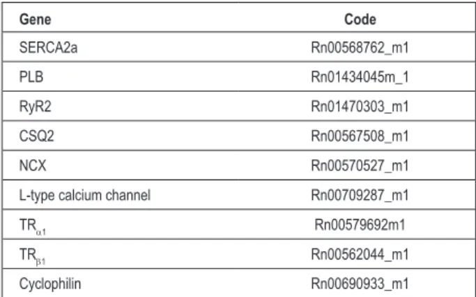
PCR. From this graph, we determined the cycle in which the reaction crosses the threshold of detection (cycle threshold - CT) based on the variability of baseline data obtained from the initial cycles of PCR. The primers for the genes analyzed (Box 1) were obtained using the software Primer Express™
(Applied Biosystems, Foster City, CA, USA) from published
sequences in the GenBank (www.pubmed.com). The relative quantification of each gene normalized by endogenous reference (cyclophilin) was performed according to the User Bulletin # 2 (Applied Biosystems, Foster City, CA, USA).
Statistical analysis
The experimental results were expressed as mean ± SD. Comparisons between groups were performed by two-way analysis of variance (ANOVA), exercise and diet, followed by the Tukey’s test. Relations between the mRNA of SERCA2a, PLB, CQS, NCX, L-channel and RYR2 and thyroid hormone receptors, TRb1 and TRa1, were measured by Pearson’s correlation coefficient. The significance level was 5%.
Results
The general characteristics of the animals are shown in Table 1. The initial body weights and the LV/FW were not different between the groups. The animals restricted, R50 and EX50, had final body weights, LV, RV, atrium and feed consumption significantly lower than their respective controls (C vs R50 and EX vs EX50). The trained animals, EX and EX50,
showed a reduction in final body weights compared to C and R50 rats, respectively. The EX rats increased their atrium weight in relation to animals C. The LV/FW ratio significantly increased in EX50 animals compared to EX animals.
Table 2 presents the results of the evaluation of mRNA by the real-time PCR technique. It was found that R50 animals increased the expression of NCX, L-calcium channel and PLB, and reduced the expression of TRa1 comparedto control rats. Animals in the EX group increased the expression of NCX, L-channel and TRb1 receptor comparedto control animals. The association of physical training and FR, in the EX50 group, increased expression of SERCA2a, NCX and PLB over animals
Box 1 - Primers employed
Gene Code
SERCA2a Rn00568762_m1
PLB Rn01434045m_1
RyR2 Rn01470303_m1
CSQ2 Rn00567508_m1
NCX Rn00570527_m1
L-type calcium channel Rn00709287_m1
TRa1 Rn00579692m1
TRb1 Rn00562044_m1
Cyclophilin Rn00690933_m1
Table 1 - General characteristics of animals
C n = 7
R50
n = 7
EX n = 7
EX50
n = 7
Food intake (g) 20.2 ± 2.3 10.1 ± 1.1 * 20.7 ± 2.5 10.4 ± 1.2 *
IW (g) 292 ± 12 291 ± 21 294 ± 10 293 ± 17
FW (g) 354 ± 19 219 ± 8* 333 ± 16 * 207 ± 8 *†‡
LV (g) 0.694 ± 0.064 0.462 ± 0.016 * 0.672 ± 0.053 0.474 ± 0.024 *
RV (g) 0.213 ± 0.030 0.126 ± 0.019 * 0.217 ± 0.050 0.149 ± 0.022 *
Atrium (g) 0.059 ± 0.006 0.042 ± 0.005 * 0.077 ± 0.013 * 0.049 ± 0.006 *‡
LV/FW (mg/g) 1.962 ± 0.139 2.135 ± 0.142 2.017 ± 0.122 2.284 ± 0.100 *‡
RV/FW (mg/g) 0.601 ± 0.069 0.571 ± 0.069 0.654 ± 0.177 0.721 ± 0.121
Values expressed as mean ± standard deviation; C - control rats; R50 - rats with restricted food (FR) of 50%; EX - trained rats; EX50 - rats trained with 50% FR; IW - initial body weight (g); FW - inal body weight (g); LV - left ventricle weight (g); RV - right ventricle weight (g) * versus C; † versus R50; ‡ versus EX - ANOVA. Tukey. p < 0.05.
EX and R50. Moreover, the EX50 animals showed increased expression of TRb1 compared to R50 rats.
Serum levels of T3, T4 and TSH are presented in Table 3. There was no change in serum concentration of T3 by the action of FR or physical training. The trained rats, EX, showed no change in serum concentrations of T4 and TSH. However, there was significant reduction of T4 and TSH in
the groups R50 and EX50 compared to respective controls (C
vs R50 and EX vs EX50).
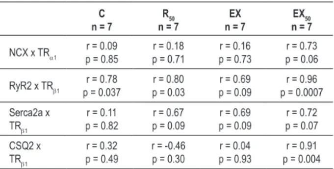
The correlation coefficients and significances between mRNA of calcium handling structures and thyroid hormone receptors are shown in table 4. Only the positive correlations (r > 0.70) in group EX50 are presented, since the objective of this study was to evaluate the association and physical training and food restriction on the gene transcription of molecular structures involved in the calcium handling and the influence of the TH receptor in the myocardium. Thus, the correlation coefficients for the groups C, R50 and EX were presented to
define the participations of the factors exercise, food restriction and association between two factors.
Table 2 - Evaluation of mRNA by real time PCR
C n = 7
R50
n = 7
EX n = 7
EX50
n = 7
L-channel 1.00 ± 0.34 2.34 ± 0.48 * 3.60 ± 0.76 * 2.74 ± 0.76*
NCX 1.00 ± 0.32 1.89 ± 0.56 * 2.48 ± 0.33* 3.13 ± 0.52 *†‡
RyR2 1.00 ± 0.18 1.08 ± 0.17 1.11 ± 0.23 1.26 ± 0.23
Serca2a 1.00 ± 0.36 0.92 ± 0.27 1.04 ± 0.24 1.42 ± 0.10 *†‡
PLB 1.00 ± 0.09 1.25 ± 0.19 * 1.14 ± 0.22 1.51 ± 0.26 *†‡
CSQ2 1.00 ± 0.29 1.03 ± 0.21 1.07 ± 0.22 1.27 ± 0.25
TRa1 1.00 ± 0.29 0.67 ± 0.16 * 0.78 ± 0.23 0.86 ± 0.26
TRb1 1.00 ± 0.29 0.96 ± 0.25 1.47 ± 0.40* 1.66 ± 0.17*†
Values expressed as mean ± standard deviation; C - control rats; R50 - rats with restricted food (FR) of 50%; EX - trained rats; EX50 - rats trained with 50% FR; RyR2 - ryanodine channel; Serca2a - sarcoplasmic reticulum calcium pump; PLB - phospholamban; CSQ2 - calsequestrin; NCX - Na+/Ca2+; TR - thyroid hormone receptor; * versus C; † versus R50 - ‡ versus EX. ANOVA. Tukey. p < 0.05.
Table 3 - Plasma concentration of thyroid hormones
C n = 7
R50
n = 7
EX n = 7
EX50
n = 7
T3 (mg/dl) 4.35 ± 1.32 3.11 ± 1.34 5.52 ± 2.72 3.96 ± 2.36
T4 (mg/dl) 242 ± 29 113 ± 17 * 191 ± 61 126 ± 6 *‡
TSH (mg/dl) 6.35 ± 1.77 1.94 ± 0.75 * 5.07 ± 3.13 1.54 ± 0.66 *‡ Values expressed as mean ± standard deviation; C - control rats; R50 - rats with restricted food (FR) of 50%; EX - trained rats; EX50 - rats trained with 50% FR; RyR2 - ryanodine channel; Serca2a - sarcoplasmic reticulum calcium pump; PLB - phospholamban; CSQ2 - calsequestrin; NCX - Na+/Ca2+ exchanger; TR - thyroid hormone receptor; * versus C; † versus R50 - ‡ versus EX. ANOVA. Tukey. p < 0.05.
It was observed that the TRb1 gene has more correlations with the molecular structures of the calcium handling than the TRa1 gene (Table 4). Although the L-channel mRNA and PLB have shown significant increase in EX50 rats, these genes did not show any correlation with the genes TRa1 or TRb1. The mRNA of ryanodine was significantly correlated with mRNA of TRb1 in all groups and was higher in the combination of FR with physical training. The SERCA2a mRNA also showed
Table 4 -Pearson correlation coeficient between mRNA of calcium
handling proteins and thyroid hormone receptors
C n = 7
R50
n = 7
EX n = 7
EX50
n = 7
NCX x TRa1 p = 0.85r = 0.09 p = 0.71r = 0.18 p = 0.73r = 0.16 p = 0.06r = 0.73
RyR2 x TRb1 p = 0.037r = 0.78 p = 0.03r = 0.80 p = 0.09r = 0.69 p = 0.0007r = 0.96
Serca2a x TRb1
r = 0.11 p = 0.82
r = 0.67 p = 0.09
r = 0.69 p = 0.09
r = 0.72 p = 0.07
CSQ2 x TRb1
r = 0.32 p = 0.49
r = -0.46 p = 0.30
r = 0.04 p = 0.93
r = 0.91 p = 0.004
a good correlation with the mRNA of TRb1 in the groups R50, EX and EX50 although it is not statistically significant, which
may be due to the low number of samples. Calsequestrin and NCX genes correlated well with the mRNA TRb1 and TRa1, respectively, only in the group EX50 indicating the dependence of the association of both factors. It is interesting to note that the mRNA of CQS and RYR2 were strongly correlated with TRb1 in EX50 animals. However, there was no significant increase in gene expression of CQS and RYR2 in this group. Conversely, the NCX gene increased but correlated only with the gene TRa1. The SERCA2a gene was the only one to correlate with the mRNA of TRb1 and increase in gene expression in animals EX50.
Discussion
The objective of this study was to evaluate the participation of thyroid hormones in the association between severe food restriction and physical training and the correlation between TH receptors and gene expression of the molecular structures involved in myocardial intracellular calcium handling. The results of this study showed that physical training associated with severe FR promoted a significant increase of mRNA of SERCA2a, L-channel, phospholamban and Na+/Ca2+ exchanger. These
changes were accompanied by an increase in the expression of thyroid hormone receptors (TRb1 despite normal serum levels of T3 and reduced serum levels of T4 and TSH.
FR and physical training protocols promoted significant morphological changes (Table 1). The reductions in body weight, RV weight and LV weight induced by FR are associated with the degree of caloric restriction. Food restriction, excessive intake or fasting state, change energy expenditure and body composition25. The reduction in energy expenditure
during food restriction has been documented and represents a mechanism of energy conservation, which prevents excessive loss of body weight25. The heart chambers were reduced in
proportion to the reduction of body weight, as observed by the relations LV/BW and RV/BW. The decrease in body weight by exercise training is associated with increased energy expenditure resulting from physical training sessions; the LV/BW and RV/BW ratios were maintained compared to the control group. The association of PT and FR enhanced reduction in body weight, but kept the LV and RV mass and maintained the LV/BW and RV/BW ratios compared with R50 rats. Thus, the results related to the cardiac chambers indicate that the FR promoted a significant reduction of cardiac mass and physical training, alone or combined with FR, did not cause cardiac hypertrophy. These results were similar to those observed in previous studies10.
FR, alone or in trained rats, reduced levels of T4 and TSH, while physical training did not change serum levels of thyroid hormones. Although FR reduces the serum level of T3 by 29% in both groups (R50 and EX50), this reduction was not of sufficient magnitude to show a statistical difference between groups. The lack of change in T3 levels may be due to total T3 rather than free T3, which is the active form of the hormone.
Literature data indicate that in states of fasting and severe calorie restriction, there is marked increase of reverse T3
13. So
it is possible that the free T3/reverse T3 ratio was reduced in animals subjected to FR. It is believed that energy deprivation
may modulate serum concentration of T3 and reduce the activity or concentration of iodothyronine deiodinase that converts T4 into T326. The significant reduction of serum levels
of T4 and TSH induced by FR indicates state of hypothyroidism, which was also observed in experimental8,13,27,28 and clinical
studies29. The mechanism responsible for decreased plasma
levels of thyroid hormones induced by FR may be related to the actual reduction in energy consumption. Reduced levels of thyroid hormones relate to the mechanism of energy conservation, since these hormones are mainly responsible for the energy expenditure25. Considering that energy expenditure
is reduced in the food restriction, reduced levels of TH would be expected. Physical training did not promote changes in plasma levels of thyroid hormones. This result differs from that reported by Iemitsu et al22 and Katzeff et al13, who found an
increase in serum levels of T4. This discrepancy in results may have been due to the type, intensity and volume of physical training. In conclusion, while the FR promoted the state of hypothyroidism, physical training seems to have no direct effect on the action of thyroid hormones.
In the heart of rats, over 70% of all TH receptors are TRa1, with TRb1 accounting for only 30%30. The thyroid hormone binds
to thyroid response elements (TH receptors) in regions that promote many genes, regulating transcription of target genes in the heart including Na+/Ca2+ exchanger, ryanodine, L-type
calcium channel and mainly SERCA2a and fosfolambam30. This
regulation by TH is an important mechanism in the process of myocardial contraction and relaxation31.
The results of this study indicate that the state of hypothyroidism induced by FR decreased the expression of the gene TRa1, a receptor prevalent in the myocardium. This result was expected since the reduction of TH causes a negative feedback in the TH receptor. Exercise training, alone or combined with FR, increased expression of TRb1 without changing the levels of TRa1. There is limited literature on the role of TRa1 and TRb1 receptors on the gene transcription of proteins of the myocardial calcium handling. Kinugawa et al21
showed that the TRa1 relates to the transcription of a-MHC and myocardial protein synthesis, while TRb1 relates to SERCA2a, TRb1 itself and inhibition of b-MCH. Thus, our data suggest that the reduction of the TRa1 gene by FR could be a mechanism associated with loss of heart mass; absence of hypertrophy in trained animals could be due to maintenance of TRa1 levels.
with studies by Kemi et al32 who reported an increase in the
reuptake of calcium in the myocardium derived from the increase of SERCA2a and phosphorylated phospholamban (PLB-thr17) in trained rats. The increased L-channel observed in EX50 animals suggests an increased influx of calcium to
improve the contractile response. The increase in the NCX gene would aim to offset the increased influx of calcium through sarcolemal slow calcium channels.
In this study, we could not relate TH receptors with the molecular structures of the myocardial calcium handling in rats trained and subjected to severe FR, considering that the correlation test showed no significant difference in the mRNA of SERCA2a, PLB, NCX, L-channel with TRa1 or TRb1 receptors. Literature data show an association of the molecular structures involved in calcium handling with TH receptors induced by training or state of hypothyroidism; Iemitsu et al22
found a significantly increased expression of TRa1 and TRb1 receptors accompanied by increased expression of SERCA2a by physical training. In conditions of hypothyroidism, thyroid failure is associated with decreased SERCA2a and increased phospholamban23, increased expression of NCX33 and
unchanged34 or increased35 levels of L-channel.
The molecular mechanisms for increasing gene transcription of SERCA2, PLB and NCX in the exercise alone or combined with FR remain unclear; as previously mentioned, our findings suggest that TH and/or TH receptors do not participate in the gene transcription of molecular structures of calcium handling in this group of animals. The process of cardiac remodeling derives from extracellular stimuli that activate complex cytosolic signals with a number of integration points, and nuclear transcriptional processes. Among the extracellular
stimuli, agonists of G protein-coupled receptors (angiotensin, endothelin, catecholamines), cytokines and growth factors (IGF, TGF, FGF) are the main candidates that could therefore stimulate the gene transcription of calcium handling structures36,37. However, there is no literature data that allow
speculating the mechanisms responsible for increased gene transcription of SERCA2, PLB and NCX in the interaction between exercise and food restriction.
Study limitations
Our findings of gene expression of mRNA obtained by quantitative PCR of SERCA2a, phospholamban, Na+/Ca2+
exchanger, L-type calcium channel, and calsequestrin and TH receptors may not be reflected in protein formation. Recent data obtained in our laboratory found a significant reduction of protein expression of L-type channel in rats with severe FR, which is contrary to our findings of increased mRNA in the same protein14. Therefore, the continuity of these findings
will be the determination of the protein expression by Western blot. Nor was it possible to establish a cause-effect relationship between thyroid hormone and/or TH receptors with the mRNA of the calcium handling; studies with TH treatment or others using specific monoclonal animals for TH would be required.
In conclusion, the findings of the study showed increased mRNA expression of the molecular structures involved in the intracellular calcium handling in animals subjected to severe restriction and physical training. Although the association between PT and FR induces a state of hypothyroidism, the TH-receptor axis does not seem to participate in the transcription of mRNA of myocardial calcium handling proteins.
References
1. Keenan KP, Laroque P, Ballam GC, Soper KA, Dixit R, Mattson BA, et al. The effects of diet, ad libitum overfeeding, and moderate dietary restriction on the rodent bioassay: the uncontrolled variable in safety assessment. Toxicol Pathol. 1996;24(6):757-68.
2. Venditti P, Di Meo S. Antioxidants, tissue damage, and endurance in trained and untrained young male rats. Arch Biochem Biophys. 1996;331(1):63-8.
3. Lesourd BM, Mazari L. Immune responses during recovery from protein-energy malnutrition. Clin Nutr. 1997;16(Suppl 1):37-46.
4. Torun B, Chew F. Protein-energy malnutrition. In: Shils ME, Ross AC. (eds). Modern nutrition in health and disease. Baltimore: Williams & Wilkins; 1999. p.936-88.
5. Liu J, Yeo HC, Overvik-Douki E, Hagen T, Doniger SJ, Chyu DW, et al. Chronically and acutely exercised rats: biomarkers of oxidative stress and endogenous antioxidants. J Appl Physiol. 2000;89(1):21-8.
6. Ramsey JJ, Harper ME, Weindruch R. Restriction of energy intake, energy expenditure, and aging. Free Radic Biol Med. 2000;29(10):946-68.
7. Perez AC, Cabral de Oliveira AC, Estevez E, Molina AJ, Prieto JG, Alvarez AI. Mitochondrial, sarcoplasmic membrane integrity and protein degradation in heart and skeletal muscle in exercised rats. Comp Biochem Physiol C Toxicol Pharmacol. 2003;134(2):199-206.
8. Haddad F, Bodell PW, McCue SA, Herrick RE, Baldwin KM. Food restriction-induced transformations in cardiac functional and biochemical properties in rats. J Appl Physiol. 1993;74(2):606-12.
9. Broderick TL, Driedzic WR, Gillis M, Jacob J, Belke T. Effects of chronic food restriction and exercise training on the recovery of cardiac function following ischemia. J Gerontol A Biol Sci Med Sci. 2001;56(1):B33-7.
10. Sugizaki MM, Dal Pai-Silva M, Carvalho RF, Padovani CR, Bruno A, Nascimento AF, et al. Exercise training increases myocardial inotropic response in food restricted rats. Int J Cardiol. 2005;112(2):191-201.
11. Opie LH, Bers DM. Excitation-contraction coupling and calcium. In: Opie LH (ed). Heart physiology: from cell to circulation. 4th ed. Philadelphia: Lippincott Williams & Wilkins; 2004. p. 159-85.
12. Vizotto VA, Carvalho RF, Sugizaki MM, Lima AP, Aragon FF, Padovani CR, et al. Down-regulation of the cardiac sarcoplasmic reticulum ryanodine channel in severely food-restricted rats. Braz J Med Biol Res. 2007;40(1):27-31.
13. Katzeff HL, Powell SR, Ojamaa K. Alterations in cardiac contractility and gene expression during low-T3 syndrome: prevention with T3. Am J Physiol. 1997;273(5 Pt 1):E951-6.
14. De Tomasi LC, Bruno A, Sugizaki MM, Lima-Leopoldo AP, Nascimento AF, Junior SA, et al. Food restriction promotes downregulation of myocardial L-type Ca2+ channels. Can J Physiol Pharmacol. 2009;87(6):426-31.
15. Buttrick PM, Malhotra A, Scheuer J. Effects of systolic overload and swim training on cardiac mechanics and biochemistry rats. J Appl Physiol. 1988;64(4):1466-71.
increased in hearts of exercise-trained old rats. Am J Physiol. 1996;271(1 Pt 2):H68-72.
17. Wisloff U, Loennechen JP, Falck G, Beisvag V, Currie S, Smith G, et al. Increased contractility and calcium sensitivity in cardiac myocytes isolated from endurance trained rats. Cardiovasc Res. 2001;50(3):495-508.
18. Stauffer B, Mitro G, Moore RL. Chronic treadmill running does not influence ryanodine binding to rat myocardium. Med Sci Sports Exerc. 1993;25(5):S98.
19. Lankford EB, Korzick DH, Palmer BM, Stauffer BL, Cheung JY, Moore RL. Endurance exercise alters the contractile responsiveness of rat heart to extracellular Na+ and Ca2+. Med Sci Sports Exerc. 1998;30(10):1502-9.
20. Mokelke EA, Palmer BM, Cheung JY, Moore RL. Endurance training does not affect intrinsic calcium current characteristics in rat myocardium. Am J Physiol Heart Circ Physiol. 1997;273(3 Pt 2):H1193-7.
21. Kinugawa K, Yonekura K, Ribeiro RC, Eto Y, Aoyagi T, Baxter JD, et al. Regulation of thyroid hormone receptor isoforms in physiological and pathological cardiac hypertrophy. Circ Res. 2001;89(7):591-8.
22. Iemitsu M, Miyauchi T, Maeda S, Tanabe T, Takanashi M, Matsuda M, et al. Exercise training improves cardiac function-related gene levels through thyroid hormone receptor signaling in aged rats. Am J Physiol Heart Circ Physiol. 2004;286(5):H1696-705.
23. Dillmann WH. Biochemical basis of thyroid hormone action in the heart. Am J Med. 1990;88(6):626-30.
24. Chomczynski P, Sacchi N. Single-step method of RNA isolation by acid guanidinium thiocyanate-phenol-chloroform extraction. Anal Bichem. 1987;162(1):156-9.
25. Passadore MD, Griggio MA, Nunes MT, Luz J. Effects of ageing on the energy balance of food-restricted rats. Acta Physiol Scand. 2004;181(2):193-8.
26. Katzeff HL, O´Connell M, Horton ES, Danforth Jr E, Young JB, Landsberg L. Metabolic studies in human obesity during overnutrition and undernutrition: thermogenic and hormonal responses to norepinephrine. Metabolism. 1986;35(2):166-75.
27. Katzeff HL, Selgrad C. Maintenance of thyroid hormone production during exercise-induced weight loss. Am J Physiol. 1991;261(3 Pt 1):E382-8.
28. Cokelaere M, Decuypere E, Flo G, Darras VM, Kühn ER. Influence of feeding pattern on thyroid hormones in long-term food-restricted rats. Horm Metab Res. 1996;28(7):315-8.
29. Fontana L, Klein S, Holloszy JO, Premachandra BN. Effect of long-term calorie restriction with adequate protein and micronutrients on thyroid hormones. J Clin Endocrinol Metab. 2006;91(8):3232-5.
30. Dillmann WH. Cellular action of thyroid hormone on the heart. Thyroid. 2002;12(6):447-52.
31. Carr AN, Kranias EG. Thyroid hormone regulation of calcium cycling proteins. Thyroid. 2002;12(6):453-7.
32. Kemi OJ, Ellingsen O, Ceci M, Grimaldi S, Smith GL, Condorelli G, et al. Aerobic interval training enhances cardiomyocyte contractility and Ca2+ cycling by phosphorylation of CaMKII and Thr-17 of phospholamban. J Mol Cell Cardiol. 2007;43(3):354-61.
33. Arai M, Otsu K, MacLennan DH, Periasamy M. Regulation of sarcoplasmic reticulum gene expression during cardiac and skeletal muscle development. Am J Physiol. 1992;262(3 Pt 1):C614-20.
34. Seppet EK, Kolar F, Dixon IM, Hata T, Dhalla NS. Regulation of cardiac sarcolemmal Ca2+ channels and Ca2+ transporters by thyroid hormone. Mol Cell Biochem. 1993;129(2):145-59.
35. Hawthorn MH, Gengo P, Wei XY, Rutledge A, Moran JF, Gallant S, et al. Effect of thyroid status on beta-adrenoceptors and calcium channels in rat cardiac and vascular tissue. Naunyn Schmiedebergs Arch Pharmacol. 1988;337(5):539-44.
36. Franchini KG. Hipertrofia cardíaca: mecanismos moleculares. Rev Bras Hipertens. 2001;8(1):125-42.