Coffee resistance to the main diseases:
leaf rust and coffee berry disease
Maria do Céu Silva1, Victor Várzea1, Leonor Guerra-Guimarães1, Helena Gil Azinheira1,
Diana Fernandez2, Anne-Sophie Petitot2, Benoit Bertrand3, Philippe Lashermes2 and Michel Nicole2
1Instituto de Investigação Científica Tropical (IICT) - Centro de Investigação das Ferrugens do Cafeeiro (CIFC), Quinta do Marquês, 2784-505
Oeiras, Portugal; 2Institut de Recherche pour le Développement (IRD), UMR DGPC, Résistance des Plantes, BP 64501 - 34394 Montpellier
Cedex 5, France; 3Centre de Coopération Internationale en Recherche pour le Développement (CIRAD), Département des Cultures Pérennes,
UMR DGPC, Résistance des Plantes, BP 64501 - 34394 Montpellier Cedex 5, France. *Corresponding author: mceusilva@hotmail.com
Considerable success has been obtained in the use of classical breeding to control economically important plant diseases, such as the coffee leaf rust and the coffee berry disease (CBD). There is a strong consensus that growing genetically resistant varieties is the most appropriate cost effective means of managing plant diseases and is one of the key components of crop improvement. It has also been recognized that a better knowledge of both, the pathogens and the plant defence mechanisms will allow the development of novel approaches to enhance the durability of resistance. After a brief description of concepts in the field of plant disease resistance, we attempt to give a view of the research progress on coffee leaf rust and CBD concerned with the pathogens infection and variability, coffee breeding for resistance and coffee resistance mechanisms.
Key words: Coffea, Colletotrichum kahawae, Hemileia vastatrix, coffee breeding, resistance.
Resistência do cafeeiro para suas principais doenças: ferrugem alaranjada das folhas e antracnose dos frutos: Sucesso considerável tem sido obtido no uso do melhoramento clássico para o controle de doenças de plantas economicamente importantes, tais como a ferrugem alaranjada das folhas e a antracnose dos frutos do cafeeiro (CBD). Há um grande consenso de que o uso de plantas geneticamente resistentes é o meio mais apropriado e eficaz em termos de custos do controle das doenças das plantas, sendo também um dos elementos chave do melhoramento da produção agrícola. Tem sido também reconhecido que um melhor conhecimento do agente patogênico e dos mecanismos de defesa das plantas permitirá o desenvolvimento de novas abordagens no sentido de aumentar a durabilidade da resistência. Após uma breve descrição de conceitos na área da resistência das plantas às doenças, nesta revisão tentou-se dar uma idéia do progresso na investigação da ferrugem alaranjada do cafeeiro e do CBD relativamente ao processo de infecção e variabilidade dos agentes patogênicos, melhoramento do cafeeiro para a resistência e mecanismos de resistência do cafeeiro.
Palavras-chave: Coffea, Colletotrichum kahawae, Hemileia vastatrix, melhoramento do cafeeiro, resistência.
1. INTRODUCTION
Although coffee is not a food crop, it represents for most coffee-growing countries the major source of revenue for foreign exchange. Limiting factors of coffee production include major diseases, such as the coffee leaf rust (or orange rust) and the coffee berry disease (CBD) caused by the fungi Hemileia vastatrix Berkeley and Broome and Colletotrichum kahawae Bridge and Waller, respectively.
occurs only in West Africa where it can be serious for some cultivars of C. canephora grown in very wet areas and it is favoured by hot humid conditions which are unsuitable for Arabica coffees.
Other fungal diseases like coffee wilt disease or tra-cheomycosis caused by Fusarium xylarioides Steyaert (tele-omorph: Gibberella xylarioides Heim and Saccas) is becom-ing important in some regions of Central and West Africa, not only in Robusta but also for Arabica. It is a vascular disease causing yellowing and wilting of the trees. Fusarium stilboides Wollenw (telemorph: Gibberella stilboides), the causal agent of the coffee bark disease, is also present in some African countries, particularly in Ethiopia, Kenya, Malawi and Tanzania. Its characteristic symptom is a scaling of the bark leading to stem cankers and a progressive dying back of the whole tree. The brown eye spot or berry blotch (Cercospora coffeícola Berk and Cooke) is a disease of widespread occur-rence in nurseries and plantations, infecting the coffee leaves and the fruits as well. The American leaf spot of coffee caused by Mycena citricolor (Berk and Curt.) Sacc. has been reported exclusively in the American continent in cool, moist areas at higher altitudes (Waller 1985; Wrigley 1988; Chen, 2002; Geiser et al., 2005).
The two known bacterial coffee diseases are the halo blight of coffee incited by Pseudomonas syringae pv. garcae Amaral et al. (Amaral et al., 1956) and the coffee leaf scorch caused by the polyphagous bacteria Xilella fastidiosa Wells et al. (Wells et al., 1987). The former disease has been described in Brazil, Kenya, Uganda and China where it is becoming of some concern due to its higher incidence and severity (Wen and Chen 1995; Chen, 2002). The bacterium infects the coffee leaves, branch-tips and young fruits, in some instances with symptoms (irregular dark necrotic lesions) identical to Phoma sp. However, it can be distinguished from the fungus by the presence of bacterial exudates in the young lesions. The bacte-ria persist, in a large number, on all healthy surfaces of the tree, and during cool and wet weather they multiply and initiate the epidemics. Coffee leaf scorch has been recorded so far only in Brazil and Costa Rica where the bacterium X. fastidiosa at-tacks several crops with high incidence, particularly in citrus and prunes. A strain of the bacterium, able to infect coffee, is transmitted by the sharpshooter leafhopper Dilobopterus cor-talimai (Cicadellidae: Cicadellinae) and was recorded for the first time in 1995 in the State of S. Paulo (Leite Jr. et al., 1999; Li et al., 2001). Symptoms in affected coffee plants include shortened internodes, premature loss of older leaves, terminal clusters of small pale green to yellow deformed leaves, lateral
shoot dieback, and overall plant stunting (De Lima et al., 1998; Li et al., 2001).
Some records of virus diseases in coffee have sporadically been made (leaf rugosity, leaf curl) in Brazil and Colombia but with no economic significance and of eventual misinterpreted diagnostic (Chen, 2002). However, the coffee ringspot virus CoRSV associated with the presence of the mite Brevipalpus phoenicis Geijskes has been reported in several Brazilian states (São Paulo, Paraná, Minas Gerais, and Distrito Federal) and recently found in Costa Rica. It causes conspicuous ringspot symptoms on leaves, berries, and less frequently on twigs. A similar disease is known in the Philippines, but no information exists about its relationship to CoRSV. Coffee ringspot had no economical significance until recently when a large-scale infection was reported in Minas Gerais that resulted in yield loss (Chagas et al., 2003).
The extremely high costs of fungicides to control dis-eases in coffee plantations, particularly leaf rust and CBD, as well as the difficulties in its application, have led the coffee experimental centres to give priority in their coffee improve-ment programmes to select for resistance to these diseases, as the main part of an integrated global protection approach (Bettencourt and Rodrigues Jr., 1988). Improvement of coffee resistance to leaf rust and CBD has been mainly focused on finding new sources of resistance and a better understanding of both the pathogens diversity and the host defence mecha-nisms. In view of thecomplexity of plant disease resistance to pathogens, which is becoming increasingly apparent as new tools in genetics, cell biology, biochemistry and molecular biology are being used, this review will focus on pathogen recognition, signal transduction pathways and subsequent defence responses, as well as on the specificity of host resist-ance. Particular attention will be given to developments that have been made in coffee leaf rust and CBD, including aspects related to pathogen infection and variability, coffee breeding for resistance and host resistance mechanisms.
2. CONCEPTS OF DISEASE RESISTANCE
patho-gen attack which involves a network of signal transduction and the rapid activation of gene expression (Dixon and Lamb 1990; Bradley et al., 1992; Heath 1997a).
2.1. Pathogen recognition and signal transduction
The perception and rapid response to the presence of most pathogens by plants determine whether or not it will survive. Perception involves the detection of molecules from the pathogen or from the wounded plant itself (Keen 1999). These molecules have come to be called elicitors (non-race-specific or race-specific) and may be peptides or proteins, fatty acid derivatives, sterols or other low molecular weight chemicals, with diverse structures and present in very low concentrations (Ebel and Scheel 1997; Somssich and Hahlbrock 1998; Yamaguchi et al., 2000; Peck, 2003). Whatever the source of the elicitor, the mechanism of signal perception appears to rely on the presence of specific receptors on the plant cell surface (Ebel and Scheel 1997; Pontier et al., 2002). Several of the receptor-like protein kinase (RLK) family are transmembrane proteins with cytoplasmic serine/threonine kinase domains and divergent extracellular domains (Morris and Walker, 2003). Some of the rapidly activated kinases have been identified as mitogen-activated protein kinases (MAPKs) (Jonak et al., 2002), calcium-dependent protein kinases (CDPK) (Romeis et al., 2001) and wall-associated kinase (WAK) (He et al., 1998). As yet, however, little is known about how these kinases transmit information to the cell (Peck, 2003).
Upon recognition, signalling events are initiated resulting in numerous cellular changes, such as an inward flux of Ca2+, the production of reactive oxygen species (ROS), actin cytoskeleton involvement, changes in the levels of a subset of transcripts (Grant and Mansfield 1999; Guest et al., 1999; Heath, 2000a, Dangl and Jones, 2001; Peck, 2003), involvement of plasma-membrane-bound NADPH oxidases, cell-wall-bound peroxidases, amino oxidases in the apoplast and phosphoproteins (Bestwick et al., 1997; Heath, 2000a, Laloi et al., 2004). The activation of signalling cascades at the plasma membrane leads to the induction of a complex network of other signal molecules, such as salicylic acid, jasmonic acid, ethylene, nitric oxide, that will induce, within minutes, a general defence response that will kill/limit the spread of the pathogen through the plant (Alvarez, 2000; Dixon et al., 2002; Turner et al., 2002; Harrison and Baldwin, 2004; Delledonne, 2005).
2.2. Defence responses
2.2.1. Preformed defences
Protection from the initial invasion of the pathogen is achieved via passive defences, such as physical and/or chem-ical barriers. Physchem-ical barriers largely involve properties of the plant surface, that is, the amount and quality of wax and cuticle that cover the epidermal cells, the structure of the epi-dermal cell walls and the size, location and shape of stomata (Agrios 1997). Some plants invest in very thick walls and/or cuticles, and bark (where present) can also provide a physi-cal impediment to infection. Chemiphysi-cal barriers include com-pounds that have antimicrobial activity and comcom-pounds that affect the vectors of plant viruses, such as, phenols, quinines, lactones, cyanogenic glucosides, saponins, terpenoids, stil-benes and tannins (Keen 1999). Evidence is accumulating to show that plant cell wall, apart from being a structural bar-rier against pathogen attack, is capable of triggering multiple signalling pathways. These signal properties are essential not only for the role of the cell wall in defence but also for the co-ordination of wall synthesis and expansion between adjacent cells (Pilling and Hofte, 2003).
2.2.2. Induced defences
reactive oxygen species (ROS) (Goodman and Novacky, 1994; Bolwell 1999; Hammerschmidt and Nicholson 1999). However, it is not at all clear that the production of ROS (oxidative burst) is required for the development of the HR for all plant-pathogen interactions (Heath 1998, 2000b). It has been suggested that ROS produced during the oxidative burst may function in the defence-signalling pathways leading to different defence responses against pathogens (Neill et al., 2002; Laloi et al., 2004). These molecules also have the potential to act directly as antimicrobial agents (Baker and Orlandi 1995).
Another early event in the interaction between a pathogen and a host plant cell is the expression of genes encoding enzymes of the phenylpropanoid pathway, such as phenylalanine ammonia-lyase - PAL (Dorey et al., 1997). PAL has been shown to play an important role in plant resistance, namely by its involvement in the biosynthetic pathway providing isoflavonoids and phenylpropanoids with phytoalexin activity and as a precursor of lignin-like compounds, suberin or other types of phenolic materials that are often deposited in the host cell walls at the point of fungal invasion (e.g. papilla formation), and appear to block the progression of the hyphae (Hammerschmidt 1999; Dixon et al., 2002; Caño-Delgado et al., 2003). PAL has also been implicated in the biosynthetic pathway for salicylic acid (SA), another defence-related compound and a key signalling component required for the activation of PR-genes, catalases, receptor-like protein kinases and transcription factors (Mauch–Mani and Slusarenko 1996; Klessig et al., 2000; Dixon et al., 2002; Way et al., 2002; Mould et al., 2003).
Resistance expression is often accompanied by the activation of the phenol-oxidising enzymes peroxidase and phenoloxidase and the lipid peroxidizing enzyme lipoxygenase (Goodman and Novacky, 1994). Peroxidase activity often increases in response to infection, and may function in defence through production of antimicrobial quantities of hydrogen peroxide as well as in cell wall lignification and crosslinking (Peng and Kúc, 1992; Rasmussen et al., 1995; Chittor et al., 1999; Do et al., 2003). Increase in activity of phenoloxidases and its interaction with endogenous phenols have been correlated with the onset of HR (Hammerschmidt and Nicholson 1999). Lipoxygenase contributes to the HR via disruption of cell membrane lipids, to defence through the formation of toxic lipid oxidation products, as well as in the formation of lipid-derived signals (Croft et al., 1993; Jalloul et al., 2002; Montillet et al., 2005).
Another induced response is the synthesis of pathogen-esis-related proteins (PR proteins), which do not only accu-mulate locally in the infected tissues, but are also induced systemically (Van Loon et al., 1994). Most of the existing 17 families include components that are exported to the extracel-lular space, where they are believed to have a role in resist-ance (Fritig et al., 1998; Christensen et al., 2002). A collec-tive set of PR proteins (e.g. chitinases and β-1,3-glucanases) may be effective in inhibiting pathogen growth, multiplica-tion and/or spread (Van Loon and Van Strien, 1999). Others like PR-9 proteins (with peroxidase activity) are enzymes with possible implications in the oxidative cross-linking of plant cell wall components to prevent the pathogen from penetrating (Thordal-Christensen et al., 1992). Furthermore, oxalate oxidase (Zhou et al., 2000) and some chitinases are PR proteins that have been suggested to be involved in the re-lease of chemical signals, from fungal cell walls that will act as elicitors of other plant-defence responses (Schlumbaum et al., 1986; Ryan, 1987; Lawrence et al., 2000). It is very likely that novel extracellular PR proteins play a role within one of these three classes of phenomena, namely antimicrobial activity, cell wall biology and signal transduction (Van Loon 1999; Tuzun, 2001; Christensen et al., 2002).
2.3. Specificity of host resistance
effective for a long period, where application is made on a large scale in an environment conducive to the pathogen (Johnson, 1981) has been achieved using single R genes. In general, the rapid evolution of new virulent pathotypes, on previously resistant cultivars, forced breeders into producing replacement cultivars with new R genes (Pink, 2002). Thus, although this type of resistance is the most frequently deployed in plant breeding, the great disadvantage is that it is often non-durable (Vale et al., 2001; Lindhout, 2002; Niks and Rubiales, 2002; Parlevliet, 2002).
Another type of host resistance is the race non-specific resistance, which appears to be specific for one pathogen species only, although more or less equally effective against all genotypes of the pathogen (Parlevliet, 1983). This type of resistance refers to plant disease resistance generated via interactions between the products of multiple plant genes, not a single R gene (Simmonds, 1991). This multigenic (or poly-genic) resistance is generally quantitative. With the onset of molecular technologies, the identification of chromosome re-gions that are involved in quantitative resistance has become feasible. These regions are designated “quantitative trait loci” (QTLs) and, as they are defined by the position on the genome and the quantitative effect on resistance, they are not informative of the function. Hence QTLs may be involved in any resistance mechanism and hence also in HR (Lindhout, 2002). The genetics of this type of resistance, also described as “field-resistance, horizontal resistance, partial resistance, etc. (Van der Plank, 1963) is hard to study, as the effect of each gene is small and often influenced by the environment or by the interaction with other genes (epistasis). As QTLs can confer a high level of resistance by their combined forc-es, the adaptation of a pathogen to render each QTL involved in the polygenic resistance ineffective is theoretically more complex than to render just one gene ineffective in the case of monogenic resistance (Lindhout, 2002). Thus, in some pathosystems with long experience of non-durable race-spe-cific monogenic resistance associated with HR, the polygenic quantitative resistance offers a good alternative, and marker assisted breeding will facilitate the exploitation of this resist-ance for commercial purposes (Lindhout, 2002)
3. COFFEE RESISTANCE TO THE MAIN DISEASES
3. 1. Coffee leaf rust
H. vastatrix, the causal agent of coffee leaf rust, pro-duces the uredinal, telial, and basidial stages, but only the
dycariotic urediospores are responsible for the disease. H. vastatrix infects the lower surface of the leaves where it produces large, orange colonies of uredosori (figures 1A-B), leading to premature leaf fall and yield losses. The leaf rust was apparently recorded for the first time in 1861; near Lake Victoria, but it was in Sri-Lanka that, in 1868, it caused the first great economic impact (Wellman, 1957). Leaf rust is spread throughout coffee-growing countries and may cause up to 10-40 % of losses.
3.1.1. Fungal infection process
Biotrophic fungi, such as rusts are entirely dependent on plant living cells for their growth and reproduction (Heath 1997b, Shulze-Lefert and Panstruga, 2003). The following properties appear to be hallmarks of these fungi: (i) highly developed infection structures; (ii) limited secretory activity, especially of lytic enzymes; (iii) carbohydrate-rich and protein-containing interfacial layers that separate fungal and plant plasma membranes; (iv) haustoria, which are specialized hyphae for nutrient absorption and metabolism; (v) both long-term suppression of host defences and induction of specific host genes for the establishment of biotrophy (Mendgen and Hahn, 2002; Shulze-Lefert and Panstruga, 2003; Voegele and Mendgen, 2003).
a uredosporic sorus protrudes like a “bouquet” through the stomata about 20 days after inoculation.
After adhesion of rust urediospores to the plant surface, the development of infection structures results from a sophisticated host-surface recognition system. The tip of the dicaryotic germ tube is able to follow topographical features of the plant cuticle and thus increase the probability of encountering a stomatal opening (Mendgen and Voegele, 2005). Host specific features, like the dimension of the outer lip of stomatal guard cells serve as inductive signals, perhaps through synergistic interaction with chemicals such as leaf alcohols (Collins et al., 2001; Wiethölter et al., 2003). To control further fungal development within the plant a successive sequence of signals is also required (Heath, 1997b, Mendgen and Voegele, 2005). In order to understand which signals and signalling pathways are involved in the differentiation of H. vastatrix infection structures, different artificial membranes were tested. With oil collodion membranes the percentages of urediospore germination and appressoria formation did not differ significantly from the values obtained on coffee leaves, indicating the role of topographic signals in the induction of pre-penetration structures. However, subsequent infection structures only developed at a very low rate (1-2 %) and in the majority of cases with no septum to delimitate the penetration hypha from the apressorium (Azinheira et al., 2001; Azinheira, 2005). Thus, it seems that for H. vastatrix a second stimulus is required to continue the fungal differentiation, although the nature of the signal is still unknown (Azinheira, 2005). Molecular studies, using RT-PCR strategy showed the presence of a gene homologue of pmk1 from Magnaporthe grisea which belongs to the MAPK family. Despite its expression in urediospores and germ tubes a slight increase observed in the expression of the MAPK during appressorium formation may suggest that this protein is important in signalling mechanisms leading to the formation of that structure (Azinheira, 2005).
H. vastatrix contains ß-1,3-glucans and chitin as major components of the cell walls of urediospores, and pre- and post-penetration structures (Maxemiuc-Naccache and Dietrich, 1981; Silva et al., 1999a). Both polymers are regularly distributed over the walls of pre-penetration fungal structures (Silva et al., unpublished). Over the cell walls of intercellular hyphae a regular labelling for ß-1,3-glucans was observed, while chitin accumulated preferentially over the internal part, likely to be less exposed to the eventual action of host chitinases (Silva et al., 1999a). For different rusts, Deising et al. (1996) infer that on the infection structures formed in the intercellular space of the leaf, chitin is difficult to detect by FITC-labelled WGA. They suggested that restriction of chitin exposition to fungal structures which are not in direct contact
with inter- and intracellular host defence enzymes may be a general rule in rust fungi.
The ultrastructure of intercellular hyphae and haustoria of H. vastatrix in susceptible coffee leaves, described by Rijkenberg and Truter (1973), Rijo and Sargent (1974), Silva and Nogueira (1977), Benac (1981) and Silva et al. (1999a) is similar to that observed in other compatible host-rust interactions (review of Littlefield and Heath, 1979). Cytochemical studies made by Silva et al. (1999a) showed that during haustorium formation, plant cell wall degradation was restricted to the site of penetration with minimal damage to host cells, a common goal of biotrophic fungi (Mendgen and Hahn, 2002; Schulze-Lefert and Panstruga, 2003).
3.1.2. Fungal variability
The first race differentiation of H. vastatrix was carried out by Mayne (1932) in India, who differentiated the local rust samples into four physiologic races. No other studies were made on the physiological specialization of H. vastatrix until D’Oliveira initiated a world survey of coffee rust races in 1952 in Portugal (D’Oliveira 1954-1957, 1965). The work carried out at the Coffee Rusts Research Center (CIFC) enabled the characterization of about 45 rust races (D’Oliveira, 1965; D’Oliveira and Rodrigues, 1961; Rodrigues et al., 1965; Bettencourt et al., 1965; Rodrigues et al., 1975; Rodrigues et al., 1993; Várzea et al., 2002a).
Molecular studies to detect genetic diversity in H. vastatrix were carried out by Nandris et al. (1998). The RAPD (Random Amplified Polymorphic DNA) method revealed polymorphism between individuals. However, a linkage between the molecular markers obtained and the pathotypes used was not established. In recent studies at CIFC, using RAPD and MSP-PCR (Microsatellite-Primed Polymerase Chain Reaction), a considerable degree of variability among the populations studied was observed, although no clear relationship was obtained between host, geographical origin and physiologic race (Gouveia et al., 2005).
3.1.3. Coffee resistance to leaf rust
3.1.3.1. Inheritance of resistance
Figures 1H-K. Coffee - H. vastatrix interaction. Figures 1H-1K - Resistance responses induced by the fungus. Figure 1H - Li-ght microscope observation, epifluorescence test using blue and u.v. liLi-ght. Infection site showing an appressorium (A) over stomata associated with autofluorescence (death) of the guard and subsidiary cells (arrow), 48h after the inoculation (x 400) Figures 1I-1J - Transmission electron microscope observations, uranyl acetate and lead citrate staining. Figure 1I - Encased haustorium (h) in a subsidiary host dead cell, 3 days after inoculation. HMC=haustorial-mother cell (x4,500). Figure 1J - Accumulation of partially crystallised material (arrows) in the intercellular areas, in close association with the host dead cells and surrounding the fungal hypha (H), 21 days after the inoculation (x3,500). Figure 1K - Scanning electron micros-cope observation. Hypertrophy of cells of the spongy parenchyma, around the fungal penetration, originating a tumefaction (arrow), 21 days after the inoculation (x2,250).
infer 9 genes of virulence (v1-v9) in H. vastatrix. (Rodrigues Jr. et al., 1975; Bettencourt and Rodrigues Jr. 1988). Major genes SH1, SH2, SH4 and SH5 were found in pure Arabicas from Ethiopia origin, the gene SH3 is considered to derive from C. liberica, and SH6, SH7, SH8 and SH9 were only found in “Hibrido de Timor” - HDT (C. arabica x C. canephora) derivatives, therefore supposedly coming from the Robusta side of the hybrid (Rodrigues Jr. et al., 1975; Bettencourt and Rodrigues Jr., 1988). Besides these SH genes it is likely that other major and minor genes might condition the coffee-rust interactions (Bettencourt and Rodrigues Jr., 1988).
E-group, characterized by susceptibility to almost all known races, includes the traditional Typica and Bourbon cultivars (Bettencourt and Rodrigues Jr., 1988).
Non-specific polygenic resistance has been assessed at CIFC and more extensively in other countries (Kushalappa and Eskes 1989), mainly under laboratory conditions using different parameters, such as latency period, percentage of sporulating lesions, and spore production per lesion. Eskes (1983), Leguizamon (1983), Várzea et al. (1985), Fagioli et al. (1990), and Holguin (1993), among others, indicated the existence of sources of this type of resistance in C. canephora and interspecific hybrids and also in some C. arabica genotypes, but the inheritance of this type of resistance remains unknown.
3.1.3.2.Coffee breeding for resistance
Since 1927 the Central Coffee Research Institute (CCRI) in Balehonnur, in India, has carried out an important national breeding programme. However, it was after the creation of CIFC that coffee breeding for rust resistance received a decisive impulse.
A major breakthrough in the CIFC´s plant breeding programme for obtaining resistant varieties was the discovery in the late 1950s of the Hibrído de Timor (HDT), a single plant found in the ex-Portuguese colony of Timor (now Timor Lorosae). Remarkably, most of the HDT offspring offered resistance to all or some of the known rust races. It has been demonstrated that HDT is supposed to be a natural hybrid between C. arabica and C. canephora and to have received from the latter the genes responsible for rust resistance. This interspecific hybrid has the same number of chromosomes as C. arabica and the crosses are fertile. The breeding programme of CIFC was essentially based on the utilization of HDT as a resistant parent. The main hybrids produced at CIFC with HDT were: HW26 = Caturra Vermelho x HDT 832/1; H 46 = Caturra Vermelho x HDT 832/2; H361 = Villa Sarchi x HDT 832/2; H528 = Catuaí Amarelo x HW26/13; H529 = Caturra Amarelo x H361/3.
The fundamental and applied research developed at CIFC on leaf rust, as well as the research it originated in several coffee-growing countries, led to substantial advances towards obtaining durable resistance to this disease in Arabica. This progress has been possible as a result of a joint co-operation with several coffee Experimental Centers in different countries, namely Kenya (Coffee Research Station), Brazil (Instituto Agronômico de Campinas, Instituto Brasileiro do Café, Sistema Estadual de Pesquisa
Agropecuária de Minas Gerais, Instituto Agronômico do Paraná, Universidade Federal de Viçosa and Universidade Federal de Lavras); Colombia (Centro Nacional de Investigaciones de Café); Central America (Mexico, Panama and the Dominican Republic) under the project Promecafe (PROMECAFE: Programa Cooperativo para la Protección, Modernización de la Caficultura en Centroamerica, México, Panama Y República Dominicana) (Bettencourt, 1981; Bettencourt, 1983; Rodrigues Jr. et al., 2000)
In the pedigree selections, the F3 and F4 generations of HW26 and H46 received the designation of Catimor by the Universidade Federal de Viçosa. The hybrids H361, H528 and H529 were introduced in the American Continent in 1970, and F3 and further generations received the designations of Sarchimor, Cavimor and Cachimor (Bettencourt, 1983). Catimor and Sarchimor, the most advanced selections, have been widely distributed in the coffee-growing countries, not only in Latin America but also in Africa (Malawi), Asia (India) and Oceania (Papua New Guinea). After local selection for several years, Catimor received regional designations such as Oeiras, Tupi, Obata, Iapar59 (Brazil), Catrenic (Nicaragua), Costa Rica 95 (Costa Rica), Ihcafe-90 and Lempira (Honduras), Catisic (el Salvador) and Mida 96 (Panama). In Colombia, HDT 1343 was crossed with Caturra to produce the original hybrid from which variety “Colombia” has derived (Rodrigues Jr. et al., 2000)
3.1.3.3. Durability of resistance
under conditions of strong infection (Bertrand et al., unpub-lished data). Moreover in a F2 population resulting from a cross between ‘Resistant x Susceptible’, the same authors observed that plants with low productivity appear with very high frequency, partially or totally resistant and plants with high productivity appeared resistant or susceptible and rarely partially resistant. Consequently, some partial resistance might be explained by the physiological status of the plant.
3.1.3.4. Cytological and biochemical resistance mechanisms
There is no evidence for the existence of preformed defences in coffee, which could limit the growth of H. vastatrix although several resistance mechanisms are induced after infection (Rodrigues et al., 1975; Kushalappa and Eskes, 1989; Rodrigues et al., 2000; Várzea et al., 2002a, Várzea et al., 2004). H. vastatrix urediospores usually germinate and differentiate the appressoria over stomata equally well on susceptible and resistant coffee plants (Silva 1996; Silva, et al., 2002). For a number of coffee (Coffea spp.) genotypes, the resistance is post-haustorial (in that the fungus ceases its growth at different stages of the infection, but more frequently after the formation of the first haustorium) and is expressed by the rapid hypersensitive cell death (HR) (figure 1H) recognized by the presence of autofluorescent and/or browning cells or by deep blue staining with Evans blue. Cell death began to be observed around 2 days post-inoculation, in the guard cells only, or in both the guard and subsidiary cells at the infection sites in which the fungus reached the stage of appressorium or penetration hypha (Silva 1996; Silva et al., 2000, 2002). Death of subsidiary and mesophyll cells invaded by a haustorium was observed from the 3rd day after inoculation. During the time course of infection, cell death spread to adjacent epidermal and mesophyll non-invaded cells, as has been generally described for other coffee resistant genotypes (Martins et al., 1985; Rijo et al., 1991) and in plants resistant to other rust fungi and to other obligate biotrophs (Heath, 2000a, Koga et al., 1990; Huang et al., 1998). In susceptible coffee plants, death of guard and subsidiary cells was observed from the 3rd day after inoculation, but only in a small percentage of infection sites (generally less than 20 %), in which the fungus had stopped its growth at early stages (Silva, 1996; Silva et al., 2002). Transmission electron microscope observation of host cells undergoing HR revealed membrane breakdown at the level of the plasma membrane and in different organelles, namely chloroplasts, nucleus and mitochondria, with a change in
the chloroplast and nucleus appearance and coagulation of cytoplasm (Silva et al., unpublished data).
Another signal of incompatibility detected early at the cytological level was the haustoria encasement (figure 1I). This host response was also observed in compatible interactions, but latter in the infection process (from the 7th day post-inoculation) and only in a small number of haustoria (Rijo and Vasconcelos, 1984; Silva, 1996; Silva et al., 1999a, 2002). The haustoria encased material in resistant or susceptible leaves reacted positively for callose and β-1,4-glucans, as indicated by the use of polyclonal antibodies raised against β-1,3-glucans and an exoglucanase-gold complex, respectively. The use of anti-galacturonic acid monoclonal antibodies (JIM7) allowed the localization of pectins in the encasing material around the penetration pegs, but not around haustorial bodies (Silva et al., 1999a, 2002). In several plants resistant to rust and other obligate biotrophs, haustorium encasement has been regarded as one expression of incompatibility (Littlefield and Heath 1979; Cohen et al., 1990; Skalamera et al., 1997). Callose, the major compound of haustorial encasements, has been reported to be less permeable to small molecules than other cell wall components (Heslop-Harrison, 1966) and may therefore restrict the passage of nutrients to the fungus and consequently to slow the fungal growth (Rijo and Vasconcelos, 1984).
Figure 1L. EST classification according to putative biological function following the Expressed gene Anatomy database cellular role classification scheme (White and Kerlavage 1996), based on homologies to sequences of known function detected by BLASTX searches.
Figure 1M. Histograms of the relative expression level of CaN-DR1, CaWRKY1 and CaR111 coffee genes monitored by real – time quantitative RT-PCR. Gene expression was measured in leaves inoculated with H. vastatrix either eliciting a com-patible or an incomcom-patible interaction. Gene expression in the treated plants was relative to that of the uninoculated control plants. Data presented are means and standard deviations of three independent replicated experiments.
endoplasmic reticulum of stomatal and spongy cells, at infec-tion and penetrainfec-tion sites. The treatment of resistant coffee leaves with 2,4-dichlorophenol, an activator of peroxidases and other oxidases, significantly increased cell death. On the contrary, salicyl hydroxamic acid, an inhibitor of the same enzymes and diphenyleneiodonium chloride, an inhibitor of NADPH oxidases decreased cell death. These results sug-gested that the peroxidases, NADPH oxidases and eventu-ally other oxidases are involved in the HR of the coffee-rust interaction. The same kind of treatments using scavengers of active oxygen species (catalase, superoxide dismutase and manitol) showed that only the superoxide dismutase signifi-cantly inhibited the cell death, also suggesting the involve-ment of the superoxide anion radical O2- in the HR (Silva et
al., 2001, 2003b). On the other hand, studies made by Rojas et al. (1993) revealed a rise in lipoxygenase activity during an incompatible coffee–rust interaction, whereas the activ-ity of the enzyme remained fairly constant in the compatible interaction.
An early increase of chitinase and glucanase activity in coffee-leaf rust incompatible interactions, but not in the compatible ones was observed by Maxemiuc-Naccache et al. (1992) using crude extracts of coffee leaves. Similar results
Incompatible
Compatible
Relative expression
were obtained when studying chitinase activity in intercel-lular fluids (IF) of incompatible coffee-rust interactions. Although basic isoforms of chitinases, from IF of coffee leaves, were present in both compatible and incompatible interactions, they were detected earlier in the incompatible ones. Immunodetection analyses performed with antibodies specific to class I chitinases revealed the importance of these isoforms in the incompatible interactions (Guerra-Guimarães et al., 2001, 2003).
Ultrastructural observations of different coffee resistant genotypes revealed the accumulation of a material partially crystallised in the intercellular spaces around the senescent hyphae, next to dead host cells and in close association with the middle lamella (figure 1J), around 5-7 days post-inocula-tion. However, such material was never detected in healthy or susceptible tissue. Cyto- and immunocytochemical tests showed that at the beginning of accumulation the material contained weakly esterified pectins. It also contained polysaccharides and phenolic-like compounds. Cellulose, hemicellulose, extensins, hydroxyproline-rich glycoproteins and proteins were not de-tected. Although the role of this material is unknown it might be the result of plant cell death associated with the slowdown of tis-sue invasion by the pathogen (Silva et al., 2002, 2005). Another response observed, around 12 days post-inoculation in differ-ent coffee resistant genotypes was the hypertrophy of the host mesophyll cells in the infection area (Rijo et al., 1990; Silva et al., 2002) suggesting the possible involvement of growth regula-tors (figure 1K). These larger cells surrounding the intercellular hyphae gave rise to a localized tumefaction and corresponded macroscopically to the reaction type flt, the most common reac-tion type of incompatible coffee-rust interacreac-tions.
3.1.3.5. Molecular disease resistance responses of C. arabica
3.1.3.5.1. Identification of coffee genes expressed during the HR to H. vastatrix
To gain insights into defence and resistance gene activation in coffee, a catalogue of genes expressed early in coffee leaves when challenged by the rust pathogen was established (Fernandez et al., 2004; Santos et al., 2004). Two subtractive cDNA libraries were constructed and Expressed Sequence Tags (ESTs) were generated by random sequencing of cDNA clones. Library 1 contained subtracted cDNAs obtained from coffee leaves inoculated with the rust fungus for 12 h. Library 2 contained subtracted cDNAs derived from a pool of mRNAs obtained from coffee leaves collected 24 and 48 h post-inoculation (Fernandez et al., 2004). Genes
associated with expression of early resistance mechanisms of coffee plants to parasites were isolated from the two cDNA libraries (Fernandez et al., 2004) (figure 1L). At least 13 % of the ESTs may represent genes involved in plant defence reactions (disease resistance proteins, stress- and defence-proteins, components of resistance signal pathways), 13 % in cell signalling processes (ionic channels, MAP kinases, receptor kinases), and 13 % in gene regulation (transcription factors, proteasome machinery). The highest proportion of cDNAs (34 %) were homologous to plant genes of unknown function and might be an additional source of genes participating in the expression of coffee defence responses to parasites. Although far from being exhaustive, the ESTs isolated may provide a significant set of data for improving our knowledge of coffee resistance to pathogens. A lot of the genes isolated showed homology to known plant genes suggesting conservation of signalling pathways and resistance mechanisms against pathogens in C. arabica and other plants.
When comparing libraries, quantitatively more ESTs were assigned to the cell/organism defence category in library 2 (15 %) than in library 1 (10 %). On the other hand, more ESTs were related to the general metabolism category in library 1 (18 %) than in library 2 (12 %). Evolution of the percentage of ESTs in these categories during infection may reflect reorganization of the plant host metabolism in order to fight off pathogen attack. It has been recently shown that a shift from housekeeping to pathogen defence metabolism in Arabidopsis thaliana correlated with induction of HR to Pseudomonas syringae (Scheideler et al., 2002). In addition to the quantitative difference, we also observed qualitative differences in the cell/organism defence category between the two libraries. As an example, ESTs with similarity to known R proteins (Hammond-Kosack and Jones, 1997) were only found in library 2.
3.1.3.5.2. Coffee gene expression in biotic and abiotic stresses
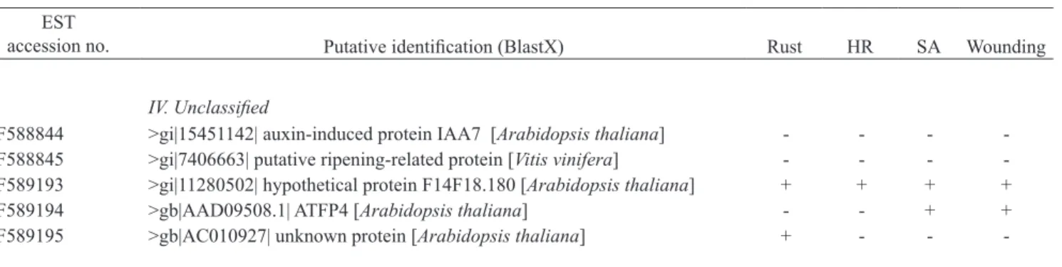
Table 1. List of coffee cDNA sequences tested and summary of gene induction upon biotic and abiotic stresses (-= noninducible; += inducible; n.t.= non tested).
EST
accession no. Putative identification (BlastX) Rust HR SA Wounding
I. Signaling
CF589181 >gi|20521419| Receptor-like kinase [Oryza sativa] + + - + CO773973 >pir||T07079| leucine-rich repeat protein LRP [Lycopersicon esculentum] + - + -CF589186 >gi 9255920| DND1 [Arabidopsis thaliana] + + - -CO773976 >ref|NP_188696.1|| NDR1 [Arabidopsis thaliana] + + + + CAME1003E12 >gb|AAT57642.1| NPR1-NIM1-like protein 1 [Helianthusannuus] - - -
-II. Defence II.1 Stress responses
CF58839 > gi|508296| metallothionein I [Coffea arabica] - - + -CF589183 >gi|15222057| cytochrome P450, putative [Arabidopsis thaliana] + - - -CF589184 >pir|D71417| Cytochrome P450 [Arabidopsis thaliana] + + nt nt CF589185 >gb|AAB97316.1| Cytosolic heat shock 70 protein [Spinacia oleracea] + + - -CF588840 >gi|15241023| Macrophage migration inhibitory factor [Arabidopsis
thaliana]
- - -
-CAH13001B12 >gb|AAT66912.1| dehydration-induced protein RD22-like protein 1 [Gossypium arboreum]
+ - nt nt
II.2. Disease resistance proteins
CF588898 >gb|AAK38727.1| importin alpha 2 [Capsicum annuum] + - - -CF589169 >gi|16974114| putative resistance protein (asc-1 gene) [L. esculentum] + - - -CF588919 >gb|AAC02202.1| resistance protein candidate [Lactuca sativa] - - - -CF588923 >gb|AAF44097.1| disease resistance-like protein [Glycine max] + - - + CF589128 >emb|CAB50786.1| Rx protein [Solanum tuberosum] - - - + CF588996 >gi|14330714| HcrVf1 protein [Malus floribunda] + - +
-II.3. Defense-related proteins
CF588677 >gi|33235471| lipoxygenase [Fragaria x ananassa] + - + + R’-8 >gi|56692180|dbj| 2-hydroxyisoflavanone dehydratase [Glycine max] - - - -CF589058 >ref|NP_176656.1| beta-1.3-glucanase [Arabidopsis thaliana] - - - -CF589187 >gi|4835584| acidic chitinase [Glycine max] + - - + CF588841 >gi|24745591| hydroxyproline-rich glycoprotein [Solanum tuberosum] - - - -CAH13001C11 >dbj|BAC77694.1| lipid transfer protein [Atriplex nummularia] - - nt Nt CO773975 >gb|AAF61647.1| UDP-glucose:salicylic acid glucosyltransferase
[Nicotiana tabacum]
+ + + +
CAME1004C07 >emb|CAA56174.1| Pathogenesis-related protein PR-1 [Medicago truncatula]
+ - + +
III.Transcription factors
CF589189 >gb|AAD55283| Putative AP2 domain transcription factor [Arabidopsis thaliana]
+ + + +
CF588842 >gi|27436378| dehydration-responsive element binding protein [Lycopersicon esculentum]
+ - -
-CO773974 >gb|AAL11009.1| WRKY31 transcription factor [Arabidopsis thaliana] + + + + CF588749 >tr|AAK96200| WRKY44 transcription factor [Arabidopsis thaliana] - - - - CF588951 >gi|22652115| BEL1-related homeotic protein 5 [Solanum tuberosum] - - - -
IV. Unclassified
CF588844 >gi|15451142| auxin-induced protein IAA7 [Arabidopsis thaliana] - - - -CF588845 >gi|7406663| putative ripening-related protein [Vitis vinifera] - - - -CF589193 >gi|11280502| hypothetical protein F14F18.180 [Arabidopsis thaliana] + + + + CF589194 >gb|AAD09508.1| ATFP4 [Arabidopsis thaliana] - - + + CF589195 >gb|AC010927| unknown protein [Arabidopsis thaliana] + - -
-EST
accession no. Putative identification (BlastX) Rust HR SA Wounding
Table 1. Continued
well as gene coding proteins of unknown function (table 1). Transcript accumulation was examined during both compat-ible and incompatcompat-ible rust interactions, and upon salicylic acid (SA) treatment (500 µM) or wounding (leaf cuttings). A lot of genes clearly showed enhanced transcript accumulation in rust inoculated plants (compatible or incompatible interaction), over the time-course experiment as compared with control plants (mock–inoculated). In addition, 9 genes also showed transient enhanced transcript accumulation during the incom-patible interaction (HR) when comparing with the comincom-patible interaction (Fernandez et al., 2004). Gene expression patterns upon abiotic treatments showed that about half of the genes examined also responded to SA or to wounding.
Based on sequence similarity to known genes, 4 out of the 9 HR-upregulated sequences putatively encoded stress-related proteins or components of disease resistance signalling pathways (table 1). Two HR-upregulated sequences matched cytochromes P450 and HSP70. Cytochrome P450 genes may be involved in the biosynthesis of defence-related compounds, such as the Arabidopsis PAD3 gene required for camalexin synthesis in the resistance response to Alternaria brassicicola (Zhou et al., 1999). Recently, Kanzaki et al. (2003) showed that members of the HSP70 and 90 families were essential components of the plant defence signal transduction pathway. With regard to HR-upregulated sequences encoding resistance-signalling components, two coffee sequences best matched the A. thaliana DND1 and NDR1 proteins. The DND1 (defence, no death) protein is a cyclic nucleotide-gated ion channel (AtCNGC2) involved in the HR signalling pathway to P. syringae (Clough et al., 2000). The NDR1 (non race-specific disease resistance) protein is a key component of the signalling pathway of many CC-NBS-LRR resistance proteins (Century et al., 1997). Isolation of ESTs displaying similar expression patterns and sequence homology to DND1 and NDR1 suggest they might have a
potential role in the resistance of C. arabica to H. vastatrix and indicate the conservation of some components of R-gene-mediated resistance signalling pathways in coffee plants.
Finally, 4 other HR-upregulated cDNA clones were of po-tential interest regarding defence mechanisms (table 1). One EST putatively encoded a receptor-like kinase. This class of signal proteins is involved in a diverse array of developmen-tal and defence functions (Du and Chen, 2000; Morris and Walker, 2003). Another gene best matched an UDP-glucose: salicylic acid glucosyltransferase. Glucosyltransferases cata-lyze the transfer of glucose residues to numerous substrates and regulate the activity of compounds that play important roles in plant defence against pathogens, such as salicylic acid (Chong et al., 2002). Two ESTs putatively encoded an AP2-type transcription factor and a WRKY transcription factor. A number of gain-of-function studies have shown the direct implication of several transcription factors in potentiat-ing the plant responses to pathogen infection. Particularly in-volved are several WRKY proteins which are implicated in the regulation of several biological processes, including pathogen defence (Dong et al., 2003; Ülker and Somssich, 2004).
3.1.3.5.3. Time-course of gene induction during coffee rust infection
For the 3 candidate genes tested, enhancement of the mRNA levels was observed at early times of pathogen infection (12-20 hpi). Analysis of time-course experiments showed that the 3 genes were transiently induced during the plant/fungus interaction. Statistically significant differences (P<0.05) in the relative expression were found between the compatible and incompatible interactions for CaR111 and CaWRKY1. CaR111 was induced by the avirulent H. vastatrix strain around 12-16 hpi but weakly activated by the virulent strain. Activation of the CaWRKY1 gene occurred during both interactions, but was higher in the incompatible samples around 16 hpi and higher in the compatible samples at 24 hpi. With regard to the CaNDR1 gene, only a poor induction (< 3-fold) was observed at 12-16 hpi and occurred in response to both virulent and avirulent pathogen (no statistically significant difference). The 3 analyzed genes returned to their basal expression level at 24 hpi for CaR111 and CaNDR1 (figure 1M), and 48 hpi for CaWRKY1 (data not shown).
Coffee gene induction that we observed around 12-18 hpi shows that recognition of the pathogen may occur soon after penetration of the fungus into the substomatal chamber. In several plant-rust interactions, host specific resistance responses are typically expressed concurrently with the formation of the first haustorium (Heath, 1997b; Mould et al., 2003). In the cowpea (Vigna unguiculata) / Uromyces vignae pathosystem, specific upregulation of cowpea genes in the resistant cultivar occurred soon after the penetration of the epidermal cell wall by the fungus (Mould et al., 2003). In the cereal rusts Puccinia graminis tritici and P. graminis avenae, activation of defence–related genes was observed in barley (Rostoks et al., 2004) and in oat when haustorial mother cells (HMCs) were formed in almost all infection sites (Lin et al., 1998). In the coffee–H. vastatrix interaction, the haustorium stage may be reached by H. vastatrix between 24-48 hpi (Martins and Moraes 1996; Silva et al., 1999a, 2002). Cytological observations of resistant coffee leaves revealed that in many infection sites (stomata) the fungus had stopped its growth at a pre-haustorium stage (HMC) (Silva et al., 2002), suggesting that early host resistance responses may be expressed. The gene activation observed around 12-18 hpi may be part of the coffee resistance responses and may determine the outcome of the coffee-rust interaction.
The genes we isolated might therefore participate in the variety of expression of the plant defence responses to H. vastatrix. With the availability of high-density cDNA filters technology, the expression profiles of hundreds ESTs will be monitored simultaneously in several coffee-rust interactions
to help determine the mechanisms of these biological processes. In addition, identification of candidate genes for disease resistance would allow allelic–mining for exploiting coffee genetic resources and the development of molecular markers to assist coffee genetics programmes.
3.2. COFFEE BERRY DISEASE (CBD)
Three species of Colletotrichum have been isolated from coffee berries, leaves and branches: C. kahawae (the only parasitic species, originally designated as C. coffeanum), C. gloeosporioiedes Penz and C. acutatum Simmond (Hindorf, 1970; Masaba and Waller, 1992; Waller et al., 1993). Colletotrichum kahawae infect all stages of the crop from flowers to ripe fruits and occasionally leaves, but maximum crop losses occur following infection of green berries with the formation of dark sunken lesions with sporulation (acervuli) (figures 2A-B), causing their premature dropping and mummification. This disease, first reported in Kenya in1922 (McDonald, 1926) may cause up to 70-80 % of losses if no control measures are adopted (McDonald, 1926; Saccas and Charpentier, 1969; Kibani, 1982; Waller, 1985). Although CBD is so far restricted to Africa, in other coffee-growing areas precautions should be taken to prevent the introduction of this disease.
3.2.1. Fungal infection process
Figures 2A-B. Coffee – C. kahawae interaction. Figure 2A - Durk sunken lesion with sporulation on the green berry, 7 days after inoculation. Figure 2B - Scanning electron micrograph. Acervulus with conidia (c) (x5,500).
cell walls (Garcia et al., 1997; Garcia, 1999; Várzea et al., 2002b, Chen, 2002; Chen et al., 2003, 2004b). Chen (2002) and Chen et al. (2004b) concluded that the C. kahawae appressorium turgor pressure of about 44 bars was 4 times higher that the osmotic pressure of the coffee green berries. On the other hand, unmelanized appressoria induced by the use of the fungicide tricyclazole showed turgor pressures as low as one quarter as melanized ones and, as a consequence, the percentage of infection on coffee leaves and green berries was much lower. They also showed that cutinase was present in C. kahawae conidial mucilage and in extracellular fluids of germinated conidia in vitro and in vivo. The use of diisopropyl fluorophosphates, a cutinase inibitor, totally inhibited cutinase activity of culture filtrates and extracellular fluids but did not prevent infection. Based on these results they concluded that turgor pressure of C. kahawae appressoria might play a major role in coffee cuticle penetration.
Following penetration, Colletotrichum species use two main strategies to successfully colonise host tissues and avoid host defence responses: subcuticular intramural coloni-zation and intracellular colonicoloni-zation (Benhamou et al., 1991; O´Connell andBailey, 1991; Bailey et al., 1992; Pring et al., 1995; Latunde-Dada et al., 1996; O´Connell et al., 1996; Dada et al., 1997; O´Connell et al., 2000; Latunde-Dada, 2001; Mendgen and Hahn, 2002; Diéguez-Uribeondo et al., 2005). For Colletotrichum species exhibiting a subcuticu-lar intramural mode of infection, penetration is followed by growth of hyphae beneath the cuticle and within the periclinal and anticlinal walls of epidermal cells in a necrotropic manner, which involve the massive secretion of cell wall-degrading enzymes. Thus, these pathogens obtain the nutrients from the host cells that they have killed, being classified as necrotrophs (e.g. C. capsici). By contrast, most Colletotrichum species, including C. kahawae (Garcia et al., 1997; Garcia, 1999; Silva et al., 1999b, Várzea et al., 2002b) are hemibiotrophs, as they exhibit a transient post-penetrative asymptomatic biotrophy that is rapidly succeeded by a phase of destructive necrotrophy culminating in the appearance of symptoms of disease and pathogen reproduction. During the symptomless biotrophic phase, the pathogen invades host cells without killing them and feeds on living cells. Subsequently, the pathogen switches to a necrotrophic mode of nutrition, feeding on dead host tis-sues. The intracellular hemibiotrophic Colletotrichum species can be further subdivided in two groups: i) those in which the biotrophic phase extends to many host cells, as for example C. lindemuthianum on Phaseolus vulgaris and C kahawae on Coffee spp., and those in which the biotrophic phase occurs
only inside the initially infected epidermal cell, for example C. destructivum on Medicago sativa. Few Colletotrichum spe-cies (mostly C. gloeosporioides and C. acutatum) are either mutualistic endophytes or quiescent endophytes which resort to necrotrophy after a spell of latency (Prusky and Plumbley, 1992; Latunde-Dada et al., 1999). Endophytic species often penetrate host leaves directly or through stomatal openings and then ramify the mesophyll intercellularly without devel-opment of symptoms. Quiescent species have a prolonged phase of penetrative growth arrested in synchrony with the physiological state of the infected organ.
oculation, depending, respectively, on the greater or lesser degree of aggressiveness of the fungal isolates (Varzea et al., 1999). At this initial stage of the infection process no mac-roscopic symptoms are visible. Transmission electron micro-scope observations showed that the intracellular infection vesicles and hyphae remained external to the plant plasma membrane, which became invaginated around the fungus. The biotrophy was followed by necrotrophic growth. In the latter, the fungus colonization was associated with severe cell wall alterations and death of the host protoplast (Figs 2E-F). In some host cells the cytoplasm was markedly reduced, and appeared often as fine strands in which membranous debris were the only recognizable features of pre-existing organelles. The biotrophic phase was repeated as the fungus started the colonisation of new host cells. Consequently, it was possible to observe hyphal growth simultaneously in dead and living host cells. As shown by light and electron microscopic observations (Garcia et al., 1997; Garcia, 1999 and Várzea et al., 2002b), during the necrotrophic phase of susceptible hypocotyls, green fruits and leaves, callose was present, but only around some intracellular hyphae (figures 2G-H) of epidermal and cortex cells. This host response in the susceptible tissues seemed to occur too late to prevent fungal growth and sporulation (Garcia et al., 1997; Garcia, 1999 and Várzea et al., 2002b).
An evaluation of the activity of several cell wall-degrad-ing enzymes (polygalacturonases, polymetylgalacturonase, pectate lyase, pectin lyase, carboxymethylcellulase) was carried out in extracts of susceptible green berries inoculated with C. kahawae (Chen, 2002; Chen et al., 2004a). The activ-ity of pectate lyase, measurable on the 3rd day after inocula-tion (with the fungus in the biotrophic phase) almost doubled by the 5th day (when necrotic lesions were already observed) and reached the maximum at the 9th day (the last day of the experiment). These results indicate that this enzyme may have a role in the pathogenicity of C. kahawae.
3.2.2. Fungal variability
Research carried out on CBD in Kenya has provided much valuable information. Many different coffee genotypes were tested with local C. kahawae isolates and differential pathogenicity was never observed in this country (van der Vossen et al., 1976). Moreover, differential interactions be-tween host and pathogen population were seldom found in Ethiopia (van der Graaff, 1981). The latter author mentions that the positive effects were, however, small and it is im-probable that they were caused by gene-for-gene specificity.
Many authors agree that the aggressiveness of the pathogen population shows variability between isolates from the same or different geographic origins (Firman and Waller, 1977; Omondi et al., 1997; Masaba and van der Vossen, 1978; van der Vossen, 1985; Manga et al., 1998; Manga, 1999; Varzea et al., 2002b, Varzea et al., 2001).
For the first time, Rodrigues et al. (1992) and Várzea et al. (1993) mention evidence for the existence of physiologic races of C. kahawae. Differential reactions on selections of Sarchimor and Catimor from CATIE were also found by Bieysse et al. (1995). In more exhaustive studies differences were only found in the aggressiveness in C. kahawae isolates (Manga et al., 1998). Omondi et al. (1997, 2000) working with local Kenyan isolates concluded that variation in pathogenic-ity among isolates of C. kahawae was predominantly due to aggressiveness, and differential pathogenicity, although small, was highly significant and could not be ignored. However they did not find positive evidence of physiologic races for C. ka-hawae. Bieysse et al. (unpublished data, INCO-project ICA4-CT-2001-10008), in a study with strains collected in East Africa and Central Africa, concluded that differences between strains were predominantly due to aggressiveness.
Molecular studies carried out in the population of C. kahawae from different geographic origins did not show the existence of polymorphism within this species (Beynon et al., 1995; Sreenivasaprasad and Mills, 1993; Manga et al., 1998; Loureiro et al., 2004, 2005). However, isoenzymatic charac-terisation using PAGE (non-denaturating electrophoresis in polyacrylamide gel) and IEF (isoelectric focusing) techniques showed polymorphism among the isolates studied (Loureiro et al., 2004; Loureiro et al., 2005). Recently, Bieysse et al. (unpublished data - INCO-project ICA4-CT-2001-10008) us-ing microsatellite primers have analysed 140 strains collected from all the geographical zones (Cameroon, Kenya, Burundi, Tanzania, Rwanda, Zimbabwe, Malawi, Angola, Ethiopia). They concluded that the strains studied might be classified into two groups - East African and Cameroon. Each geographical population showed a strong homogeneity between the isolates suggesting a clonal multiplication of the pathogen.
3.2.3. Coffee resistance to CBD
3.2.3.1. Inheritance of resistance
be controlled by major genes on three different loci. The highly resistant variety Rume Sudan carries the dominant R- and the recessive K-genes. The variety Pretoria also has the K-gene. The moderately resistant variety K7 carries only the recessive K-gene. Hibrido de Timor carries one gene for CBD resistance on the T-locus with intermediate gene action. Robinson (1974) and van der Graaff (1981, 1985) suggested that CBD resistance is horizontal/quantitative. Ameha and Belachew (1982) mentioned that 3-5 recessive genes control resistance to CBD in non-introgressed Arabicas.
3.2.3.2. Coffee breeding for resistance
Differences in resistance of coffee trees to CBD are frequently observed under field and laboratory conditions. In Kenya, “Geisha 10” and “Blue Mountain” K7, Rume Sudan, and also some progenies of “Hibrido de Timor” (HDT) have more resistance than “Harar” and “Bourbon”. The high levels of resistance were found in Rume Sudan (Rayner, 1952; Nutman and Roberts, 1960; Firman, 1964; Gibs, 1968; Firman and Waller, 1977; van der Vossen, 1985; van der Vossen et al., 1976; van der Vossen and Walyaro, 1980). The rapid outbreaks of CBD in Eastern Africa prompted some countries to carry out hybridisation programmes with the objective of combining yield with resistance to CBD and coffee leaf rust (CLR) as in Kenya and Tanzania. In 1989, a new research line aimed at supporting breeding programmes on CBD resistance in different coffee- growing countries was initiated at CIFC. Thousand of progenies of different coffee genotypes were tested against CBD isolates from different geographic origins. No coffee genotypes showed 100 % resistance to all the isolates used in the hypocotyl pre-screening tests. However some lines of Rume Sudan showed high levels of resistance to the majority of the studied isolates as well as some derivatives of HDT CIFC 1343. Intermediate levels of resistance can also be found in some derivatives of interspecific tetraploid hybrids from different origins.
In the coffee-growing countries, among progenitors for disease resistance were Hibrido de Timor, Rume Sudan, Kaffa and Geisha which were crossed to varieties SL 28, SL 34, N 39, KP 423 and H 66 (Walyaro, 1983; van der Vossen and Walyaro, 1980, 1981). In Zambia, a pure line, likely derived from the Colombia variety, exhibited a high level of resistance compared with Caturra or Catuai. With these latter varieties, 80 % of crop losses were observed when chemical control was not applied, while losses corresponded only to 15-20 % for the pure line derived from the Colombia variety (C. Hemmings, personal communication). In Tanzania, 8
new clones with high levels of resistance have been selected and multiplied vegetatively by somatic embryogenesis (Teri et al., 2004). In Kenya, the complex hybrid Ruiru 11 revealed a good level of resistance. In Ethiopia (based on ‘the coffee land races programme’ developed by the EARO Institute) results were obtained very quickly and around twenty pure coffee lines displaying good tolerance to CBD, under conditions of strong parasite pressure, were selected and distributed to farmers from 1978 onwards throughout the coffee-growing areas in the country.
3.2.3.3. Cytological and biochemical resistance mechanisms
In Arabica coffees resistance mechanisms to C. kahawae are both preformed and induced, and operate at different stages of pathogenesis (Gichuru, 1997). The coffee berry cu-ticle could act as a physical barrier to the penetrating patho-gen. Nutman and Roberts (1960) among others observed that removal of the epidermis of green coffee berries, in compari-son with unwounded ones, transformed resistant varieties, such as Blue Mountain and Kent, as susceptible as the var. Harar. Wounding allows direct leaching of nutrients to the infection peg and also the direct access of the fungus to the internal tissues (Nutman ad Roberts, 1960; Gichuru, 1997).
According to Masaba and van der Vossen (1982), the resistance to CBD in Arabica coffee may, to a certain extent, be based on the formation of cork barriers. Phellogen was rapidly formed in some cells below the infection site and the progress of the fungal invasion was blocked by a complete barrier of suberised cells. These cork barriers corresponded macroscopically to the scab lesion, the common macroscopic expression of resistance to CBD (Gichuru, 1997). Such a re-sistance mechanism is likely to be stable (race-nonspecific) and depends apparently on actively metabolizing plant tis-sues (Masaba and van der Vossen, 1982; Masaba and Waller, 1992). In fact, cork barrier formation was observed in both attached berries and hypocotyls but not in detached berries, which quickly lost their ability to respond to C. kahawae infection. The formation of a cork barrier is temperature dependent (Masaba, 1982). Differences in cork barrier for-mation in resistant and susceptible varieties were greatest be-tween 19ºC and 22ºC. On the other hand, temperatures below 16ºC and over 28ºC depressed barrier formation.
Biochemical studies showed a significant increase in proteolytic enzymes and in peroxidase activity in coffee varieties after inoculation with C. kahawae (Gichuru, 1993; Gichuru et al., 1997). Peroxidase activity of extracts obtained from non-inoculated hypocotyls and detached and attached berries did not reveal differences between the resistant and susceptible varieties, but the highest increases in enzyme activity were associated with susceptibility rather than with resistance (Gichuru et al., 1997).
Histological studies made with resistant hypocotyls of Hibrido de Timor (HDT) derivatives, in comparison with those of the susceptible cultivar Caturra revealed no differences in conidia germination and in differentiation of melanised ap-pressoria, but the hyphal length inside the host tissues was significantly lower in the resistant genotypes, from 41h after inoculation. (Silva et al., 1999b; Várzea et al., 2002b). In the resistant coffee genotypes the fungal hyphae were confined to epidermal cells or to the first layers of the cortex cells. At these sites, signs of incompatibility were observed: (i) hypersensi-tive host cell death (monitored by cell autofluorescence and/or browning); (ii) modifications in the cell walls (thickness and autofluorescence) and (iii) early accumulation of phenolic compounds (indicated by the epifluorescence test) (figures 2I-K). These host responses corresponded macroscopically to the scab lesion. The same pattern of responses was observed in resistant hypocotyls of C. racemosa (Silva et al., unpublished data), as well as in C. canephora, in which no symptoms were detected in the majority (>90 %) of the hypocotyls tested (Loureiro et al., unpublished data).
The rapid hypersensitive death of host infected cells is typical of incompatible interactions of C. lindemuthianum with P.vulgaris and Vigna unguiculata, C. trifolii with Medicago sativa and Medicago truncatula and, some interactions involving C. gloeosporioides and Stylosanthes spp. (O´Connell and Bailey, 1986; Trevorrow et al., 1988; Bailey et al., 1990; Mould et al., 1991; Torregrosa et al., 2004). In the bean anthracnose pathosystem, this early HR does not occur in the compatible interactions, thereby indicating a high correlation between the HR and the race-cultivar specificity (Esquerré-Tugayé et al., 1992). The host cell wall modifications, namely the increase in the levels of hydroxyproline-rich glycoproteins (HRGPs), as well as the early accumulation of phenolic compounds in the infected host cells have also been associated with host resistance to Colletotrichum spp. (Esquerré-Tugayé et al., 1992; Skipp et al., 1995; Torregrosa et al., 2004). In the M. truncatula - C. trifolli incompatible interaction, one of the earlier phenolic compounds accumulated is the phytoalexin medicarpin, which is synthesized through the mobilization of several enzymes of the phenylpropanoid pathway. Several genes of this pathway were induced in the resistant varieties before the maximum level of autoflourescence (indicative of the accumulation of phenolic compounds) (Torregrosa et al., 2004). The identification of the phenolic compounds involved in the coffee - C. kahawae interaction is currently under study and, preliminary results revealed an accumulation of flavonoides and hydroxycinnamic acid derivatives at infection sites. These compounds were detected earlier in resistant and partially resistant genotypes than in susceptible ones.