AR
TIGO ORIGINAL / ORIGINAL AR
TICLE
INTRODUCTION
Gastrointestinal polyps are frequent in children and adolescentes, given that, historically, the majority are considered solitary, located in the rectosigmoid colon, presenting a juvenile hamartomatous histology with a minimal risk of developing into a malignant tumor(1). However, with the progress in endoscopic
techniques, a change in the comprehension of co-lonic polyps has occurred. The application of the pancolonoscopy has demonstrated that, though the colonic polyps can more commonly be found in the rectosigmoid colon, these can also be found along the entire large intestine; multiple, with the possibility of recurrence in some children; and associated with pre-malign alterations(26).
CLINICAL, EPIDEMIOLOGIC, AND
ENDOSCOPIC PROFILE IN CHILDREN AND
ADOLESCENTS WITH COLONIC POLYPS IN
TWO REFERENCE CENTERS
Denise O
ANDRADE
1, Alexandre Rodrigues
FERREIRA
1,
Paulo Fernando S
BITTENCOURT
1, Daniela F P
RIBEIRO
1, Rodrigo Gomes da
SILVA
1and Luiz Ronaldo
ALBERTI
2Received 8/6/2015 Accepted 22/6/2015
ABSTRACT-Background- The main goal of this paper is to investigate the frequency, clinical proile, and endoscopic indings of children and teenagers submitted to colonoscopies. Methods- Patients of below 18 years of age, diagnosed with polyps by means of colonoscopies at two reference centers of pediatric endoscopy were followed-up between 2002 and 2012. The clinical variables evaluated in this study included: gender, recommendation of colonoscopy, associated signs and symptoms, age of onset of symptoms, age at identiication of the polyp, interval of time between the onset of symptoms and the endoscopic diagnosis of colonic polyps, and family history of intestinal polyposis and/or colorectal cancer. The characteristics of the polyps also included: number, mor-phological type, histology, and distribution. Polyposis syndromes were also investigated. Results- From the 233 patients submitted to colonoscopies, polyps were found in 74 (31.7%) patients, with a median age of 6.6 years, of which 61% were male. Juvenile polyps were identiied in 55 (74%) patients, with 7 (9%) characterized within the criteria for juvenile polyposis. Patients with intestinal polyposis syndromes were diagnosed in 35% of the patients. The most frequent clinical presentation was hematochezia. Abdominal pain with acute episodes of intestinal partial obstruction or intussusception with emergency laparotomy was observed in the majority of Peutz-Jeghers syndrome patients leading to an increased morbidity. Conclusions- Even though juvenile colonic polyps are the most frequent type of diagnosed polyps, the present study identiied a signiicant level of children with polyposis syndromes (35%), associated with a higher morbidity of these individuals.
HEADINGS- Colonic polyps. Child. Adenomatous polyposis. Peutz-Jeghers syndrome. Colonoscopy.
Declared conflict of interest of all authors: none
Disclosure of funding: Fundação de Amparo à Pesquisa do estado de Minas Gerais - FAPEMIG.
1 Departamento de Pediatria, Universidade Federal de Minas Gerais, Belo Horizonte, MG; 2 Departamento de Cirurgia, Universidade Federal de Minas Gerais, Belo Horizonte, MG, Brasil.
Correspondence: Prof. Luiz Ronaldo Alberti. Departamento de Cirurgia, Universidade Federal de Minas Gerais. Av. Prof. Alfredo Balena, 190 - Santa Efigênia - CEP: 30130-000 - Belo Horizonte, MG, Brasil. E-mail: [email protected]
Reports in prior literature describe the occurrence of multiple polyps in 20%-50% of the children submitted to a colonoscopy with polypectomy(3, 9, 14, 15, 22-24, 26), in which
10%-50% of the polyps can be found near the sigmoid colon(17, 23, 27, 31), while 3%-15% can be found exclusively
in the right colon (including the transverse colon, the ascending colon, and the cecum)(3, 9, 17). Children with
multiple juvenile polyps, especially those with Juvenile Polyposis syndrome (JPS), can develop adenomatous transformations or even carcinomas in situ in the juve-nile polyp in 26%-47% of the patients(5, 15, 23). The
age range, in addition to the increased risk of colorectal cancer and various types of cancer in other organs. Thus, laws in the correct evaluation of these patients can lead to improper treat-ment. In this context, the absence of screening and check-ups on the part of at-risk individuals can lead to a diminishing of one’s quality of life and early death.
Few studies can be found in literature regarding pediatric case series on polyps and intestinal polyposis. The present study aims to determine frequency, clinical characteristics, and colonoscopic indings in children and adolescents sub-mitted to colonoscopies at two references centers in Gas-troenterology and Pediatric Endoscopy in the city of Belo Horizonte, Brazil.
METHODS
This study was carried out in accordance with recommen-dations from the Declaration of Helsinki and the Resolution 196/96 from the Brazilian Ministry of Health concerning research involving human beings, and was approved by the Research and Ethics Committees from the Hospital Foun-dation of the State of Minas Gerais (protocol number 016-B/2010) and from the Federal University of Minas Gerais (UFMG) (CAAE Project– 01882612.3.1001.5149).
This paper is a descriptive study of children and adoles-cents of up to 18 years of age who have been diagnosed with colonic polyps, identiied though colonscopy and in medical procedures carried out in the Pediatric Endoscopy Services of the João Paulo II Children’s Hospital from the Hospital Foundation of the State of Minas Gerais and the Alfa Insti-tute of Gastroenterology from the UFMG Clinical Hospital, during the period of January 2002 to December 2012.
The data were retrieved from patient medical records, registries of endoscopic and histological reports, as well as interviews with the families during doctor’s appointments or by telephone contact with the parents or responsible guardians. Patients diagnosed with pseudopolyps caused by intestinal inlammatory disease or polyps of infectious etiology, as well as those whose medical procedures and search for data were incomplete were excluded.
The clinical variables evaluated in this study included: gender, indication for the procedure , associated signs and symptoms, age of onset of symptoms, age at identiication of the polyp, interval of time between the onset of symp-toms and the endoscopic diagnosis of colonic polyps, and family history of intestinal polyposis and/or colorectal cancer (CRC).
Regarding the characteristics of the polyps, this study described: the number of polyps viewed by colonoscopy, morphological (sessile/pediculated) and histological types, and distribution along the large intestine. The anatomic distribution of the polyps was recorded as rectal, sig-moid, descending colon, left colon (when located up to the splenic lexure, with no speciication of the location), right colon (when located proximal to the splenic lexure, with no speciication of the location), transverse colon, ascending colon and cecum, pancolonic if located both
proximally and distally to the splenic lexure, and ileum. The juvenile hamartomatous polyps were categorized as solitary and multiple (between two and ive polyps). The Juvenile Polyposis syndrome (JPS) was defined by the presence of at least one of the following characteristics: more than ive colorectal juvenile hamartomatous polyps, the presence of these polyps along the gastrointestinal tract, and the presence of any number of colorectal juvenile polyps associated with a family history of juvenile polyposis. The Peutz-Jeghers syndrome (PJS) was defined by the presence of one of the following criteria: two or more polyps conirmed histologically (hamartomatous-type polyp with an extension branching from the smooth muscles throughout the polyp); any number of typical polyps from the PJS confirmed histologically in an individual with a positive family history of PJS (irst-degree relatives), mucocutaneous pigmentations that are characteristic of an individual with a positive family history of PJS (irst-degree relatives), any number of histologically conirmed polyps from the PJS in an individual who presents characteristic mucocutaneous pigmentations. The Familial Adenomatous Polyposis (FAP) Syndrome was deined by the presence of multiple adenomatous colorectal polyps despite its size and location throughout the large bowel.
The data banks were created in the SPSS for Windows program. The descriptive analysis, including the average, median, standard deviation, interquartile range, and per-centages, was used to characterize the studied group. The continuous variables with no normal distribution were expressed through medians and an interquartile range of 25%-75% (IQ25-75%). The continuous variables with normal distribution were expressed through averages and standard deviation. Differences were considered significant for values corresponding to P<0.05.
RESULTS
During the period of the study, 233 patients were submit-ted to colonoscopies and 74 (31.7%) children and adolescents were diagnosed with colorectal polyps. Patients with no polyps were submitted to only one colonoscopy. Among a total of 158 colonoscopies and 537 polypectomies carried out in 74 children diagnosed with polyps (non-inlammatory or infectious), complications were found in one patient, who had undergone a polypectomy, with a possible perforation. In this case, endoscopic therapy was successful with the use of an endoclip and antibiotics.
The age of the patients when diagnosed with a polyp presented a median of 6.6 years (IQ25-75% - 3.9-10.7 years); 45 (61%) of the patients were male.
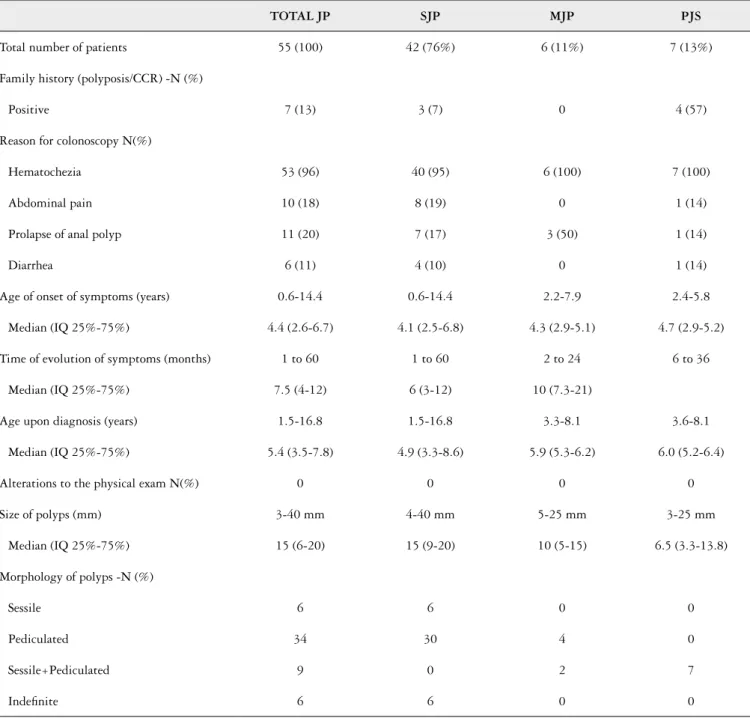
A predominance of larger and pediculated polyps could be observed in patients with solitary juvenile polyps, occur-ring most commonly in younger patients, as compared to those with polyposis/multiple polyps (Table 3).
PJS was found in six patients. The clinical characteristics and treatment of these patients is summarized in Tables 2 and 3. Three patients (50%) were submitted to emergency laparotomy due to intestinal intussusception. The histo-logical evaluation of the dry colonic polyps identiied both Peutz-Jeghers hamartomatous polyps and hyperplasic
TABLE 1. Clinical and endoscopic characteristics of the patients with juvenile polyps
TOTAL JP SJP MJP PJS
Total number of patients 55 (100) 42 (76%) 6 (11%) 7 (13%)
Family history (polyposis/CCR) -N (%)
Positive 7 (13) 3 (7) 0 4 (57)
Reason for colonoscopy N(%)
Hematochezia 53 (96) 40 (95) 6 (100) 7 (100)
Abdominal pain 10 (18) 8 (19) 0 1 (14)
Prolapse of anal polyp 11 (20) 7 (17) 3 (50) 1 (14)
Diarrhea 6 (11) 4 (10) 0 1 (14)
Age of onset of symptoms (years) 0.6-14.4 0.6-14.4 2.2-7.9 2.4-5.8
Median (IQ 25%-75%) 4.4 (2.6-6.7) 4.1 (2.5-6.8) 4.3 (2.9-5.1) 4.7 (2.9-5.2)
Time of evolution of symptoms (months) 1 to 60 1 to 60 2 to 24 6 to 36
Median (IQ 25%-75%) 7.5 (4-12) 6 (3-12) 10 (7.3-21)
Age upon diagnosis (years) 1.5-16.8 1.5-16.8 3.3-8.1 3.6-8.1
Median (IQ 25%-75%) 5.4 (3.5-7.8) 4.9 (3.3-8.6) 5.9 (5.3-6.2) 6.0 (5.2-6.4)
Alterations to the physical exam N(%) 0 0 0 0
Size of polyps (mm) 3-40 mm 4-40 mm 5-25 mm 3-25 mm
Median (IQ 25%-75%) 15 (6-20) 15 (9-20) 10 (5-15) 6.5 (3.3-13.8)
Morphology of polyps -N (%)
Sessile 6 6 0 0
Pediculated 34 30 4 0
Sessile+Pediculated 9 0 2 7
Indeinite 6 6 0 0
SJP: solitary juvenile polyp; MJP: multiple juvenile polyp; PJS: Peutz Jeguers syndrome; N: number of patients.
polyps. The gastric polyps presented a varied topographic distribution and a predominance of the hyperplasic type when submitted to histology (Figure 1). All of the polyps resected by enteroscopy presented a typically Peutz-Jeghers type of hamartomatous histology.
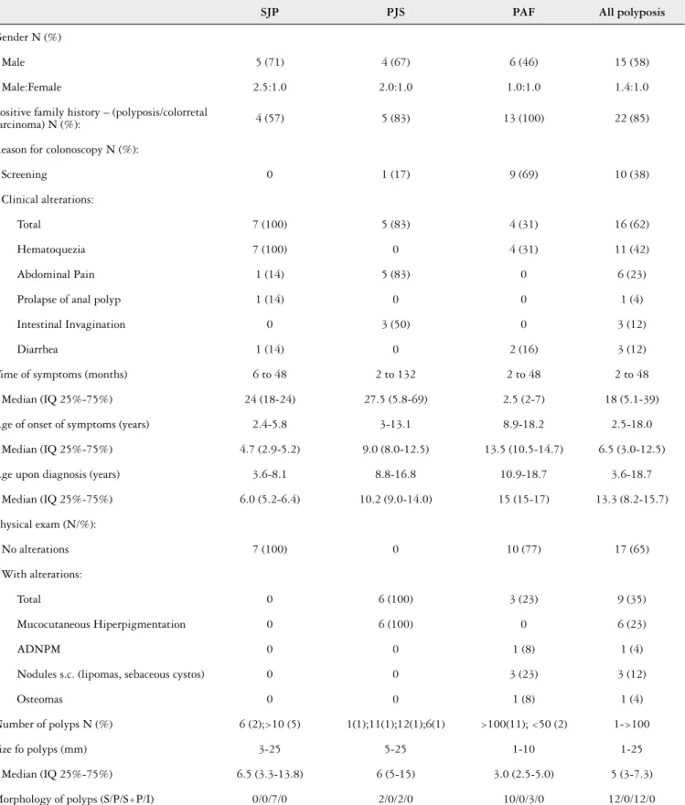
TABLE 2. Clinical and endoscopic characteristics of patients with polyposis
SJP PJS PAF All polyposis
Gender N (%)
Male 5 (71) 4 (67) 6 (46) 15 (58)
Male:Female 2.5:1.0 2.0:1.0 1.0:1.0 1.4:1.0
Positive family history – (polyposis/colorretal
carcinoma) N (%): 4 (57) 5 (83) 13 (100) 22 (85)
Reason for colonoscopy N (%):
Screening 0 1 (17) 9 (69) 10 (38)
Clinical alterations:
Total 7 (100) 5 (83) 4 (31) 16 (62)
Hematoquezia 7 (100) 0 4 (31) 11 (42)
Abdominal Pain 1 (14) 5 (83) 0 6 (23)
Prolapse of anal polyp 1 (14) 0 0 1 (4)
Intestinal Invagination 0 3 (50) 0 3 (12)
Diarrhea 1 (14) 0 2 (16) 3 (12)
Time of symptoms (months) 6 to 48 2 to 132 2 to 48 2 to 48
Median (IQ 25%-75%) 24 (18-24) 27.5 (5.8-69) 2.5 (2-7) 18 (5.1-39)
Age of onset of symptoms (years) 2.4-5.8 3-13.1 8.9-18.2 2.5-18.0
Median (IQ 25%-75%) 4.7 (2.9-5.2) 9.0 (8.0-12.5) 13.5 (10.5-14.7) 6.5 (3.0-12.5)
Age upon diagnosis (years) 3.6-8.1 8.8-16.8 10.9-18.7 3.6-18.7
Median (IQ 25%-75%) 6.0 (5.2-6.4) 10.2 (9.0-14.0) 15 (15-17) 13.3 (8.2-15.7)
Physical exam (N/%):
No alterations 7 (100) 0 10 (77) 17 (65)
With alterations:
Total 0 6 (100) 3 (23) 9 (35)
Mucocutaneous Hiperpigmentation 0 6 (100) 0 6 (23)
ADNPM 0 0 1 (8) 1 (4)
Nodules s.c. (lipomas, sebaceous cystos) 0 0 3 (23) 3 (12)
Osteomas 0 0 1 (8) 1 (4)
Number of polyps N (%) 6 (2);>10 (5) 1(1);11(1);12(1);6(1) >100(11); <50 (2) 1->100
Size fo polyps (mm) 3-25 5-25 1-10 1-25
Median (IQ 25%-75%) 6.5 (3.3-13.8) 6 (5-15) 3.0 (2.5-5.0) 5 (3-7.3)
Morphology of polyps (S/P/S+P/I) 0/0/7/0 2/0/2/0 10/0/3/0 12/0/12/0
DISCUSSION
Gastrointestinal polyps are common in the pediatric age group, with lesions presenting juvenile harmatomatous histol-ogy identiied in 88%-100% of the polyps removed by colons-copy(3, 9, 24, 30). The polyposis syndromes are quite rare, varying
between 12% and 17%(14, 19, 21), while the JPS is predominant in
the majority of pediatric cases(9, 19, 21, 30). The greater prevalence
in this paper is due to the fact that the two centers where the exams were carried out are reference centers for the treatment of complex pediatric diseases. The predominance of males in this series (61%) is in accordance with the majority of prior publications(14, 17, 21, 25, 26), with the tendency of solitary juvenile
polyps occurring at earlier ages as compared to children with multiple polyps or juvenile polyposis(9, 17, 19, 21, 24, 26, 32).
It could be observed that the majority of children with juvenile polyps, presented hematochezia associated or not with other symptoms, including the prolapse of the rectal mucous membrane or an anal polyp, abdominal pain, and diarrhea, which is similar to indings from other pediatric case series(9, 31).
This study identiied no signs of protein-losing enteropathy, which commonly presents a worse prognosis and mortality at early ages, generally in children of less than ive years of age(33).
In the majority of patients with JPS, the irst symptoms and complications appear in the irst two decades of life(8, 34, 37).
Abdominal pain associated or not with an acute obstructive condition is the most common symptom and was present in 93%-100% of the patients, with one case reported in the neo-natal period(6). Utsunomiya et al. observed an acute obstructive
condition of the abdomen in 43% of the patients(34), which is
similar to other case series(2, 8, 37), and could also be observed in
this sample. The mucocutaneous hyperpigmentation, present in 95% of the patients with JPS(13), appears most commonly in the
irst two years of life, tapering off at puberty, with a tendency of disappearing in adulthood(34, 37). In the present study, one
patient presented these symptoms as of the irst month of life. In the diagnosis of adenomatous polyps in this case series, the patients were asymptomatic in most cases and were submitted to a colonoscopy due to a family history of PAF, which is in accordance with indings reported in the literature(12, 28). Patients with PAF were asymptomatic within
the pediatric age group, with only rare symptomatic cases before the second decade of life(10, 11),when the main
mani-festation appears as intermittent rectal bleeding, followed by abdominal pain, anemia, and diarrhea, which is similar to that found in the present case series. Benign extracolonic manifestations, such as congenital hypertrophy of the retina, epidermoid cysts, and osteomas, may also be present(15, 35), a
fact also proven in the present case series. Desmoid tumors can be found in 10%-15% of the patients with PAF(4, 18).
Al-though these are histologically benign lesions, they can be aggressive and dificult to cure, with signiicant morbidity and mortality rates.
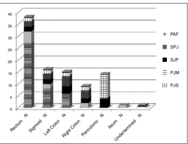
Although the majority of polyps in children appeared in a distal location, the frequency of the proximal location varied from 5%-32%(1, 3, 9, 14, 17, 24, 27, 32). In the present study, polyps
located proximal do the splenic lexure was found in 35% of the cases.
In the JPS, the hamartomatous polyps, which are most common in the colorectal region, predominate, a fact which could be observed in this study, which are also associated with hyperplasic polyps. Hofting et al.(20) reported the involvement
of the large intestine in 98%, the stomach in 14%, the jejunum and ileum in 7%, and the duodenum in 2% of the patients. In rare cases, these can also occur in an isolated form in the stomach in the absence of colonic polyposis(36). The risk of
cancer in patients with juvenile polyps, mainly in patients with multiple polyps, including the presence of adenomatous alterations in the juvenile polyp (26%-47%) and the coexist-ence of adenomas with no characteristics of the juvenile polyp TABLE 3. Comparison of patients with solitary juvenile polyps and the
others (multiple juvenile polyps and polyposis)
SJP Non SJP
Age of onset of symptoms (years) 0.8-12 2.2-18.2
Median (IQ 25%-75%) 4.2 (2.8-6.2) 6.2 (4.5-12.4)
Age upon diagnosis (years) 1.5-16.8 3.7-18.7
Median (IQ 25%-75%) 4.9 (3.3-8.6) 11.3 (6.7-11.5)
Size of polyps (mm) 4-40 MM 1-25 MM
Median (IQ 25%-75%) 15 (9-20) 5 (3-10)
Morphology of polyps (N)
Sessile 6 13
Pediculated 31 4
Sessile + Pediculated 0 11
Indiscriminate 6 3
SJP: solitary juvenile polyp; MJP: multiple juvenile polyp; PJS: polyposis juvenile syndrome; N: number of patients.
SPJ: solitary juvenile polyp; PJM: multiple juvenile polyp; SJP: polyposis juvenile syndrome; PJS: Peutz-Jeghers syndrome; FAP: familial adenomatous polyposis.
FIGURE 1. Patients and distribution of polyps according to topography
40 35 30 25 20 15 10 5 0
N N N N N N N
Rectum Sigmoid Left C
or even of adenocarcinoma associated with the presence of juvenile polyp (2%-15%)(15), has been reported. In the group
of patients with JPS in this study, no dysplastic alterations nor cases of adenomas or adenocarcinomas could be observed.
In the individuals with PJS, the polyps can be detected in 88% of the patients distributed along the entire diges-tive tract, with the exception of the esophagus(34), located
predominantly in the small intestine (65%-97%) and colon (50%-64%), and with a greater prevalence in the stomach (24%-38%), duodenum (37%), and rectum (32%)(2, 8, 23, 34).
Other extra-intestinal sites, present in up to 5.5% of the patients, include the gallbladder, the respiratory tract, the urinary bladder, and the ureter(34). In the present case series,
hamartomatous polyps, which are typical of the syndrome, were detected in all of the patients in the colon, small intes-tine, and gastroduodenal topographies.
In patients with PAF, the classic phenotypical manifesta-tion is that of innumerous adenomatous colonic polyps, which, by deinition, are more than 100, but can reach thousands, distributed along the entire gastrointestinal tract, commonly detected at an average age of 15(1), reaching a 90% prevalence
in patients of 40 years of age(1). However, in 8%-15% of the
patients, the syndrome can appear in a lighter form, with a smaller number of colonic adenomas. The present study ob-served that 13 patients presented innumerous adenomatous polyps (more than 100) in a pancolonic distribution, while two (15%) patients presented a lighter phenotypical manifestation of the disease. All of the polyps were less than 1 cm, with a median size of 3 mm, with a tubular or tubular-villous histol-ogy, which was in accordance with the literature(1).
The polyps can also develop in the upper gastrointestinal tract, being the fundic gland polyps in the stomach the most common, both in children and in adults(1). The duodenum is
the second most common location for the development of adenomas in patients with PAF, occurring in 90%-100% of the patients throughout their lifetimes(5, 35). The patients submitted
to upper endoscopy in this case series presented gastroduode-nal alterations, which was in accordance with prior reports.
The risk of malign transformation in a solitary juvenile polyp appears to be insigniicant. Nugent, in a review of 82 patients with solitary juvenile polyp, observed no increased risk of cancer or greater mortality related to the polyp(29). However,
considerations regarding the risk of cancer in patients with
juvenile polyps have been reported concerning those patients with multiple polyps, in which the presence of adenomatous alterations in the juvenile polyp (26%-47% of the patients with JPS), the coexistence of adenomas with no characteristics of juvenile polyps, or even of adenocarcinoma associated with the presence of juvenile polyps (2%-15% of the cases of JPS) have been reported(37). Moreover, the patient must be aware that
polyps can occasionally appear in the context of a hereditary polyposis disease characterized by signiicant morbidity in this age group and with an increased risk of cancer in the gastro-intestinal tract and in other organs, especially in adulthood(16).
Based on these data, the majority of specialists currently recommend an initial investigation with a complete colo-noscopy in all children suspected of having colonic polyps. All polyps should be removed and submitted to histological studies. Upon obtaining the endoscopic and histological data (number, distribution, and histological type), associated with the information relevant to the family history of polyposis and/or CCR, as well as the clinical history of the patient, the doctor can make more precise decisions concerning the therapy and follow-up to for the patients and at-risk family members.
CONCLUSION
Although the solitary juvenile colonic polyps are the most common, a signiicant percentage of children with polyposis syndromes (35%), associated with a greater morbidity could be observed. In this light, the establishment of diagnostic pro-tocols and effective follow-up treatment with affected patients and at-risk family members is of utmost importance.
ACKNOWLEDGEMENTS
The authors gratefully acknowledge the Foundation for the Assistance to Research of Minas Gerais State (FAPEMIG) for inancial support.
Authors’ contributions
REFERENCES
1. Attard TM, Cuffari C, Tajouri T, Stoner JA, Eisenberg MT, Yardley JH, et al. Multicenter experience with upper gastrointestinal polyps in pediatric patients with familial adenomatous polyposis. Am J Gastroenterol. 2004;99(4):681-6. 2. Bartholomew LG, Moore CE, Dahlin DC, Waugh JM. Intestinal polyposis
associated with mucocutaneous pigmentation. Surg Gynecol Obstet. 1962;115:1-11.
3. Bartnik W, Butruk E, Ryzko J, Rondio H, Rasinski A, Orlowska J. Short- and long-term results of colonoscopic polypectomy in children. Gastrointest Endosc. Dec. 1986;32(6):389-92.
4. Bertario L, Russo A, Sala P, et al. Genotype and phenotype factors as determinants of desmoid tumors in patients with familial adenomatous polyposis. Int J Cancer. 2001;95(2):102-7.
5. Bulow S, Bjork J, Christensen IJ, et al. Duodenal adenomatosis in familial adenomatous polyposis. Gut. 2004;53(3):381-6.
6. Burgmeier C, Schier F, Staatz G. Gastric outlet obstruction in a neonate because of Peutz-Jeghers syndrome. J Pediatr Surg. 2012;47(8):e1-3.
7. Campos FG, Habr-Gama A, Kiss DR, Atui FC, Katayama F, Gama-Rodrigues J. [Extracolonic manifestations of familial adenomatous polyposis: incidence and impact on the disease outcome]. Arq Gastroenterol. 2003;40(2):92-8.
8. Choi HS, Park YJ, Youk EG, et al. Clinical characteristics of Peutz-Jeghers syndrome in Korean polyposis patients. Int J Colorectal Dis. 2000;15(1):35-8.
Andrade DO, Ferreira AR, Bittencourt PFS, Ribeiro DFP, Silva RG, Alberti LR. Peril clínico, epidemiológico e endoscópico em crianças e adolescentes com pólipos colônicos em dois centros de referência. Arq Gastroenterol. 2015,52(4):xxx.
RESUMO - Objetivos - Conhecer a frequência, o peril clínico, os achados endoscópicos, de crianças e adolescentes submetidos à colonoscopia em dois centros de referência em gastroenterologia e endoscopia pediátrica. Métodos- Foram avaliados e acompanhados pacientes com idade menor ou igual a 18 anos com diagnóstico de pólipos identiicados à colonoscopia em dois centros de referência em endoscopia pediátrica no período de 2002 a 2012. As variáveis clínicas avaliadas foram: gênero, indicação da colonoscopia, sinais e sintomas associados, idade de início dos sintomas, idade à identiicação do pólipo, intervalo de tempo entre início dos sintomas e diagnóstico endoscópico do pólipo colônico, história familiar de polipose intestinal e/ou câncer coloretal. Em relação às características dos pólipos foram descritos: número, tipo morfológico, histológico e distribuição. Foram estudadas também as síndromes poliposas (síndrome de Peutz-Jeghers, síndrome juvenil poliposa, síndrome poliposa adenomatosa familiar). Resultados- Dos 233 pacientes submetidos à colonoscopia, foram encontrados 74 (31,7%) pacientes com pólipos, com mediana de idade de 6,6 anos, 61% do gênero masculino. Pólipos juvenis foram identiicados em 55 (74%) dos pacientes, sendo 7 (9%) com critérios diagnósticos de polipose juvenil. Pacientes com síndromes poliposas intestinais foram diagnosticados em 35% dos pacientes. Destes, 12% com diagnóstico de polipose adenomatosa familiar, 9% com síndrome juvenil poliposa e 8% com diagnóstico de Síndrome de Peutz-Jeghers. A apresentação clínica mais frequente foi o sangramento retal indolor. Nos pacientes com polipose adenomatosa familiar o principal motivo da indicação da colonoscopia foi para rastreamento da doença devido história familiar da síndrome poliposa. Um paciente apresentou adenocarcinomacoloretal, simultâneo ao diagnóstico da polipose adenomatosa aos 15 anos de idade. Dor abdominal com episódios agudos de semiobstrução ou intussuscepção intestinal com laparotomia de urgência foi observado nos pacientes com Peutz-Jeghers. Conclusões - Embora os pólipos colônicos juvenis sejam os mais frequentemente diagnosticados, foi observado um percentual signiicativo de crianças com síndromes poliposas (35%) associado a uma maior morbidade destas crianças. Desta forma concluímos ser importante estabelecimento de um protocolo de diagnóstico e seguimento dos pacientes afetados e familiares de risco.
DESCRITORES - Pólipos do colo. Criança. Polipose adenomatosa. Síndrome de Peutz-Jeghers. Colonoscopia.
9. Cynamon HA, Milov DE, Andres JM. Diagnosis and management of colonic polyps in children. J Pediatr. 1989;114(4 Pt 1):593-6.
10. Dukes CE. Cancer control in familial polyposis of the colon. Dis Colon Rectum. 1958;1(6):413-23.
11. Dukes CE. Familial intestinal polyposis. Ann Eugen. 1952;17(Part 1):1-29. 12. Durno CA. Colonic polyps in children and adolescents. Can J Gastroenterol.
2007;21(4):233-9.
13. Fernandez Seara MJ, Martinez Soto MI, Fernandez Lorenzo JR, Trabazo S, Gamborino E, Forteza Vila J. Peutz-Jeghers syndrome in a neonate. J Pediatr. 1995;126(6):965-7.
14. Fox VL, Perros S, Jiang H, Goldsmith JD. Juvenile polyps: recurrence in patients with multiple and solitary polyps. Clin Gastroenterol Hepatol. 2010;8(9):795-9. 15. Giardiello FM, Hamilton SR, Kern SE, et al. Colorectal neoplasia in juvenile
polyposis or juvenile polyps. Arch Dis Child. 1991;66(8):971-5.
16. Giardiello FM, Offerhaus GJ, Traboulsi EI, et al. Value of combined phenotypic markers in identifying inheritance of familial adenomatous polyposis. Gut. 1991;32(10):1170-4.
17. Gupta SK, Fitzgerald JF, Crofie JM, et al. Experience with juvenile polyps in North American children: the need for pancolonoscopy. Am J Gastroenterol. 2001;96(6):1695-7.
18. Gurbuz AK, Giardiello FM, Petersen GM, et al. Desmoid tumours in familial adenomatous polyposis. Gut. 1994;35(3):377-81.
20. Hofting I, Pott G, Stolte M. [The syndrome of juvenile polyposis]. Leber Magen Darm. 1993;23(3):107-8, 111-102.
21. Hood B, Bigler S, Bishop P, et al. Juvenile polyps and juvenile polyp syndromes in children: a clinical and endoscopic survey. Clin Pediatr (Phila). 2011;50(10):910-5. 22. Hueting WE, Buskens E, van der Tweel I, Gooszen HG, van Laarhoven CJ.
Results and complications after ileal pouch anal anastomosis: a meta-analysis of 43 observational studies comprising 9,317 patients. Dig Surg. 2005;22(1-2):69-79. 23. Hyer W, Beveridge I, Domizio P, Phillips R. Clinical management and genetics of gastrointestinal polyps in children. J Pediatr Gastroenterol Nutr. 2000;31(5):469-79. 24. Jalihal A, Misra SP, Arvind AS, Kamath PS. Colonoscopic polypectomy in
children. J Pediatr Surg. 1992;27(9):1220-2.
25. Ko FY, Wu TC, Hwang B. Intestinal polyps in children and adolescents--a review of 103 cases. Zhonghua Min Guo Xiao Er Ke Yi Xue Hui Za Zhi. 1995;36(3):197-202.
26. Mestre JR. The changing pattern of juvenile polyps. Am J Gastroenterol. 1986;81(5):312-4.
27. Mougenot JF, Baldassarre ME, Mashako LM, Hanteclair GC, Dupont C, Leluyer B. [Recto-colic polyps in the child. Analysis of 183 cases]. Arch Fr Pediatr. 1989;46(4):245-8.
28. Munck A, Gargouri L, Alberti C, et al. Evaluation of guidelines for management of familial adenomatous polyposis in a multicenter pediatric cohort. J Pediatr Gastroenterol Nutr. 2011;53(3):296-302.
29. Nugent KP, Spigelman AD, Phillips RK. Life expectancy after colectomy and ileorectal anastomosis for familial adenomatous polyposis. Dis Colon Rectum. 1993;36(11):1059-62.
30. Perisic VN. Colorectal polyps: an important cause of rectal bleeding. Arch Dis Child. 1987;62(2):188-9.
31. Pillai RB, Tolia V. Colonic polyps in children: frequently multiple and recurrent. Clin Pediatr (Phila). 1998;37(4):253-7.
32. Poddar U, Thapa BR, Vaiphei K, Singh K. Colonic polyps: experience of 236 Indian children. Am J Gastroenterol. 1998;93(4):619-22.
33. Sachatello CR, Griffen WO, Jr. Hereditary polypoid diseases of the gastrointestinal tract: a working classiication. Am J Surg. 1975;129(2):198-203.
34. Utsunomiya J, Gocho H, Miyanaga T, Hamaguchi E, Kashimure A. Peutz-Jeghers syndrome: its natural course and management. Johns Hopkins Med J. 1975;136(2):71-82.
35. Valanzano R, Cama A, Volpe R, et al. Congenital hypertrophy of the retinal pigment epithelium in familial adenomatous polyposis. Novel criteria of assessment and correlations with constitutional adenomatous polyposis coli gene mutations. Cancer. 1996;78(11):2400-10.
36. Watanabe A, Nagashima H, Motoi M, Ogawa K. Familial juvenile polyposis of the stomach. Gastroenterology. 1979;77(1):148-51.