PhD Thesis in Medicine
(Sub-specialization Biomedicine)
Clinical decisions in breast cancer, a heterogenous disease
Student: Sofia Braga,
Faculdade de Ciências Médicas Instituto Gulbenkian de Ciência José de Mello Saúde
Supervisor: José Pereira Leal,
Instituto Gulbenkian de CiênciaI dedicate my work to breast cancer patients
I want to thank,
My family and my patients for the desire to learn
Table of contents
Glossary
4
Publications that are part of this work
5
Synopsis
6
Synopsis in Portuguese
10
Introduction: Breast cancer care in the past
14
Part 1: Who to treat with adjuvant systemic therapy?
18
Chapter 1: TNM is not dead in breast cancer
18
Chapter 2: Redefining breast cancer prognosis: The predictive power and
mechanism of centrosome alterations in breast cancer
34
Part 2: How to treat breast cancer subtypes?
53
Chapter 3: How many diseases is triple negative breast cancer?
53
Chapter 4: Systemic treatment for triple negative breast cancer
65
Chapter 5: Randomized Phase II Study of the Anti-Epidermal Growth Factor
Receptor Monoclonal Antibody Cetuximab With Cisplatin Versus Cisplatin Alone
in Patients With Metastatic Triple-Negative Breast Cancer
82
Chapter 6: Blocking angiogenesis to treat breast cancer
90
Chapter 7: Does hypoxic response mediate primary resistance to sunitinib in
untreated locally advanced breast cancer?
103
Part 3: When to stop systemic treatment of breast cancer patients?
123
Chapter 8: The aggressiveness of cancer care in the last three months of life: a
retrospective single centre analysis
123
Chapter 9: Is breast cancer treatment in the end of life less aggressive?
130
Chapter 10: Why do our patients get chemotherapy until the end of life?
139
Glossary
ABC Advanced breast cancer
AC Doxorubicin cyclophosphamide
ALND Axillary lymph node dissection
BC Breast cancer
BRCA Breast cancer associated
CMF Cyclophosphamide, methotrexate and 5 fluouracil
CT Chemotherapy
DDCT Dose dense chemotherapy
DDFS Distant disease free survival
DFS Disease free survival
DMFS Distant metastasis free survival
EGF Epidermal growth factor
EGFR Epidermal growth factor receptor
ER Estrogen receptor
EoL End of life
Her2 Epidermal growth factor receptor type 2
HR Hazard ratio
IDFS Invasive disease free survival
IGF Insulin growth factor
IGFR Insulin growth factor receptor
LHRH Luteinizing hormone releasing hormone
MBC Metastatic breast cancer
OS Overall survival
ORR Overall response rate
pCR Pathologic complete response
PFS Progression free survival
RR Response rate
SLN Sentinel lymph node
TN Triple negative
TNBC Triple negative breast cancer
TK Tirosine kinase
TKI Tirosine kinase inhibitor
VEGF Vascular endothelial growth factor
Publications that are part of this work
Published
Chapter 5: Randomized Phase II Study of the Anti-Epidermal Growth Factor Receptor
Monoclonal Antibody Cetuximab With Cisplatin Versus Cisplatin Alone in Patients With
Metastatic Triple-Negative Breast Cancer
Chapter 8:
The aggressiveness of cancer care in the last three months of life: a
retrospective single centre analysis.
Chapter 10:
Why do our patients get chemotherapy until the end of life?
Submitted/Undergoing rebubmisson
Chapter 3: How many diseases is triple negative breast cancer?
Chapter 4: Systemic treatment for triple negative breast cancer
Chapter 6: Blocking angiogenesis to treat breast cancer
Chapter 7:
Does hypoxic response mediate primary resistance to sunitinib in untreated
locally advanced breast cancer?
Chapter 9:
Is breast cancer treatment in the end of life changing?
Authorship review
Chapter 1: TNM is not dead in breast cancer
FInal paper under discussion
Synopsis
Introduction: Breast cancer care in the past
This work starts with an overview of the treatment of breast cancer (BC). From the first reports of patients ill with BC until 1950. From 1950 until 2000, there is a more detailed account on how BC patients were treated with emphasis on the different modalities, local, regional and systemic treatments and their evolution.
Part 1: Who to treat with adjuvant systemic therapy?
Chapter 1: TNM is not dead in breast cancer
It has been said that the current TNM staging system might not be suitable for predicting breast cancer (BC) outcomes and for making therapeutic decisions, especially for patients with screen detected BC which is smaller. The reason for this is also due to the non inclusion of tumor biology parameters in the current TNM system. We hypothesize that in a population where there is still a large abundance of non screen detected BC, with a low median age of incidence and abundance of high TNM staged lesions, biology is still second to classical staging in predicting prognosis.
We analyzed a population of consecutive BC patients from a single institution during ten years. We characterized current established prognostic factors, classical staging variables included in the current TNM staging system and biological variables, currently not included in the TNM system. We quantified the capacity of individual prognostic factors to predict survival. We analyzed a population of 1699 consecutive BC patients. We found that individually both the TNM system prognostic factors and the biological prognostic factors are differing among BC survivors and dead patients in a statistically significant distribution. Explicitly, patients with larger tumors, positive nodes, higher stage lesions, ER negative, HER2 positive, TN or lower differentiation tumors show decreased survival.
In the multivariate analysis we can conclude that in a population such as ours classical TNM staging variables, irrespective of tumor biological features, are still the most powerful outcome predictors.
Chapter 2: Defining breast cancer prognosis: The predictive power and mechanism of centrosome alterations in breast cancer
We performed a systematic analysis of the literature and compiled an extensive data set of gene expression data originated in primary tumours of BC patients with prognostic information. We analysed this data seeking for genes consistently up or down regulated in poor prognosis BC, i.e. that relapsed after initial treatment. In the course this bioinformatics analysis our lab identified 65 genes statistically significant across multiple datasets that can discriminate between relapsed and non-relapsed BC patients. Among the identified genes, we have detected genes such as MKI67, a marker of mitotic activity which is routinely used in the clinic. Unexpectedly, we also discovered several genes found to be involved in centrosome clustering, The most prominent of these is the kinesin KIFC1, also called HSET, and previously identified as regulator of centrosome clustering. Centrosome abnormalities (numerical, structural) have been observed in cancer. Indeed, compelling data has shown that cells from many cancers have multiple and abnormal centrosomes, that are either correlated with tumour malignancy or considered an early tumorigenesis event. However, extra centrosomes come at a cost and cells must be able to handle such abnormalities or otherwise die. Thus our results suggested a new mechanism of breast cancer progression with negative prognostic value. We aimed at quantifying the predictive power of centrosome clustering in BC clinical setting and at detecting this process in BC patient material. We validated the centrosome clustering genes KIFC1 and TACC3 in formalin fixed paraffin embedded (FFPE) BC patient material, using quantitative real-time PCR (RT-qPCR) technology. Our results indicate that the tested KIFC1 has a clear IHC signal (1) and that the protein expression patterns and levels correlate with prognosis, with relapsing patients having increased expression and nuclear localisation of this kinesin (2). Next we were able to show that centrosome clustering does occur in vivo. We identified centrosome amplification and clustering in breast cancer samples, and we established a fluorescence microscopy-based IHC approach by staining FFPE samples with centrosomal markers. Using this approach we have observed centrosome amplification and clustering in a small set of poor prognosis samples. By expanding the number of samples in which we have characterised the number of centrosomes, we were able to confirm our preliminary observation that centrosomes are clustered in relapsed BC.
Part 2: How to treat breast cancer subtypes?
Chapter 3: How many diseases is triple negative breast cancer? (review)
treated with targeted therapies probably because no positive markers have been described to reliably classify them - they are described for what they are not. Perhaps for this reason, they are among the most aggressive of breast carcinomas, albeit with very heterogenous clinical behavior. The clinical observation that these patients do not carry a uniformly dismal prognosis, coupled with data coming from pathology and epidemiology, suggests that this negative definition is not capturing a single clinical entity, but several. We critically evaluate this evidence in this paper, reviewing clinical and epidemiological data, as well as molecular data. There is evidence for heterogeneity, but it is not clear how many diseases are grouped into triple negative breast cancer. Answering this question, and identifying the molecular basis of heterogeneity will help define prognosis and, eventually, the identification of new targeted therapies.
Chapter 4: Systemic treatment for triple negative breast cancer (review)
Chemotherapy remains the backbone of treatment for triple negative breast cancer (TNBC). Despite the appearance of new targeted and biologic agents there has been no targeted therapy validated for TNBC, possibly because the biology of TNBC has not been conclusively elucidated. Many studies have shown that TNBC derive significant benefit of chemotherapy in the neoadjuvant, adjuvant and metastatic treatment, possibly more benefit than other BC subtypes. Neoadjuvant chemotherapy studies have repeatedly shown higher response rates in TNBC than non-TNBC. Pathologic complete response has been shown to predict improved long term outcomes in BC. Although specific adjuvant regimens for TNBC are under study, third generation chemotherapy regimens utilizing dose dense or metronomic polychemotherapy are among the most effective tools presently available. The role of specific chemotherapy agents, namely platinum salts, in the treatment of TNBC remains undefined. Taxanes and anthracyclines are active in TNBC and remain important agents, but have not shown specific benefit over non-TNBC. TNBC is itself a heterogeneous group in which subgroups like basal like BC defined by higher proliferation and including those TNBC arising in BRCA1 mutation carriers may be more sensitive to platinum agents and relatively less sensitive to taxanes. The molecular characterization of TNBC is lacking and therefore the search for targeted therapy is still ongoing.
Chapter 5: Randomized phase II study of the anti-epidermal growth factor receptor monoclonal antibody cetuximab with cisplatin versus cisplatin alone in patients with metastatic triple-negative breast cancer
Epidermal growth factor receptor is overexpressed in metastatic triple-negative breast cancers, an aggressive subtype of breast cancer. Our randomized phase II study investigated cisplatin with or without cetuximab in this setting.
Patients who had received no more than one previous chemotherapy regimen were randomly assigned on a 2:1 schedule to receive no more than six cycles of cisplatin plus cetuximab or cisplatin alone. Patients receiving cisplatin alone could switch to cisplatin plus cetuximab or cetuximab alone on disease progression. The primary end point was overall response rate (ORR). Secondary end points studied included progression-free survival (PFS), overall survival (OS), and safety profiles. The full analysis set comprised 115 patients receiving cisplatin plus cetuximab and 58 receiving cisplatin alone; 31 patients whose disease progressed on cisplatin alone switched to cetuximab-containing therapy. The ORR was 20% with cisplatin plus cetuximab and 10% with cisplatin alone (odds ratio, 2.13). Cisplatin plus cetuximab resulted in longer PFS compared with cisplatin alone (median, 3.7 v 1.5 months; hazard ratio, 0.67. Corresponding median OS was 12.9 versus 9.4 months. While the primary study end point was not met, adding cetuximab to cisplatin doubled the ORR and appeared to prolong PFS and OS, warranting further investigation in mTNBC.
Chapter 6: Blocking angiogenesis to treat breast cancer (review)
Angiogenesis is a hallmark of cancer because tumors larger than 1mm need new vessels to sustain their growth. Since the discovery of the molecular players of this process and some inhibitors, that angiogenesis became a promising therapeutic target. Bevacizumab was the first molecular-targeted antiangiogenic therapy approved by the FDA and is used as first-line therapy in metastatic breast cancer. A second class of approved inhibitors (sunitinib, sorafenib, pazopanib and axitinib) include oral small-molecule tyrosine kinase inhibitors that target vascular endothelial growth factor receptors, platelet-derived growth factor receptors, and other kinases including KIT, Ret, BRAF and Flt-3, but none of these have gained approval to treat breast cancer.
some outcomes. Endostatin has been tested in neoadjuvant clinical trials in combination with anthracycline-based chemotherapy in treatment-naive patients and has increased the clinical response rate, but more trials are needed to establish this drug. Most trials of anti-angiogenic agents in BC have reported improved RR and PFS but no increase in OS compared to chemotherapy alone, leading to skepticism towards blocking angiogenesis. Selected trials in selected BC populations with translational endpoints related to harvested tumor tissue and other biological material samples, preferentially at several timepoints, will be crucial if antiangiogenesis is to survive as a strategy to treat BC.
Chapter 7: Does hypoxic response mediate primary resistance to sunitinib in untreated locally advanced breast cancer?
The antiangiogenic drug sunitinib has never been evaluated as single agent in untreated BC patients. We aimed to characterize the activity of sunitinib, alone and with docetaxel, in untreated locally advanced or operable BC, and, to uncover the mechanisms of response. Twelve patients were treated with an upfront window of sunitinib followed by four cycles of sunitinib plus docetaxel. Response, resistance and toxicity were evaluated according to standard clinical parameters, magnetic resonance imaging, positron emission tomography, pathology characterization and gene expression profiling. We detected primary resistance to sunitinib upfront window in untreated BC, as evidenced by four non-responding patients. At surgery, five patients had viable disease in the breast and axilla, four had viable tumor cells in the breast alone and three were taken off study due to unacceptable toxicity and thus not evaluated. Early functional imaging was useful in predicting response. There were no pathologic complete responses (pCR). Comparison of gene expression profiling tumor data between early responders and non-responders allowed us to identify up-regulation of VEGF and angiogenic pathways in non responders. Specifically, in tumors resistant to the single-agent sunitinib we detected a transcriptional response to hypoxia characterized by over-expression of several HIF1α target genes. In this report of single-agent sunitinib treatment of untreated localized BC patients, we found molecular evidence of primary resistance to sunitinib likely mediated by up-regulation of hypoxia responsive genes.
Part 3: When to stop systemic treatment of breast cancer patients?
Chapter 8: The aggressiveness of cancer care in the last three months of life: a retrospective single centre analysis.
All adult patients with solid tumors who died in our hospital in 2003 and received chemotherapy for advanced cancer, were included. Detailed data concerning chemotherapy and toxicity, in the last three months of life, were collected from patientsʼ clinical charts. A total of 319 patients were included. Median age was 61 years. Median time from diagnosis of metastatic disease to death was 11 months. The proportion of patients who received chemotherapy in the last three months of life was 66% (n=211), in the last month 37% and in the last two weeks 21%. Among patients who received chemotherapy in the last three months of life, 50% started a new chemotherapy regimen in this period and 14% in the last month. There was an increased probability of receiving chemotherapy in the last three months of life in younger patients and in patients with breast, ovarian and pancreatic carcinomas. There was a large proportion of patients who received chemotherapy in the last three months of life, including initiation of a new regimen within the last 30 days. Thus, further study is needed to evaluate if such aggressive attitude results in better palliation of symptoms at the end of life.
Chapter 9: Is breast cancer treatment in the end of life changing?
Chapter 10: Why do our patients get chemotherapy until the end of life? (editorial)
The editorial starts with a clinical case of a 21 year old patient that lives three months after starting palliative chemotherapy for the first time, a case that illustrates therapeutic futility at the end of life. Why are we not ceasing chemotherapy when it is useless, toxic, logistically complex and expensive? Are we prescribing chemotherapy until too late in solid tumor patientsʼ lives? Medical oncologists have overly optimistic predictions and, excessive, treatment-prone attitude and they are criticized by other health care providers for this. Increasingly, patients, their families, advocacy groups, policy makers, journalists and society at large dwell on this topic, which is a perplexing conundrum, because sometimes they are the ones demanding not to stop aggressive systemic anticancer treatments, when it comes to their loved ones. There is a growing culture of awareness toward preserving quality of life, palliative care, symptom-directed care, hospice referral and end of life issues regarding terminal cancer patients. Sadly, this issue is gaining momentum, not because oncologists are questioning their practice but because health care costs are soaring. Whatever the motive, the reasons for administering chemotherapy at the end of life should be known. There are few and conflicting scientific data to guide treatments in this delicate setting and we review this evidence in this paper.
Conclusion: What is the future of breast cancer care?
Synopsis in Portuguese
Introdução: Tratamento do carcinoma da mama
Este trabalho inicia-se com a história do tratamento do carcinoma da mama, desde os primeiros documentos que descrevem doentes com carcinoma da mama até 1950. Desde 1950 até 2000 o diagnóstico, risco e as modalidades terapêuticas usadas no tratamento das doentes são mais detalhadas com ênfase nas terapêuticas locais, regionais e sistémicas.
Parte 1:Quem tratar com terapêutica sistémica adjuvante
Capítulo 1: A classificação TNM não está morta no carcinoma da mama
Tem sido dito que a classificação TNM não é adequada para usar como ferramenta de prognóstico e decisão terapêutica no carcinoma da mama, especialmente em doentes com carcinoma detectado através de rastreio, que tem geralmente menores dimensões. A razão desta classificação não ser adequada prende-se com o facto de não estarem incluidos parâmetros biológicos na classificação TNM atual. Puprende-semos a hipótese de que numa população com alta percentagem de carcinoma da mama não detectado em exames de rastreio, com uma mediana de idade baixa e com alta percentagem de estadios II e III, o estadiamento clássico, pela classificação TNM, é mais descriminatório que as características biológicas na determinação do prognóstico.
Para isto analisámos uma população de doentes com carcinoma da mama tratados consecutivamente na mesma instituição, durante 10 anos. Caracterizámos os fatores de prognóstico do estadiamento clássico incluídos na classificação TNM e as variantes biológicas, presentemente não incluídas na classificação TNM. Quantificámos a capacidade de cada um dos factores de prognóstico para para prever a sobrevivência. A população é de 1699 doentes com carcinoma da mama que foram tratádos com terapêutica sistémica adjuvante. Individualmente, cada um dos fatores de prognostico, clássicos ou biológicos, diferem significativamente entre doentes que sobrevivem e que não sobrevivem. Explicitamente, como previsto, doentes com tumores maiores, envolvimento dos gânglios axilares, estadios TNM mais avançados, que não expressam recetor de esrogéneo, com amplificação do gene Her2, triplos negativos ou de menor diferenciação têm menor sobrevida. Na análise multivariada, só os fatores de prognostico da classificação TNM, o grau histológico e a amplificação do gene Her2, esta última com menos significância estatistica são preditores independentes de sobrevivência.
Capítulo 2: Em busca de novos factores de prognostico: Poder preditivo e mecanismo das alterações de centrossomas em carcinoma da mama
Parte 2: Como tratar com terapêutica sistémica os vários subtipos de carcinoma da mama
Capítulo 3: Quantas doenças estão englobadas na definição carcinoma da mama triplo negativo? (revisão)
O carcinoma da mama triplo negativo é um tumor que não expressa três proteínas: recetor de estrogénio, recetor de progesterona e o recetor do fator de crescimento epidermico tipo 2 (Her2). As doentes com estes tumores não são ainda tratadas com terapêutica dirigida, possivelmente porque esta definição negativa não tem ajudado. Sabemos apenas as alterações genéticas que estes tumores não têm, não as que eles têm. Talvez por esta razão, estes tumores são o subtipo mais agressivo de carcinoma da mama. No entanto, na prática clínica observamos que estas doentes não têm sempre mau prognóstico, além de que dados de histopatologia e epidemiologia sugerem que esta definição negativa não está a capturar um único subtipo de carcinoma da mama, mas vários. Avaliámos criticamente esta evidência, clínica, histopatológica, epidemiológica e molecular. Há evidência de heterogeneidade, mas não é claro quantos subtipos estão englobados nesta definição de carcinoma da mama triplo negativo. A resposta a esta pergunta, e a identificação do fundamento molecular desta heterogeneidade vai ajudar a melhor definir o prognóstico e eventualmente a definir novos alvos terapêuticos nesta população difícil.
Capítulo 4: Terapêuica sistémica em carcinoma da mama triplo negativo (revisão)
A quimioterapia é a única terapêutica sistémica disponível para as doentes com carcinoma da mama triplo negativo, ao contrário dos outros dois subtipo de carcinoma da mama que têm com a terapêutica antiestrogénica e anti Her2, importantes benefícios. Apesar de terem surgido várias opções terapêuticas para estes doentes nennhuma terapêutica dirigida foi validada pelos ensaios clínicos conduzidos, possivelmente porque a biologia deste carcinoma ainda não foi elucidada. Muitos ensaios demonstram que os tumores triplos negativos beneficiam com quimioterapia e que as mais altas taxas de resposta patológica completa à terapêutica neoadjuvante são observadas precisamente nestes tumors. A resposta patológica completa correlaciona-se com a sobrevivência. Estamos a estudar regimes adjuvantes específicos para doentes com estes tumors, mas, neste momento, regimes de terceira geração com taxanos e antraciclinas são os mais promissores. O papel de subgrupos de fármacos específicos, como os sais de platina, mantém-se mal definido. Quanto às antraciclinas e taxanos, estes grupos não mostraram beneficio específico em carcinoma da mama triplo negativo quando comparado com os outros subtipos. Os próprios carcinomas da mama triplos negativos são heterogéneos e carcinomas da mama basais triplos negativos com elevada taxa de proliferação e carcinomas da mama triplos negativos surgidos em doentes com mutação germinal BRCA1 poderão ser mais sensíveis a sais de platino e menos sensíveis a taxanos. Como a definição molecular ainda não foi explicada a busca de terapêutica dirigida vai continuar.
Capítulo 5: Ensaio randomizado de fase II do anticorpo monoclonal contra o recetor do fator de crescimento epidérmico tipo 1 combinado com cisplatino versus cisplatino em monoterapia em doentes com carcinoma da mama triplo negativo metastizado
O recetor do fator de crescimento epidérmico tipo 1 está sobre expresso nos tumores das doentes com carcinoma da mama triplo negativo metastizado, um subtipo agressivo de carcinoma da mama. Este ensaio investigou a combinação de cetuximab e cisplatino versus cisplatino isolado em doentes deste tipo.
Doentes em primeira ou segunda linha de terapêutica para doença metastizada foram randomizadas, num sistema de 2 para 1, para receber até 6 ciclos da combinação de cisplatino e cetuximab ou cisplatino isolado. Às doentes randomizadas para o braço de monoterapia podiamos, após progressão, acrescentar cetuximab ou tratá-las com cetuximab isolado. O objetivo primário foi a taxa de resposta global. Os objetivos secundários foram a sobrevivência livre de doença, a sobrevivência global e o perfil de segurança dos fármacos.
A população em análise foram 115 doentes tratadas com a combinação e 58 doentes tratadas com cisplatino em monoterapia, 31 destas em quem se documentou progressão passaram a ser tratadas com um regime que incluía cetuximab, isolado ou em combinação. A taxa de resposta global foi de 20% no braço da combinaçao e de 10% no braço da monoterapia (odds ratio, 2.13). A sobrevivência livre de doença foi de 3.7 meses no braço da combinação e de 1.5 meses no braço em monoterapia (hazard ratio, 0.67). A sobrevivência global foi de 12.9 meses no braço da combinação versus 9.4 meses no braço de cisplatino. Conclui-se que, apesar de não ter sido alcançado o objectivo primário, acrescentar cetuximab, duplica a resposta e prolonga tanto a sobrevivência livre de doença como a sobrevivência global.
Capítulo 6: Bloquear a angiogénese para tratar o carcinoma da mama (revisão)
carcinoma da mama o bevacizumab foi o primeiro fármaco aprovado pela FDA em primeira linha para tratar doença metastática. Depois foram estudados um grupo de inibidores de tirosina cinase associados aos recetores transmembranares envolvidos na angiogénese como o VEGFR, PDGFR, KIT, RET, BRAF e Flt3: sunitinib, sorafenib, pazopanib e axitinib
Neste capítulo, analisaram-se e resumiram-se os dados dos ensaios clínicos das drogas anti-angiogénicas no tratamaneto do carcinoma da mama. Os ensaios de fase III do bevacizumab em carcinoma da mama mostraram uma redução na progressão de doença de 22 a 52% e aumento da sobrevivência livre de doença de 1.2 a 5.5 meses mas nunca foi demonstrado prolongamento de sobrevivência. Os ensaios de fase III em carcinoma da mama adjuvante com bevacizumab são dois e foram ambos negativos.
O ensaio de fase III com o inibidor da tirosina cinase, sunitinib foi negativo, enquanto que os ensaios de fase II com os inibidores da tirosina cinase sorafenib e pazopanib melhoraram alguns indicadores de resposta e sobrevivência. A endostatina foi testada no contexto neoadjuvante com antraciclinas e melhorou a taxa de resposta, mas, mais ensaios são necessários para estabelecer este fármaco. A maioria dos ensaios clínicos dos agentes antiangiogénicos em carcinoma da mama reportaram aumento da taxa de resposta e de sobrevivência livre de doença mas nunca aumento da sobrevivência global quando comparado com quimioterapia isolada o que levou ao cepticismo a que assistimos atualmente em relação ao bloqueio da angiogénese.
Ensaios clínicos selecionados em doentes específicas com objetivos translacionais relacionados com material biológico colhido, preferefencialmente em diferentes intervalos da terapêutica, serão cruciais para o bloqueio da angiogénese sobreviver como estratégia terapêutica em carcinoma da mama.
Capítulo 7: A resposta à hipoxia medeia a resistência primária ao sunitinib em carcinoma da mama localmente avançado
O sunitinib é um fármaco antiangiogénico que nunca foi avaliado isolado em doentes com carcinoma da mama não tratadas. O nosso objetivo foi caracaterizar a atividade do sunitinib isolado e em combinação com o docetaxel em carcinoma da mama não tratado, localmente avançado ou operável, mas de dimensão superior a 2 cm, para compreender os mecanismos de resposta. Doze doentes foram tratadas com duas semanas iniciais de sunitinib seguido de quatro ciclos de combinação de sunitinib e docetaxel. A resposta, a reistência e a toxicidade foram avaliadas de acordo com parametros clínicos, ressonância magnética nuclear, tomografia de emissão de positrões, histopatologia e perfis de expressão genómica.
Detetámos resistência primária ao sunitinib na janela inicial de duas semanas, evidenciada em quatro doentes que não responderam. À data da cirurgia, cinco doentes tinham tumor viável na mama e axila, quatro tinahm tumor viável na mama e três foram retiradas do ensaio. Não houve respostas patológicas completas.
A comparação dos perfis de expressão genómica entre os respondedores e os não respondedores, aos quinze dias iniciais, permitiu-nos identificar sobre expressão de VEGF e outras vias angiogénicas nos não respondedores. Especificamente, em tumores resistentes ao sunitinib isolado detectámos uma resposta transcricional à hipoxia caracterizada por sobre expressão de vários dos genes alvo do HIF1α. Neste ensaio de sunitinib isolado em doentes não tratadas com carcinoma da mama localmente avançado, encontrámos evidência molecular de resistência primária ao sunitinib possivelmente mediada por sobre expressão de genes que respondem à hipoxia.
Parte 3: Quando parar a terapêutica sistémica às doentes com carcinoma da mama
Capítulo 8: Agressividade terapêutica ns últimos três meses de vida num estudo retrospetivo dum centro único
Incluímos todos os adultos que morreram com tumores sólidos na instituição em 2003 e foram tratados com quimioterapia para tratar neoplaias metastizadas. Colhemos dados detalhados relacionados com quimioterapia e toxicidade nos últimos três meses de vida a partir do processo clínico. Trezentas e dezanove doentes foram incluídos, a mediana de idade foi 61 anos. A mediana de sobrevivência de doença metastática foi de 11 meses. 66% (211) dos doentes foram tratados com QT nos últimos 3 meses de vida, 37% foram tratados com QT no úlimo mês de vida e 21% nas últimas duas semanas. Nos doentes que foram tratados com QT nos últimos três meses de vida, 50% começaram um novo regime terapêutico neste período e 14% começaram um novo regime no último mês. Identificámos como determinantes de tratamento com QT no fim de vida a idade jovem, o carcinoma da mama, do ovário e do pâncreas.
Capítulo 9: O tratamento do carcinoma da mama no fim de vida está a mudar?
Quisémos caracterizar a modificação da tendência no uso de QT e de estratégias paliativas no fim de vida das doentes com carcinoma da mama em diferentes instituições e em intervalos de tempo diferentes. Para isto selecionámos doentes que morreram de carcinoma da mama durante 6 anos, entre 2007 e 2012, num hospital geral e comparámos com as doentes que morreram de carcinoma da mama em 2003 num centro oncológico. Avaliámos um total de 232 doentes. O grupo mais recente tem 114 doentes e o grupo anterior tem 118 doentes. Usámos estatística descritiva para caracterizar QT no fim de vida e o uso de estratégias paliativas. Ambas as coortes são comparáveis em termos das características do carcinoma da mama. Observámos aumento do uso de estatégias paliativas: consulta da dor, consulta de cuidados paliativos e radioterapia paliativa no cuidado das doentes com carcinoma da mama metastizado. Evidenciámos aumento do número de mortes em serviços de cuidados paliativos. No entanto, a QT paliativa continua a ser prolongada até aos últimos meses de vida, embora tenhamos mostrado uma diminuição desta prática. Outros indicadores de agressividade como a admissão hospitalar também mostraram diminuição. Confirmámos a nossa hipótese de que há maior integração da medicina paliativa multidisciplinar e menos agressividade na terapêutica sistémica das doentes com carcinoma da mama nos últimos meses de vida.
Chapter 10: Porque é que os nossos doentes são tratados com quimioterapia até ao fim da vida? (editorial)
Este capítulo começa por dar o exmeplo duma jovem de 22 anos que viveu três meses após começar QT paliatva. Este caso epitomiza a futilidade terapêutica e é usado como ponto de partida para explorar as razões pelas quais administramos QT no fim de vida aos doentes quando é inútil, tóxica, logisticamente complexa e cara. Será que estamos a prescrever QT até tarde demais? Os oncologistas fazem previsões excessivamente otimistas e têm uma atitude pró terapêutica excessiva e são criticados por outros intervenientes nas instituições de saúde por isto. Crescentemente doentes, familiares, associações de doentes, definidores de políticas de saúde, jornalistas e a sociedade em geral afloram este tema mas tornam-se inconsistentes quando se trata dum doente próximo em que se modifica o discurso para que se façam terapêuticas sitémicas agressivas. Há uma crescente cultura de preservação da qualidade de vida, paliação, abordagem sintomática, referenciação a unidades de cuidados paliativos e outros temas do fim de vida dos doentes oncológicos terminais. Infelizmente, este tema tem ganhado momentum não porque os oncologistas estejam a refletir criticamente sobre a sua prática, mas porque os custos dos cuidados de saúde são crescentes e incomportáveis. Seja qual fôr o motivo, as razões que levam os oncologistas a administrar QT no fim de vida devem ser criticamente elucidadas. Mas há poucos dados para nos guiar nesta fase delicada da vida dos doentes e os que existem são por vezes irreconciliáveis, é uma revisão destes dados que foi feita neste capítulo.
Conclusão: A abordagem do carcinoma da mama no futuro?
Introduction: Breast cancer care in the past
Egypt, Mesopotamia, India, Greece, Rome and Medieval Europe
The Edwin Smith surgical papyrus, which dates from 3000–2500 BC, is believed to be the first report of breast cancer, it describes forty eight cases of tumors or ulcers of the breast for which there was no treatment. It collects the teachings of Imhotep a great Egyptian physician, a “Rennaissance” man at the center of Egyptian “Rennaissance”, he knew medicine architecture and astronomy. The Greeks, centuries later, in Egypt, fused Imhotep into their medical god Asclepius. Imhotep writes with surgical clarity about broken bones, collapsed vertebrae, shattered skulls and abscesses of the skin, that he describes to be cool, with no fever, granulations or fluid, they are large, spreading and hard. Imhotep advises no treatment, only ointments. Assyrian cuneiform tables from 2000 BC, also mention breast cancer and Indian reports mention surgery and cautery as treatment approaches. The Greek historian Herodotus (400 BC), describes a surgical cure when he reports the story of the queen of Persia who had a red swollen mass in her breast with axillary lymph node invasion. Hippocrates only mentions breast cancer twice, and advises no treatment. Hippocrates coined the word cancer from the Greek word karkinos, that he described as large, superficial tumors, visible to the eye, like breast, epidermal or head and neck cancers. Karkinos means, in fact, crab. A better word is onkos, also greek, but meaning burden, mass or load. This word has a less physical tone and denotes a burden carried by the body, more concordant with the systemic nature of cancer.
The early Romans performed extensive surgery with removal of the pectoralis major muscle, but Celsus (40 AD), advised against surgery. Galen (200 AD), was very pessimistic and attributed breast cancer to a particular humor that prevails in the body, “an excess of black bile, without boiling”. Despite this systemic theory, he advocated surgical removal of early lesions. This black bile without boiling is reminiscent of chronic inflammation or a cortisol or catecholamine response to aggression, curiously in unison to modern thinking. In the Middle Ages, surgical approaches were discouraged by the church. It was in the Middle Ages that the high incidence of breast cancer in nuns was first described; today, we understand the higher incidence of breast cancer in nulliparous women. In the Middle Ages this phenomenon was called the convent plague and breast cancer in nuns was seen as a punishment.
Renaissance to XXth Century
Ambroise Paré (1510-90), excised small tumors and treated advanced lesions with ointments. He was the first to advocate removal of the axillary lymph nodes en bloc with the breast, recognizing they were part of the malignant process. In the nineteenth century, great advances were made in surgery: the introduction of general anesthesia, antisepsis and microscopic pathology.
A memorable legacy of the nineteenth century was the discovery that breast cancer was a hormone dependent cancer. The growth of breast lesions, in certain premenopausal patients, was observed to increase in certain phases of the menstrual cycle, and, it was observed that the disease grew more slowly in postmenopausal women. Beatson, in 1896, showed that breast cancer was hormonally dependent in a very famous paper where he describes the regression of breast lesions in two premenopausal breast cancer patients after removal of the ovaries. Today, ovariectomy remains a very useful and cheap treatment for adjuvant and advanced breast cancer with the added benefit of lowering ovarian carcinoma risk.
The results of surgery for breast cancer at this time were still poor, partly because of a high operative mortality of 20%. The patients that survived, rarely lived longer than two years. Paget, the famous British surgeon, confessed to never having seen a cure. German pathologists, a very important school before the World Wars, with microscopists dedicated to pathology and cell biology like Virchow and Boveri, were instrumental in documenting involvement of the axillary nodes and the pectoralis fascia in mastectomy samples. One of Vichowʼs students, Muller, was the first to report that cancers were composed of living cells. More interestingly, he reported the similarity of cells in a breast lesion and its metastases in the ribs and noted that cancer cells had lost the proportions of normal cells. Theodor Boveriʼs prescient depth of understanding of cell biology of cancer led one of his colleagues to write ten years after his death that,
ʻʻBoveri was the greatest cytologist of his generation, a man so keen, so careful and so cautious, that any least suggestion from him deserves most thorough consideration. Boveriʼs work should be the starting point for any studies of causes, inheritance or cure of cancerʼʼ. Remarkably, this is still true today.
mortality and morbidity, and similar results to Halsteadʼs radical mastectomy. Halstead wrote that radical mastectomy was performed in “old” patients: “their average age is 55 years, and, they are no longer very active members of society”. This is shocking today but, at the time, the average life expectancy was 47 years.
In the end of the century, X-rays and radium were discovered. Marie Curie won the Nobel Prize in chemistry in 1911, for the discovery of radium and polonium and the characterization of their chemical properties. In 1937, a british surgeon, implanted radium needles into inoperable tumors and reported on a series of 200 patients. The five-year survival was 29%, a result as good as that of radical mastectomy, but, the limited availability of radium, handling problems and fibrosis, precluded its wide use. In 1932, a report was published of 1022 patients that were irradiated, either because of early disease, frailty or surgery refusal. The 5-year survival of 80%, was better than in historical surgical controls. With this in mind, researchers started adding radiotherapy to the supraclavicular, internal mammary and axillary nodes to patients that had simple mastectomy - adjuvant radiotherapy. The long term toxic effects of radiotherapy, that were less predictable than those of surgery, were the reason for less enthusiasm for radiotherapy, as well as the increased availability of surgery. It was successfully demonstrated that inoperable cancers could be eliminated by 70 Gy of radiotherapy over 3 months. We still have patients alive and in follow up that had inoperable breast cancers treated by radiation alone.
1950-2000
The pace of change of breast cancer treatment in these 50 years is only surpassed by the even greater pace of change in the years 2000 to 2014. The evolution witnessed is truly more like a revolution. In 1954, the International Union Against Cancer devised the TNM classification, an integral part of cancer care. But two other changes were even more relevant: First, the discovery of x-rays which provided the basis for diagnosis by mammography and for treatment by radiotherapy. Second, the discovery of hormones by Starling in 1905. The discovery of estrogen changed breast cancer. It changed the concepts of carcinogenesis, incidence, risk factors and, most fundamentally, it changed breast cancer treatment. Estrogen had been responsible for Beatsonʼs surgical success while performing oophorectomy, and surgeons entered into a growing belief in endocrine surgery for controlling metastatic breast cancer. However, the second half of the twentieth century brought the rise and fall of endocrine surgery for metastatic disease. The importance of the hormonal milieu was subsequently confirmed by the use of adrenalectomy in 1951 and hypophysectomy in 1953. In the one-third of patients who benefited, the mechanism by which this occurred was thought to be oestrogen deprivation, and the scientific foundation for this, was confirmed by the discovery of the oestrogen receptor (ER) in breast tumours, by Jensen, in 1967. Ablative endocrine surgery has now largely been superseded by the development of medical endocrine therapies. Thus, the estrogen antagonist, tamoxifen, has mostly replaced surgical oophorectomy, the aromatase inhibitors (which block peripheral synthesis of estrogen) have replaced adrenalectomy, and the luteinizing hormone releasing hormone (LHRH) agonists have replaced hypophysectomy, in the management of patients with adjuvant and metastatic breast cancer.
Regarding surgery, Patey recommended preserving the pectoralis major muscle, unless it was directly involved by cancer, an operation called “conservative” radical mastectomy. This operation eventually prevailed, since 1979, as the “modified” radical mastectomy, and is still a surgical standard today. But the change from radical to modified mastectomy was not the only change. The intellectual leadership of Bernard Fisher and Umberto Veronesi dominated the landscape of breast cancer since 1970. They convincingly showed that lymph nodes were not an effective barrier to the spread of cancer and the seed for breast conservation was planted. Fisher wrote that “either the original surgical principles have become anachronistic or, if they are still valid, they were conceived originally for the wrong reasons”. This was written only 70 years after the reign of Halsteadʼs radical mastectomy swept the world. Fisher asserted that breast cancer was a systemic disease and viable cancer cells always, or almost always, disseminated before diagnosis. If this was true (1) variations in local treatment will not influence cure, and (2) effective systemic treatment was necessary to improve cure rates. Beginning at Guyʼs Hospital, in London, controlled clinical trials of breast conservation, would change breast surgery. These were also started in Milan, in 1973, and by the NSABP, in 1976, in the USA. These trials established that “lumpectomy”, followed by whole breast irradiation was as effective as total mastectomy for both local and distant disease control of most patients with early-stage breast cancers, and this was an obvious cosmetic improvement. Based on these outcomes, since 1990, breast-conserving surgery is the preferred treatment of stage I and II breast cancer.
is indicated in every single breast conserving surgery as well as for DCIS and there are still mastectomy patients that must undergo radiotherapy.
What about chemotherapy? The first experiments with mustard gas to treat humans were done, in Yale, in 1942, but wartime secrecy prevented itʼs dissemination until 1946. To most, the birth of chemotherapy was in December 1943, in Bari harbor, Italy, during the II World War, when there was an explosion of mustard gas. The sailors were later diagnosed with bone marrow aplasia showing that the gas interfered with hematopoietic cell proliferation. This was thought to be useful in the treatment of acute leukemia, and, it gave mustard gas the wide dissemination that was lacking in the experiments done in small labs. Continued research produced alkylating agents such as busulfan, cyclophosphamide and chlorambucil, all still used today. None proved toxic exclusively for cancer cells or free of undesirable side effects, and none cured clinical breast cancers, but their judicious use proved clinically useful. These drugs administered systemically often produced temporary regression and occasionally complete disappearance of advanced breast cancers. Initial trials of intravenous, perioperative triethlylene-thiophos-phoramide (Thio-TEPA) in the late 1950s, intended to destroy tumor cells released during mastectomy, were failures, but extended adjuvant treatment with L-phenylalanine mustard, directed against occult micrometastases, improved the survival of patients with early stage breast cancer. These studies were published in early sixties and established the rationale for adjuvant treatment. The 1970s brought the concept of combination chemotherapy for adjuvant treatment, joining several drugs with different mechanisms of action, cyclophosphamide, fluorouracil, and methotrexate in the acronym CMF, that we still use today. The end of the 1970s brought doxorubicin. Anthracyclines are still essential drugs in the adjuvant treatment of breast cancer today. Despite the long term toxicity profile ,that includes cardiac failure and secondary leukemias. After several clinical trials asking questions about anthracycline sparing regimens, this group of drugs is still unavoidable. In the early 1960s, paclitaxel was discovered as part of the North American National Cancer Institute (NCI) program in which extracts of thousands of plants and natural products were screened. In vitro, the crude extract of the bark of the Pacific yew, Taxus brevifolia, a slow growing evergreen, was found to have broad antineoplastic activity. The active component was only isolated in 1969, and, the structure, essential for synthesizing the molecule, was elucidated in 1971. Clinical trials started in 1977, and, because of its broad but not impressive activity, it was not given high priority. To make matters worse, paclitaxel, as we know today, is poorly soluble in water, and, at the time, due to its origin, the molecule was scarce. This meant procurement and preparation of quantities sufficient for large-scale clinical development would be difficult. In the 1980s paclitaxel was shown to have a 60% response rate in metastatic breast cancer and the race started. Academia, pharmaceutical companies, chemists, botanists and environmentalists discussed how to move forward with such an effective drug in breast cancer that was menacing Pacific northwest forests. In the initial trials, and, due to its scarcity, paclitaxel was purified from the urine and feces of one patient to give to the next, as had happened with antibiotics in the II World War. In Europe, Rohne Poulenc, a chemical company, screened Taxus baccata, and was able to produce by semi synthesis an analogue of paclitaxel prepared from a non cytotoxic precursor extracted from the needles of the yew. This drug, docetaxel, entered trials in the begining of the 1990s. One of the major toxicities of docetaxel are several effusions, pleural, pericardial and peritoneal. This fact could have jeopardized an efficacious drug, because, in the Phase I studies, the effusions were at first thought to be disease progression, and, only after closer scrutiny, were they finally interpreted as toxicity. This story is important, because, in drug development, we should be aware of serendipitous, unexpected toxicity; it may jeopardize the development of promising drugs.
In the 1980s, in the lab of a formidable cancer scientist named Robert Weinberg, researchers were changing the paradigm in the new direction of drug discovery: identify the oncogene then design the drug. They cloned oncogenes from cell lines and animal models. There, in 1982, an oncogene was isolated in a rat neuroblastoma and named neu after neuroblastoma. In 1983, in a paper in Science, Dennis Slamon demonstrated that Her2/neu amplification was a negative prognostic factor in breast cancer. Her2 is an enormous transmembrane receptor protein securely fastened to the cell membrane, not unpredictably motile in the cytoplasm, like the untargetable oncogene, myc. Soon after, it was shown that an antibody could bind and inactivate this molecule. This ignited the effort to synthesize antibodies against Her2 blocking its action. Genentech, synthesized trastuzumab, and, in the beginning of the 1990s, trials in metastatic trials performed with this monoclonal antibody showed unquestionable activity.
70% of the affected individuals had mutations in BRCA1. Mutations in BRCA2 were later associated with inherited female and male breast cancer. But half of the families with aggregation of breast cancer cases remain without identifiable mutations. Since then, families with affected young patients and with multiple cases in the family, undergo genetic testing for BRCA1 and BRCA2, and, if a mutation is found, an intensive screening program, consisting of mammography, sonography and MRI is recommended, and, interventions such as chemoprevention with tamoxifen or prophylactic surgery with oophorectomy or mastectomy are discussed. In fact tamoxifen was validated as a preventive strategy in a trial with 14000 women who had increased risk of breast cancer. The eligibility criteria allowed the randomization of patients over 60 years of age, or aged 35 to 59 years but with increased risk based on the Gail model, or women diagnosed with lobular breast cancer. Despite the 50% reduction in incidence of invasive breast cancer, the widespread use of tamoxifen in every women aged 60 has not been implemented.
Part 1: Who to treat with adjuvant systemic therapy?
Chapter 1: TNM is not dead in breast cancer
Conception and design: Sofia Braga
Provision of study materials or patients: Instituto Português de Oncologia, Lisboa
Collection and assembly of data: Nuno Guerra-Pereira, Jorge Fernandes, Joana Monteiro Dias, Sofia Braga
Data analysis and interpretation: Nuno Guerra-Pereira, Jorge Fernandes, Joana Monteiro Dias, Sofia Braga
Manuscript writing: Sofia Braga
Final approval of manuscript: All authors
Funding: Sofia Braga was funded during three years (from October 2008 until October 2011) by the Fundação Calouste Gulbenkian and worked at the Computational Genomics Laboratoty (CGL) at Instituto Gulbenkian de Ciência (IGC)
TNM is not dead in breast cancer
Nuno Guerra-Pereira¹, Jorge Fernandes¹, Joana Monteiro Dias¹, António Moreira², Sofia Braga¹²³
1- Departamento de Ciências Biomédicas e Medicina, Universidade do Algarve, Faro, Portugal 2- Instituto Português de Oncologia, Lisboa, Portugal
3- Currently at José de Mello Saúde, Lisboa, Portugal. Corresponding author
sofia.braga@jmellosaude.pt
Av. do Forte 3, Ed Suecia 3, piso 2, 2790-073 Lisboa
Summary
Background:
It has been said that the current TNM staging system might not be suitable for predicting breast cancer (BC) outcomes and for making therapeutic decisions, especially for patients with screen detected BC which is smaller. The main reason being the non inclusion of tumor biology parameters in the current TNM system. We hypothesize that in a population where there is still a large number of non screen detected BC, with a low median age of incidence and frequent high TNM stage lesions, biology is still second to classical staging in predicting prognosis.
Patients and methods:
We analyzed a population of consecutive BC patients from a single institution during ten years. The patients were treated according to institutional protocols, and followed indefinitely. We characterized current established prognostic factors, classical staging variables included in the current TNM staging system and biological variables, currently not included in the TNM system. We quantified the capacity of individual prognostic factors to predict survival.
Furthermore, we performed multivariate regression analysis to study the power to predict survival of each individual prognostic factor in a model where all other factors are taken into account.
Results:
We analyzed a population of 1699 consecutive patients diagnosed between January 1st 2000 and 31st December 2009. In the univariate analysis, increased tumor size, involved axillary nodes, higher stage lesions, estrogen receptor negativity, HER2 positivity, triple negative status or lower differentiation tumors are significant predictors of decreased survival. However, in the multivariate regression analysis, only axillary lymph node involvement, TNM stage and histological grade are independent predictors of survival.
Conclusion:
Introduction
Breast cancer (BC) is a heterogenous disease {Polyak, 2011, The Journal of Clinical Investigation, 121, 3786}. It is still an important public health problem since it is the most frequent cancer in women and the second cause of death by cancer in women {Igene, 2008, Breast J, 14, 428-34}. Screening mammography allows the diagnosis of earlier stage breast cancer with smaller lesions and less frequent axillary lymph node involvement, leading to lower mortality {Puliti and Zappa, 2012, BMC Med, 10, 106} {Bleyer and Welch, 2012, The New England Journal of Medicine, 367, 1998-2005}. BC originates in the glandular tissue of the mammary gland, the primary tumor (T), then, usually disseminates, first to the axillary nodes (N), and then to distant organs, where it forms metastases (M). With this in mind, sixty years ago, a French surgeon proposed that these three characteristics should be stratified in categories, integrated into a staging system to predict prognosis and tailor therapy. During the second half of the last century the TNM system was validated as a predictor disease free and overall survival {Gospodarowicz et al., 1998, Cancer Prev Control, 2, 262-8}. The utility of the TNM system for predicting survival has been more useful in patients treated with curative intent, i.e., stage I-III BC, because stage IV BC is generally fatal with an expected median survival of 2 to 3 years {Chung and Carlson, 2003, Oncologist, 8, 514-20}.
For the last half century we have known there are active systemic therapies for metastatic and adjuvant BC {Fisher et al., 1968, Ann Surg, 168, 337-56}{Fisher et al., 1975, The New England Journal of Medicine, 292, 117-22}. Since 1970, several discoveries changed BC and showed that it is a heterogenous disease in treatment and prognosis. These include the unequivocal demonstration of the prognostic and predictive power of the estrogen receptor (ER) {Jensen, 1962, Perspect Biol Med, 6, 47-59} and antiestrogenic therapy {Jordan, 1976, Eur J Cancer, 12, 419-24} and the unravelling of epidermal growth factor biology as oncogenic in BC, namely Her2 {Slamon et al., 1987, Science, 235, 177-82}. Endocrine therapy is extremely relevant because ER is the main therapeutic target in BC. Her2 amplification is prognostic and predictive to Her2-directed therapy and its strength is independent of ER {Piccart-Gebhart et al., 2005, N Engl J Med, 353, 1659-72}. Since 2005, trastuzumab is standard adjuvant treatment for Her2-amplified BC. About fifteen percent of BC that do not express ER and do not have Her2 amplification are identified as Triple negative (TN). Despite several years of effort, no targeted therapy for TNBC has emerged, and, possibly because of this, it is associated with worse prognosis. The evaluation of the proliferative rate of BC cells, through histological grade or Ki67 nuclear staining, has also been integrated into patient care. The relevance of all these determinants, collectively termed BC biology, have aroused criticism of TNM {Veronesi et al., 2006, Breast (Edinburgh, Scotland), 15, 3-8; Veronesi et al., 2009, The Breast Journal, 15, 291-5}{Yi et al., 2011, Journal of Clinical Oncology, 29, 4654}{Park et al., 2011, Ann Oncol, 22, 1554-60}. It has been proposed that TNM should be modified to include BC biology.
Others, however, recognize the utility of TNM in BC even without incorporating biologic features {Uehiro et al., 2013, Breast cancer (Tokyo, Japan)}. Work from Japan, looking at smaller tumors and the identification of micrometastatic axillary disease, as reflected in the 7th edition of the TNM {Greene FL., 2009, TNM classification of malignant tumours. 7th ed., p. 181–93.} concludes that the TNM classification is still useful in predicting prognosis. For the present work, different from the Japanese series, that focuses on small tumors, we have hypothesized that TNM is still the most important prognostic indicator when axillary involvement is present (stage II and III BC).
To test this hypothesis, we analyzed a consecutive population of BC patients from a single institution, treated with curative intent, mainly constituted by stage II and III BC. In the comprehensive cancer center, patients are treated according to institutional protocols for local and systemic therapy, with all treatment modalities delivered in the institution where patients are followed indefinitely. In the analysis, with the included TNM stage variables as well as standard biologic prognostic variables, we performed univariate survival analysis followed by multivariate regression analysis where we studied the contribution of each factor for survival.
Patients and methods
primary care physicians, in the rare cases where it was necessary. Vital status was confirmed in the national online database system of death certificates when there was no such information in the institution medical records.
We collected demographic data on age at diagnosis and sex. The disease was classified as localized, locally advanced or metastatic at diagnosis. Data on clinical and pathological T and N staging were recorded. Tumor samples were classified according to standard pathology criteria for histological subtype, differentiation, hormonal receptor expression and Her2 expression and/or amplification. Primary antibodies used here were estrogen receptor (clone SPI; Ventana Roche cat. 790-4324), progesterone receptor (clone 1E2; Ventana Roche cat. 790-2223) and Her2 expression or amplification was evaluated by fluorescence in situ hybridization (FISH) with pathway HER-2/neu, clone 4B5; Ventana Roche cat. 780-001, in Ventana BENCHAMRK ULTRA instrument. Ki67 was not systematically evaluated. ER end PgR were evaluated by immunohistochemistry and the cutoff for positivity is 10%. Her2 positivity is considered when the IHC score is 3 or, in case of IHC score 2, if there is amplification of the gene by FISH.
Patients were treated with systemic therapy, in the neoadjuvant or adjuvant setting, as recommended by the institutional multidisciplinary clinic, according to institutional, national, european and international guidelines. Endocrine therapy was administered to ER or PgR positive BC cases. Since 2005, Her2 positive BC patients were treated with the monoclonal antibody trastuzumab. The data on therapy for advanced disease were not collected because it was not part of the study objective which was to evaluate the ability of the TNM classification to predict survival of patients with early stage BC and also due to its individuality and variability.
We collected data on the systemic neoadjuvant or adjuvant treatment administered. But this is not included in this report. In general, all but T1N0M0 BC patients were routinely treated with adjuvant chemotherapy. Some high risk patients with T1b or T1cN0 carcinomas were equally treated. Most patients were treated with 6 cycles of FEC: 5 fluouracil 500 mg per square meter, epirubicin 100 mg per square meter and cyclophosphamide 500 mg per square meter every three weeks for 6 cycles, in the majority of neoadjuvant and adjuvant cases. For low risk patients, without involved axillary nodes, 4 cycles of AC: doxorubicin 60 mg per square meter and cyclophosphamide 600 mg per square meter every three weeks for four cycles. Since 2005, with the demonstration of the value of adjuvant taxanes in node positive patients, we changed the systemic chemotherapy protocol for node positive patients, to incorporate taxanes. Several options were available: 80 mg of paclitaxel per square meter by intravenous infusion weekly for 12 doses or 3 cycles of docetaxel 100 mg per square meter, after 3FEC. Regarding outcome variables, we collected data regarding date on local relapse and distant recurrence, location of distant disease which we do not present in this report, death from BC or other causes and date of last follow-up.
Statistical analysis was performed with PASW statistics version 19 (SPSS Inc., Chicago, IL, USA). We used T-tests to compare continuous variables and the Chi-squared test to compare categorical variables. All tests were two-sided and the results were considred significant if the p value was lower than 0.05.
Patient survival analysis, was performed the Kaplan-Meier (KM) method. Comparison of survival curves was evaluated by the log rank test. We performed multivariate analysis with categorical and continuos variables with logistic regression model.
Results
One thousand nine hundred (N=1900) patients were treated between 1st of January 2000 and 31st of December 2009, of which 41 are men (2%). The median age of the population is 56 years with a range between 15 and 87 years, but 75% of the sample had between 40 and 70 years at diagnosis, with a similar distribution of patients between these three decades i.e. 25% of the subjects in each decade. In 49 patients (2.5%) the disease was disseminated ab initio, these patients did not undergo BC surgery. Two hundred and twelve (212 - 11%) other patients did not undergo surgery for various reasons that included inoperability, refusal, comorbidities and old age. These patients were treated with several combinations of systemic treatments, mainly endocrine therapy in patients with comorbidities and old age and radiotherapy was used for local control in more aggressive local disease.
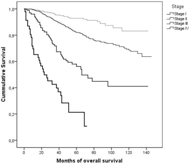
Histologically, invasive ductal carcinoma currently denominated carcinoma not otherwise specified, was the most frequent histological subtype, comprising 92% of the cases. We collected pTNM and biological profile data of 1639 patients who underwent BC surgery (Table 1). Regarding tumor size, the sample has the same amount of T1 and T2 lesions, it has 61% of axillary lymph node involvement. Survival analysis according to TNM variables is shown graphically with KM survival curves (Figures 1 to 4). The survival analysis according to biology variables is similarly depicted with KM curves (Figures 5 to 8). The p value of the Log Rank test performed to show that the survival curves are not superimposable is similarly shown. TNM staging variables and biology variables are predictive of survival.
Two thirds of the cases express ER, 9% were not evaluated. Nearly half of the cases had no PgR evaluation. Regarding HER2, in 16% of the cases had higher abundance of this oncogene, either at the protein or gene level, however, 30% of the cases were not evaluated. There were 13% of TN cases.
Regarding outcome variables, the median follow-up is 67 months for the whole population and 73 months for living patients. Four hundred and twenty one patients died, 361 deaths (22%) from metastatic breast carcinoma, 3,6% from other causes. The causes of death, other than BC, are known, but, we did not collect the data.
The logistic regression analysis contains data from the three TNM variables and the four biological variables included in the model. We conducted logistic regression to determine which variables are independent predictors of survival. In the model, for each survival predictor, we have a statistic and associated probabilities. These provide an index of the significance of each predictor in the equation. For assessing each predictor, we take the significance values, and, if the value of p is lower than 0.5 we reject the null hypothesis. We conclude the variable contributes significantly for predicting survival. The third column of table 2 shows the change in odds. If the value exceeds 1 then the odds of dying increase, if the figure is less than 1 any increase in the predictor leads to a drop in the odds of death, this is the case for Her2 negative status. The odds ratio (OR), estimates the change in the odds of survival if any given patient has BC with the characteristic in study. The OR is calculated by using the regression coefficient of the variable as exponent. In the case of TNM variables, the regression coefficient of stage is 2, thus the OR is exp², or 7.4. The odds of dying from BC are 7.4 times greater for a patient who has higher stage disease. In case of the other significant TNM variables in the model, the regression coefficient of involved axillary nodes is 1.42, therefore the odds of dying is 4 times greater for a patient with involved axillary nodes. Regarding the biological variables, in our model, built with the data from our population, we show that the odds of survival are doubled in patients with Her2 negative BC and that increasing histological grade increases the odds of death 4 times. We stress that adjuvant trastuzumab was administered since 2005.
Discussion
The cohort has a majority (61%) of stage II disease, i.e., with axillary lymph node involvement, this value is high {Turner and Leo, 2013, 2013 ASCO Educational Book, 3-8}. There are two possible reasons: First, the median age of the cohort is lower in the patients of this institution, when compared to the median age of incidence of breast cancer in the Western world {Leong et al., 2010, World J Surg, 34, 2308-24} {Turner and Leo, 2013, 2013 ASCO Educational Book, 3-8}. As has been said, this institution recruits younger patients with more advanced disease. Second, on a socioeconomic perspective, this institution serves mainly the region of southern portugal that has a less mature screening program than the north and center, and mainly serves patients without private insurance {Dourado et al., 2012, Eur J Public Health}. The high axillary lymph node involvement, and the low median age of the cohort, are the two most important findings of our work. These influence decisively of our results and conclusions.