online | memorias.ioc.fiocruz.br
Clinical and pathological evidence implicating abnor-mal cytokine release as the main mediator of disease and an increased risk of severe dengue disease have been de-scribed during secondary dengue virus (DENV) infections (Kurane & Ennis 1992). Indeed elevated levels of tumour
necrosis factor (TNF)-α have been detected in dengue
haemorrhagic fever/dengue shock syndrome (DHF/DSS) patients (Vitarana et al. 1991, Hober et al. 1993, Nguyen et al. 2004) and the production of this cytokine was corre-lated with increased numbers of macrophages/monocytes (de-Oliveira-Pinto et al. 2012), which are potent phagocyt-ic cells and primary targets for DENV infection
(Kang-wanpong et al. 1995, Jessie et al. 2004). These cells express
all three classes of FCγR making them especially prone
to DENV entry in the form of virus-antibody immune
complexes (Mathew & Rothman 2008). In addition, recent
studies have demonstrated that DENV-specific memory T-cells from a prior infection respond to heterologous DENV serotypes with an altered cytokine profile and that the level of activation and expansion of these memory cells during acute DENV infection correlates with the disease severity
(Rothman 2010). In addition, significant cross reactivity between DENV serotypes was shown in CD8(+) T-cells
after a naturally acquired primary infection with differ-ential T-cell receptor signalling after stimulation with ho-mologous and heterologous peptides (Friberg et al. 2011). DENV entry is facilitated by the phagocytosis of immune complexes via Fc receptors on monocytes (Daughaday et
al. 1981) and studies of immune responses have reported
circulating immune complexes and complement activation during the fever-to-defervescence period in which haem-orrhage, plasma leakage and/or circulatory failure occur
in patients with DHF and/or OSS (Ruangjirachuporn et al.
1979, Wang et al. 2006).
In endemic areas, where DHF incidence remains high, mosquito vectors are abundant and multiple
in-Financial support: CNPq (300460/2005-8, 301955/2007-7, 471444/2006-5, 454737/2010-6), INCT-FHV/CNPq/CAPES/ FAPESPA (573739/2008-0), FINEP/FADESP (01.04.0043.00), PROPESP-UFPA-FADESP
SMMC, EVPS, JAPD, MRTN, CWPD and PFCV are supported by INCT-FHV/CNPq. CWPD is supported by IBNnet/FINEP. + Corresponding author: [email protected] Received 15 May 2012
Accepted 2 October 2012
Environmental influences on
antibody-enhanced dengue disease outcomes
Daniel Guerreiro Diniz1, César Augusto Raiol Fôro1, Maíra C Pereira Turiel1,
Marcia CK Sosthenes1, Sâmia Demachki1, Giovanni Freitas Gomes1, Carla M Damasceno Rego1, Marina Cutrim Magalhães1, Brunno Gomes Pinho2, Juliana Pastana Ramos3,
Samir M Moraes Casseb4, Maysa de Vasconcelos Brito4, Eliana Vieira Pinto da Silva4, Marcio Roberto Teixeira Nunes4, José Antonio Picanço Diniz4, Colm Cunningham5,
Victor Hugh Perry6, Pedro F Costa Vasconcelos3,4/+, Cristovam W Picanço Diniz1
1Laboratório de Investigações em Neurodegeneração e Infecção, Hospital Universitário João de Barros Barreto, Instituto de Ciências Biológicas, Universidade Federal do Pará, Belém, PA, Brasil 2Centro de Estudos Superiores do Estado do Pará, Belém, PA, Brasil
3Universidade do Estado do Pará, Belém, PA, Brasil 4Instituto Evandro Chagas, Ananindeua, PA, Brasil 5Trinity College, Dublin 2, Ireland 6School of Biological Sciences, University of Southampton, Southampton, UK
Because an enriched environment (EE) enhances T-cell activity and T-lymphocytes contribute to immunopatho-genesis during heterologous dengue virus (DENV) infections, we hypothesised that an EE increases dengue severity. To compare single serotype (SS) and antibody-enhanced disease (AED) infections regimens, serial intraperitoneal were performed with DENV3 (genotype III) infected brain homogenate or anti-DENV2 hyperimmune serum followed 24 h later by DENV3 (genotype III) infected brain homogenate. Compared AED for which significant differences were detected between the EE and impoverished environmental (IE) groups (Kaplan-Meyer log-rank test, p = 0.0025), no significant differences were detected between the SS experimental groups (Kaplan-Meyer log-rank test, p = 0.089). Survival curves from EE and IE animals infected with the AED regimen were extended after corticoid injection and this effect was greater in the EE than in the IE group (Kaplan-Meyer log-rank test, p = 0.0162). Under the AED regimen the EE group showed more intense clinical signs than the IE group. Dyspnoea, tremor, hunched posture, ruffled fur, immobility, pre-terminal paralysis, shock and death were associated with dominant T-lympho-cytic hyperplasia and presence of viral antigens in the liver and lungs. We propose that the increased expansion of these memory T-cells and serotype cross-reactive antibodies facilitates the infection of these cells by DENV and that these events correlate with disease severity in an EE.
fections are often thought to involve different serotypes (Gubler & Meltzer 1999, Thomas et al. 2003, Suaya et al. 2009). In addition, an analysis of repeated hospital admissions for dengue showed that at least 10% of re-peat dengue admission patients may correspond to a third or fourth infection with all secondary admissions due to secondary dengue infection (Gibbons et al. 2007). Because most DENV infections are asymptomatic, it is reasonable to assume that a number of patients from endemic areas hospitalised with severe dengue disease may have been exposed to multiple infections. Thus, we have compared the outcomes of two different models of DENV infection: one induced by serial infections with a single serotype (SS) and one induced by multiple sero-types of DENV to enhance the disease through antibody cross reactivity. In this report, we used albino Swiss mice to model antibody-enhanced dengue disease after multiple infections and tested the influence of enhanced T-cell mobilisation on dengue disease severity.
An enriched environment (EE) has been defined as that which offers social interactions with con-specifics and the stimulation of exploratory and motor behaviour with periodic changes in the variety of toys, ladders, tunnels, ropes, bridges and running wheels available for voluntary physical exercise. In contrast, an impov-erished environment offers standard cages with reduced sensorial, motor and cognitive stimulation (van Praag et
al. 2000). Recent studies have demonstrated that an EE
enhances T-cell mobilisation during viral infections (de Sousa et al. 2011) and a greater number of infected target T-cells may contribute to higher viraemia and the ex-cess cytokine levels observed in severe dengue disease (Boonnak et al. 2011). Therefore, we hypothesised that environmental enrichment would exacerbate the clinical symptoms and mortality of DENV infection.
MATERIALS AND METHODS
We used adult female albino Swiss mice obtained from an outbred colony at the Animal Care Facility of the Evandro Chagas Institute and the mice were handled in accordance with the Principles of Laboratory Animal Care (National Institute of Health). All studies were ap-proved by the institutional animal care committee of the Evandro Chagas Institute, protocol 0061/2009. The mice were maintained with 12-h dark and light cycles, at a room temperature of 22 ± 2ºC and had free access to water and food.
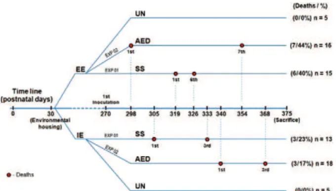
Experimental groups and inoculation - Fig. 1 shows the experimental time line. We performed serial
intra-peritoneal (i.p.) injections following one of two experi
-mental protocols. Under the first protocol [impoverished environmental (IE): n = 13; EE: n = 15] all subjects re
-ceived i.p. injections every other day with infected brain homogenate containing 0.014-0.087 viral copies/mL of
DENV3 (genotype III) (SS group) until the day of
sac-rifice. Under the second protocol (IE: n = 18; EE: n = 16), all subjects received i.p. injections with infected
brain homogenate containing a similar number of viral copies of DENV3 (genotype III) once per day for six days. The animals were i.p. inoculated with diluted
anti-DENV2 hyperimmune serum containing anti-anti-DENV2
antibodies (1:32 dilution) every other day. Each injec
-tion was followed 24 h later by injec-tion with DENV3
(genotype III) infected brain homogenate until the day of sacrifice to simulate antibody-enhanced DENV dis-ease (AED group). Animals were sacrificed when they exhibited severe clinical symptoms including dyspnoea, tremor, hunched posture, ruffled fur, immobility, pre-terminal paralysis, shock and death or when burrowing activity was below 40% for two consecutive weekly
mea-surements. Control subjects received equal volumes and
dilution of anti-DENV2 hyperimmune serum followed
24 h later by uninfected brain homogenate. On the 38th
days post-inoculation(d.p.i.), the subjects in the AED
group received a single intramuscular (i.m.) injection of
glucocorticoid (26 mg/kg, 1:1 mixture of acetate and be-tamethasone sodium phosphate) to reduce T-lymphocyte proliferation and function and to determine the impact of glucocorticoids on the Kaplan-Meyer survival curves. This combination of glucocorticoids becomes effective
30 min after injection and remains active for four weeks.
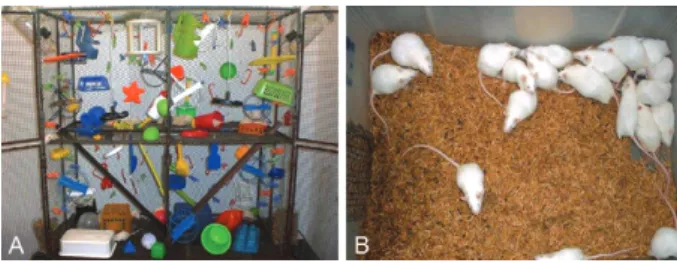
All animals from the two experiments were nine month old, female albino Swiss mice housed from wean-ing under either impoverished or enriched conditions
(Fig. 2, movie S1). Enriched conditions comprised of two-level wire cages (100 × 50 × 100 cm) with toys made of different forms of plastic, wood and metal of different colours, that were changed periodically. Each EE cage housed 15-20 young mice. Water and food were delivered to the top and bottom levels, respectively. Impoverished conditions consisted of plastic cages (32 × 39 × 100 cm) without equipment or toys. Each IE cage housed 15-20 young mice. All mice had free access to water and food.
Infected brain homogenates - Neonatal (2 day old) mice were intracerebrally infected with 10 µL of su-pernatant from C6/36 cells infected with either 1.33 × 10³ or 7.78 × 10³ cp/mL of DENV3 (genotype III)
ob-tained from human serum samples (cases ROND 2929 and ROND 3115, respectively). Upon presenting clini -cal signs of infection, the animals were sacrificed and immediately stored at -70ºC. Later, the brain tissue (0.2
g/animal) was macerated and mixed with 0.8 mL of phosphate-buffered saline (PBS) containing 100 U/mL
penicillin and 100 µg/mL streptomycin. The suspension was centrifuged at 10,000 g for 15 min at room tempera-ture (16ºC). The viral load (VL) in different tissues of the adult mice from the different experimental groups was
estimated by real time-polymerase chain reaction (RT-PCR) when clinical symptoms became apparent.
Behavioural analysis - Burrowing - For 2 h per day (from 09:00 am-11:00 am) on two non-consecutive days the week before inoculation and two non-consecutive days between 2 d.p.i. and the day of sacrifice, each ani-mal was placed in a plastic cage (32 cm × 39 cm × 16.5 cm) containing a PVC tube (20 cm long, 7.2 cm diam-eter) filled with 100 g of normal diet food pellets with the open end supported 3 cm above the floor. After the testing period, the remaining food in the cylinder was weighed and the mouse was returned to its collective cage. Burrowing is an ethological task highly sensitive to systemic inflammation (Cunningham et al. 2009).
Open field - The apparatus consisted of a grey wood box (chipboard coated) (30 cm × 30 cm × 40 cm) with the floor divided into 10-cm squares. Once a week before inoculation and between d.p.i. 2 and the day of sacrifice, each animal was placed in the centre of the arena and
kept in the apparatus for 5 min, as previously described (Kinoshita et al. 2009). The open field test is a sensitive test for detecting sickness behaviour following a vari-ety of viral infections, including dengue (de Miranda et al. 2012). A video camera connected to a computer was located 1 m above the open field and used to record each training session for later analysis with the Any-Maze software (Stöelting). The following parameters were analysed: distance travelled (m), mean speed (m/s), crossed lines and immobility time (s). After each test, the open field was cleaned with 70% ethanol.
Online Supplementary data is provided in video for-mat to illustrate some aspects of the methodology and the main effects of the environmental influences on AED in our murine model (videos 1, 2).
After the behavioural tests, all diseased animals that showed a reduction of more than 60% in burrowing ac-tivity for two consecutive measurements (4 sessions)
clinical disease signs (see Results) or clinical symptoms
were sacrificed with i.p. 1% 2,2,2-tribromoethanol (0.01 mL/g of body weight). Three animals from each group were processed for histopathology.
Histology and immunohistochemistry - Mice were transcardially perfused with heparinised saline followed by 4% paraformaldehyde in 0.1 M phosphate buffer (pH 7.2-7.4). Livers and lungs, from five animals in each group were embedded in paraffin and sections (3 µm thick) were dewaxed in xylene, rehydrated with alcohols and stained with haematoxylin-eosin or immunolabelled to detect DENV3, CD-3, CD-15 and CD-20 antigens.
T and B lymphocytes - The specimens were fixed in 4% paraformaldehyde and embedded in paraffin. Serial 3-µm cross-sections were made and the fragments were placed on glass slides previously treated with
poly-D-lysine (Sigma Chemical Co, USA) and deparaffinised in
xylene and descending ethanol series. Antigen retrieval was performed using pressure cooker heating (PAS-CAL®, DakoCytomation, USA) in citrate buffer (10 mM,
pH 6.0). After washing in PBS, the sections were im-mersed in 0.3% hydrogen peroxide for 5 min to block endogenous peroxidase activity and blocked with pro-tein for 10 min. The sections were then incubated in a dark wet chamber with primary monoclonal antibodies against CD3 (SP7 clone, dilution 1:400,
DakoCytoma-tion, USA) or CD20 (L26 clone, dilution 1:600, Dako
-Cytomation, USA) for 30 min then a secondary antibody
followed by a streptavidin-biotin-peroxidase complex, each for 20 min at room temperature (DakoCytomation,
USA). The slides were visualised with diaminobenzidine
and counterstained with Harry’s haematoxylin.
Microglia immunolabelling - Free-floating vibratome (70 µm thick) brain sections were pre-treated with 0.2 M boric acid (pH 9) at 65-70ºC for 60 min to improve anti-gen retrieval. The sections were incubated with a poly-clonal antibody against ionised calcium binding adapter molecule 1 (2 µg/mL in PBS) to detect microglia and/or macrophages (anti-Iba1, #019-19741; Wako Pure Chemi-cal Industries Ltd, Osaka, Japan). Washed sections were then incubated overnight with a secondary antibody
(goat anti-rabbit, 1:250 in PBS, Vector Laboratories). En-dogenous peroxidases were inactivated by immersion in 3% H2O2 in PBS. The PBS washed sections were trans-ferred to a solution of avidin-biotin-peroxidase complex (VECTASTAIN ABC kit; Vector Laboratories) for 1 h. The DAB/Nickel/Glucose Oxidase protocol was used to
reveal immunolabelled antigenic sites (Shu et al. 1988).
We confirmed the specificity of the immunohistochemi-cal pattern by omitting the primary antibody (Saper & Sawchenko 2003).
RNA extraction - Viral RNA was extracted from the
supernatant of infected C6/36 cells using a QIAquick
RNA extraction kit (Qiagen, Hilden, Germany) accord -ing to the manufacturer’s instructions.
SYBR green quantitative RT-PCR (SYBR-qPCR) -
qRT-PCR was performed using a commercial kit (Super
-Script III Platinum SYBR® Green One-step qRT-PCR,
Invitrogen, Carlsbad, CA, USA) and the ABI Prism 7500 Real Time PCR System (Applied Biosystem, Carlsbad, CA, USA). The reaction were performed in a final vol
-ume of 25 µL containing 5 µL of viral RNA (1-2 ng/ µL), 0.5 µL of SuperScript III RT Platinum Taq Mix, 0.2 µM of each DENV primer (dos Santos et al. 2008), 12.5 µL of 2 × SYBR Green fluorescent dye and 1 µL of ROX dye. The amplification was performed as fol -lows: 50ºC for 20 min, 95ºC for 5 min, 45 cycles of 95ºC for 15 s, 55ºC for 40 s and 72ºC for 30 s. The melting
curve was calculated during the incubation period from 60-90ºC with a capture speed of 0.2ºC/s. Each sample
was analysed in duplicate wells. The SYBR-qRT-PCR
results were expressed as genome copies/mL based on the standard curve constructed from the amplification
of RNA extracted from C6/36 cells infected with the DENV-3 strain BH 692808 (stock in concentration: 3.1
× 106 genome copies/mL) and diluted from 10-1 to 10-6.
Estimations of the VL (genome copies/mL) in the
C6-36 cell culture supernatants corresponding to the ROND 2929 and ROND 3115 human cases were 1.30 × 10³-1.33
× 103 and 7.77 × 10³-7.82 × 10³, respectively.
Standard curve - External standard curves were
generated from serial dilutions of RNA derived from
aliquots with known viral titres covering a range from 1 × 10⁻¹-1×10⁻⁸. A standard curve with eight points was
amplified in duplicate along with non-template controls to determine the threshold value. Cycle threshold values
from each RNA sample were compared with external
standard curves to determine the corresponding virus loads in each tissue.
RESULTS
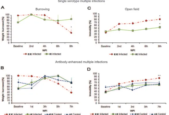
Clinical signs and survival - Burrowing and open field tests (movie S3) were performed in control animals until significant changes in both tests became apparent at 56 d.p.i. (Fig. 3A-D). After serial DENV3 single-sero-type inoculations, these changes were detected at eight
weeks post-inoculation (wpi) with a significant decrease in burrowed food for the EE animals (EE baseline =
96.82 ± 8.21% vs. EE 8 wpi = 35.04 ± 9.31%; one-way
ANOVA, Bonferroni a priori test, p < 0.05), but not for the IE animals (IE baseline = 72.31 ± 6.94% vs. 83.75 ±
9.9%; p > 0.05) (Fig. 3A). In the antibody-enhanced den-gue disease group at 7 wpi, the EE mice showed a sig-nificant decrease in burrowed food (EE baseline = 100%
± 2.74% vs. EE 8 wpi = 34.23 ± 16.95), with a reduction
of 65.77% (one-way ANOVA, Bonferroni a priori test, p < 0.05) whereas the IE mice did not change burrow-ing activity (Fig. 3B). In the open field tests at the same time points, all multiple single-serotype infected (Fig. 3C) and multiply infected, antibody-enhanced (Fig. 3D) groups showed a significant increase in immobility time. The multiple single-serotype infected groups housed an
EE (57%) and IE (28%) as well as multiple infected AED
housed in EE (45%) and IE (25.35%) showed increases in immobility time compared with base line (one-way ANOVA, Bonferroni a priori test, p < 0.05). None of the control groups showed any significant changes. Al-though these clinical signs could be associated with cen-tral nervous system (CNS) changes, we did not observe any microglial morphological changes that could be in-terpreted as CNS inflammation (Supplementary data).
Fig. 4A illustrates the survival curves after multiple infections with a SS of DENV3. Although the EE mice presented higher rates of mortality and disease symp-toms (40%) compared with the IE (23.1%) after multiple single-serotype infections, the Kaplan-Meyer survival plot did not show any significant differences between the survival curves in this experiment (Kaplan-Meyer log
rank test p = 0.089). Compared with the mice subjected
to the AED protocol, the mice that received multiple in-fections with a SS not only survived for longer periods post-challenge, but exhibited fewer clinical signs.
Fol-lowing the AED protocol (Fig. 4B), the EE mice showed earlier and more intense clinical signs, including dys-pnoea, tremor, hunched posture, ruffled fur, immobil-ity, pre-terminal paralysis, shock and deaths compared with the mice in the IE (Supplementary data, video 2). Kaplan-Meyer analysis revealed significant differences in the survival probability curves of the AED experi-mental groups (uninfected, EE and IE; log-rank test, p = 0.0025) (Fig. 4B).
To prevent death and reduce clinical symptoms in
the AED subjects, we injected the mice in this group with glucocorticoids at 38 d.p.i. After a single dose of
glucocorticoids (arrow in Fig. 4C), clinical signs and deaths were reduced and the survival probability curve stabilized four-six weeks in the IE and EE experimental groups despite continued efforts to induce AED during this period by maintaining the regime of multiple inocu-lations. Approximately 50 days after the glucocorticoids
injection, however, a progressive decline in the survival
probability was observed. In the EE and IE groups, the
AED animals injected with corticoids showed increased
survival probabilities (log-rank test, p = 0.016).
There were no significant differences in the survival probability curves between the control and infected
sub-jects treated with the SS protocol (p = 0.089), but the
AED protocol did produce significant differences (p = 0.0025). In addition, a significant difference was ob-served between the AED regimen involving infection with multiple serotypes and the animals that were
in-fected with multiple doses of a SS (SS = 68.13 ± 2.93 vs.
AED = 54.37 ± 5.96 d.p.i.; two-tail T-test p = 0.029).
None of the control animals injected with uninfected
brain homogenate showed any significant changes in survival probability.
Pathological changes after AED infection - The his-tological analysis of livers and lungs used a
tative protocol to assess lesion severity. After treatment with the antibody-enhanced regimen involving multiple infections, the tissue samples showed frequent
inflam-matory infiltrates, dominated by T-lymphocytes (++++) and rare (+) B-lymphocytes (CD20+), macrophages (CD68+) and neutrophils (CD15+). The subjects housed in the enriched environments showed intense (++++)
hyperplasia of T lymphocytes in the liver (Fig. 5B, D),
whereas this was moderate (++) in the IE mice (Figs 4C, 5A) and rare (+) in the uninfected subjects (Fig. 5H). In
addition, discrete tumefaction, microvesicular steatosis and Kupffer cell hypertrophy were detected in various liver lobules with the previously mentioned T lympho-cytic hyperplasia around the efferent veins. Liver vascu-lar congestion was mainly found in EE mice (Fig. 5F). These pathological features were rare and less intense in
the uninfected subjects (Fig. 5G, H).
Similar inflammatory infiltrates with dominant T-lymphocytic hyperplasia were also presented in the peri-bronchial space (Fig. 6). Animals housed in the IE (Fig. 6C, D) showed a lesser degree of T-lymphocytic hyper-plasia compared with those housed in the EE (Fig. 6E, F). These changes were less intense and rarely detected in
the lungs of the uninfected control subjects (Fig. 6A, B).
To illustrate the presence of viral infection in the parenchyma we selected photomicrographs of sections
from lungs (Fig. 7) and livers (Fig. 8) of infected animals
under the AED multiple infections regime immunola-belled for viral antigens.
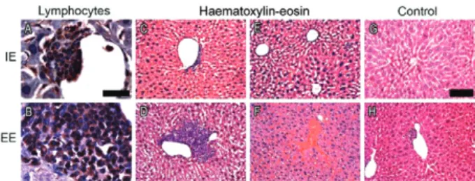
Fig. 5: liver pathology after antibody-enhanced dengue disease. Pho-tomicrographics from immunolabelled (A-B) or haematoxylin-eosin (C-H) stained sections to illustrate pathological changes in the liver of impoverished environment (IE) (n = 4), enriched environment (EE) (n = 4) and control uninfected (n = 4) subjects. A, B: immunohistochem -istry for T (CD3+) lymphocytes counterstained by Harry’s haematox-ylin. Note that the inflammatory mononuclear infiltration in the sub-endothelial space of peri-efferent veins was less intense (++) in IE (A, C) as compared with EE (++++) (B, D). Intense vascular congestion was found in EE (F). Steatosis, vascular congestion and mononuclear infiltration was rare and less intense (+) in the uninfected control sub-jects (G, H). [Bars = high power (25 µm); low power (125 µm)].
Fig. 6: lung pathology after antibody-enhanced dengue disease (AED). Photomicrographics from immunolabelled (A, C, E) and haematoxylin-eosin (H&E) (B, D, F) stained sections to illustrate pathological changes in the lungs of control uninfected (n = 4), im-poverished environment (IE) (n = 4) and enriched environment (EE) (n = 4) subjects, after AED infection. A, C, E: immunohistochemistry for T (CD3+) lymphocytes counterstained by Harry’s haematoxylin. Note that the inflammatory T (CD3) lymphocytic hyperplasia in the peribronchical space was less intense in IE (C) than in EE (E). Con-sistent with this, mononuclear infiltration in the peribronchical space, as revealed by H&E staining, was less intense in IE (D) as compared to EE (F). Mononuclear infiltration was not significant in the lungs of uninfected control subjects (A, B).
Fig. 7: photomicrography of lung immunolabelled section from antibody-enhanced dengue disease individual obtained by indi-rect immunofluorescence to illustrate the presence of dengue virus (DENV)3 fluorescein labelled viral antigens (green), counterstained by 4’-6-diamidino-2-phenylindole fluorescence for DNA (blue) in the peribronchical space. Arrows indicate immunopositive cells for DENV3 antigens.
qRT-PCRto detect DENV3 - Low viral titres were detected 24 h after the last DENV3 inoculation, at which time clinical signs became apparent. The highest con-centrations of viral DNA were found in the livers of
subjects housed in the EE (3.03 copies/mL). This was
significantly different from the concentrations observed in the spleen (0.26 copies/mL) and lungs (0.003 copies/ mL) of the same animals, but no significant differences were observed between the number of viral genome
cop-ies/mL in the EE and IE subjects. DISCUSSION
In this study we tested the effects of environmental enrichment on dengue disease progression in a model of AED in adult female mice, in which serial inoculations of anti-DENV2 antibodies were followed 24 h later by inoculation with DENV3 (genotype III) infected brain
homogenate. We found that subjects housed in EE with
AED presented more intense clinical symptoms and
higher mortality than subjects housed in impoverished
environment and clinical signs were associated with T lymphocytic hyperplasia in the liver and lungs where we found cells expressing DENV antigens.
Under the of antibody-enhanced response regime,
multiple inoculations with anti-DENV2 antibodies were followed 24 h later by inoculation with DENV3 geno- type III. This regimen was associated with a higher number of deaths in the EE group than in the IE group. One hour after virus infection under this regime a num-ber of the AED animals developed intense clinical signs with exuberant dyspnoea followed by death. TNF de-rived from DENV infected monocytes and activated
T-cells has been proposed as a major mediator responsible
for the deregulation of endothelial cells, which lead to plasma leakage (Halstead 2007). In addition, DENV in-fection causes mast-cell to trigger endothelial cell acti-vation (Brown et al. 2011) and the release of vascular endothelial growth factor and proteases during dengue shock syndrome (Furuta et al. 2012); mast-cell numbers are also increased in peribronchiolar spaces and areas
adjacent to the inter-alveolar septa (Barreto et al. 2007).
Taken together these data suggest that, during serial AED, environmental influences on disease progression might contribute to the disease severity by inducing an exacerbated inflammatory response. Indeed an EE has been shown to enhances T-cell activity during viral infec-tions (de Sousa et al. 2011) and DENV is associated with
increasing frequencies of interferon (IFN)-γ and
TNF-α-producing T-cells after primary infection with all DENV serotypes; secondary infection enhances these responses (Beaumier et al. 2010). In a previous report, T-cell re-sponses to sequential infections were measured after mice were immunized with different DENV serotypes and the frequency of peptide-specific T-cells after infection was
determined (Beaumier et al. 2008). After heterologous
secondary infection in BALB/c mice the acute response was enhanced compared with the acute response after
primary infection (Beaumier et al. 2008). However, the
passive administration of anti-DENV antibodies against one DENV serotype followed 24 h later by inoculation
of another serotype of DENV was sufficient to enhance DENV infection and disease in AG129 mice. This was the first model of AED lethal in vivo (Balsitis et al. 2010).
Because we did not assess any inflammatory media-tors, it remains to be investigated whether increased
cy-tokine production in subjects housed in EE may contrib -utes to the differences in disease severity and mortality
rates from subjects housed in impoverished environments.
In line with this hypothesis, the anti-inflammatory effects
glucocorticoids injection stabilized the survival curves of
both experimental groups for 50 days (see plateau
follow-ing corticoid injection in Fig. 4C) which not occur in the
individuals that did not received corticosteroids (Fig. 4B). Because corticosteroids reduce T-lymphocyte prolifera-tion (Lu et al. 2010), we suggest that a relative decrease in the number of T-lymphocytes following
glucocorti-coid injection may explain the reduction in mortality and
disease symptoms. A previous report demonstrated that dexamethasone completely inhibits cytokine produc-tion after a peripheral challenge with lipopolysaccharide (Teeling et al. 2010). Delayed but not early glucocorti-coid treatment has also been shown to protects the host, increase survival and decrease cerebral damage during experimental herpes simplex virus encephalitis in mice (Sergerie et al. 2007). Because delayed administration of glucocorticoids reduced death and disease severity after serial AED it is reasonable to suggest that the cytokine storm previously observed in secondary DENV infection
(Rothman 2009, Guabiraba et al. 2010, Tan et al. 2010)
and AED (Balsitis et al. 2010) may be inhibited by corti-coids (Sergerie et al. 2007).
A previous report showed a single intracerebral inoc-ulation with DENV-3 (genotype I) induced neurological disease and death in mice, whereas DENV-3 (genotype III) did not (Ferreira et al. 2010). Consistent with this result, we showed that neither neurological clinical signs such as pre-terminal paralysis nor behavioural changes, such as a 60% reduction in burrowing activity were accompanied by evi-dent pathology in the CNS (Supplementary data).
However, the results obtained with the singly-sero-type regimen suggest that the adaptive immune response may have induced efficient protection against primary DENV infection. Indeed no significant differences were found between the experimental groups under the SS multiple infections protocol. The resistance to dengue infection after primary infection may be consistent with previous results showing that the resistance was not due to a defect in the recruitment of effectors lymphocytes, but rather to the antiviral activity of those cells, which promoted viral clearance (Ip & Liao 2010). Consistent with this hypothesis, antiviral activity that promotes
viral clearance in a manner that depends on IFN-γ and TNF-α-producing T-cells directed at H-2Db-restricted
epitopes (Friberg et al. 2011) may be responsible for the resistance of mice to primary infection by dengue virus. We suggested that a serial SS infection may mimic a se-vere primary dengue infection in which others have de-scribed a cytokine storm associated with a T-lymphocyte response, that is mostly responsible for the development of disease rather than protection against severe infection
We report for the first time that after a regimen of multiple infections associated with and AED model, ani-mals maintained in an EE showed more intense clinical symptoms and behavioural changes with lower disease resolution compared with animals maintained in impov-erished housing.
REFERENCES
Balsitis SJ, Williams KL, Lachica R, Flores D, Kyle JL, Mehlhop E, Johnson S, Diamond MS, Beatty PR, Harris E 2010. Lethal anti-body enhancement of dengue disease in mice is prevented by Fc modification. PLoS Pathog6: e1000790.
Barreto DF, Takiya CM, Schatzmayr HG, Nogueira RMR, Farias-Fil -ho J da C, Barth OM 2007. Histopat-hological and ultrastructural aspects of mice lungs experimentally infected with dengue virus serotype 2. Mem Inst Oswaldo Cruz102: 175-182.
Beaumier CM, Mathew A, Bashyam HS, Rothman AL 2008. Cross-reactive memory CD8(+) T cells alter the immune response to heterologous secondary dengue virus infections in mice in a sequence-specific manner. J Infect Dis197: 608-617.
Beaumier CM, Jaiswal S, West KY, Friberg H, Mathew A, Rothman AL 2010. Differential in vivo clearance and response to second-ary heterologous infections by H2(b)-restricted dengue virus-specific CD8+ T cells. Viral Immunol23: 477-485.
Boonnak K, Dambach KM, Donofrio GC, Tassaneetrithep B, Ma-rovich MA 2011. Cell type specificity and host genetic polymor-phisms influence antibody-dependent enhancement of dengue virus infection. J Virol85: 1671-1683.
Brown MG, Hermann LL, Issekutz AC, Marshall JS, Rowter D, Al-Afif A, Anderson R 2011. dengue virus infection of mast cells triggers endothelial cell activation. J Virol85: 1145-1150.
Cunningham C, Campion S, Lunnon K, Murray CL, Woods JFC, Deacon RMJ, Rawlins JNP, Perry VH 2009. Systemic inflamma-tion induces acute behavioral and cognitive changes and acceler-ates neurodegenerative disease. Biol Psychiatry65: 304-312. Daughaday CC, Brandt WE, McCown JM, Russell PK 1981. Evidence
for two mechanisms of dengue virus infection of adherent human monocytes: trypsin-sensitive virus receptors and trypsin-resis-tant immune complex receptors. Infect Immun32: 469-473.
de Miranda AS, Rodrigues DH, Amaral DC, de Lima Campos RD, Cisalpino D, Vilela MC, Lacerda-Queiroz N, de Souza KP, Vago JP, Campos MA, Kroon EG, da Gloria de Souza D, Teixeira MM, Teixeira AL, Rachid MA 2012. Dengue-3 encephalitis promotes anxiety-like behavior in mice. Behav Brain Res230: 237-242.
de-Oliveira-Pinto LM, Gandini M, Freitas LP, Siqueira MM, Marinho CF, Setúbal S, Kubelka CF, Cruz OG, de Oliveira SA 2012. Pro-file of circulating levels of IL-1Ra, CXCL10/IP-10, CCL4/MIP-1β and CCL2/MCP-1 in dengue fever and parvovirosis. Mem Inst Oswaldo Cruz 107: 48-56.
de Sousa AA, Reis R, Bento-Torres J, Trevia N, Lins NA, Passos A, Santos Z, Diniz JA, Vasconcelos PF, Cunningham C, Perry VH, Diniz CW 2011. Influence of enriched environment on viral en-cephalitis outcomes: behavioral and neuropathological changes in albino Swiss mice. PLoS ONE6: e15597.
dos Santos HW, Poloni TR, Souza KP, Muller VD, Tremeschin F, Nali LC, Fantinatti LR, Amarilla AA, Castro HL, Nunes MR, Casseb SM, Vasconcelos PF, Badra SJ, Figueiredo LT, Aquino VH 2008. A simple one-step real-time RT-PCR for diagnosis of dengue vi-rus infection. J Med Virol 80: 1426-1433.
Ferreira GP, Figueiredo LB, Coelho LF, PA Jr S, Cecilio AB, Ferreira PC, Bonjardim CA, Arantes RM, Campos MA, Kroon EG 2010. Dengue virus 3 clinical isolates show different patterns of virulence in experimental mice infection. Microbes Infect12: 546-554.
Friberg H, Burns L, Woda M, Kalayanarooj S, Endy TP, Stephens HA, Green S, Rothman AL, Mathew A 2011. Memory CD8+ T
cells from naturally acquired primary dengue virus infection are highly cross-reactive. Immunol Cell Biol 89: 122-129.
Furuta T, Murao LA, Lan NT, Huy NT, Huong VT, Thuy TT, Tham VD, Nga CT, Ha TT, Ohmoto Y, Kikuchi M, Morita K, Yasunami M, Hirayama K, Watanabe N 2012. Association of mast cell-de-rived VEGF and proteases in dengue shock syndrome. PLoS Negl Trop Dis6: e1505.
Gibbons RV, Kalanarooj S, Jarman RG, Nisalak A, Vaughn DW, Endy TP, Mammen Jr MP, Srikiatkhachorn A 2007. Analysis of repeat hospital admissions for dengue to estimate the frequency of third or fourth dengue infections resulting in admissions and dengue hemorrhagic fever and serotype sequences. Am J Trop Med Hyg 77: 910-913.
Guabiraba R, Marques RE, Besnard AG, Fagundes CT, Souza DG, Ryffel B, Teixeira MM 2010. Role of the chemokine receptors CCR1, CCR2 and CCR4 in the pathogenesis of experimental den-gue infection in mice. PLoS ONE5: e15680.
Gubler DJ, Meltzer M 1999. Impact of dengue/dengue hemorrhagic fever on the developing world. Adv Virus Res53: 35-70.
Halstead SB 2007. Dengue. Lancet370: 1644-1652.
Hober D, Poli L, Roblin B, Gestas P, Chungue E, Granic G, Imbert P, Pecarere JL, Vergez-Pascal R, Wattre P, et al. 1993. Serum levels of tumor necrosis factor-alpha (TNF-alpha), interleukin-6 (IL-6), and interleukin-1 beta (IL-1 beta) in dengue-infected patients.
Am J Trop Med Hyg48: 324-331.
Ip PP, Liao F 2010. Resistance to dengue virus infection in mice is potentiated by CXCL10 and is independent of CXCL10-mediated leukocyte recruitment. J Immunol184: 5705-5714.
Jessie K, Fong MY, Devi S, Lam SK, Wong KT 2004. Localization of dengue virus in naturally infected human tissues by immunohis-tochemistry and in situ hybridization. J Infect Dis 189: 1411-1418.
Kangwanpong D, Bhamarapravati N, Lucia HL 1995. Diagnosing den-gue virus infection in archived autopsy tissues by means of the in situ PCR method: a case report. Clin Diagn Virol3: 165-172. Kinoshita D, Cohn DW, Costa-Pinto FA, de Sa-Rocha LC 2009.
Be-havioral effects of LPS in adult, middle-aged and aged mice.
Physiol Behav96: 328-332.
Kurane I, Ennis FE 1992. Immunity and immunopathology in dengue virus infections. Semin Immunol 4: 121-127.
Lu YS, Pu LY, Li XC, Wang XH 2010. Methylprednisolone inhibits activated CD4+ T cell survival promoted by toll-like receptor li-gands. Hepatobiliary Pancreat Dis Int9: 376-383.
Mathew A, Rothman AL 2008. Understanding the contribution of cellular immunity to dengue disease pathogenesis. Immunol Rev 225: 300-313.
Nguyen TH, Lei HY, Nguyen TL, Lin YS, Huang KJ, Le BL, Lin CF, Yeh TM, Do QH, Vu TQ, Chen LC, Huang JH, Lam TM, Liu CC, Halstead SB 2004. Dengue hemorrhagic fever in infants: a study of clinical and cytokine profiles. J Infect Dis189: 221-232. Rothman AL 2009. T lymphocyte responses to heterologous
second-ary dengue virus infections. Ann NY Acad Sci1171: e36-41. Rothman AL 2010. Cellular immunology of sequential dengue virus
infection and its role in disease pathogenesis. Curr Top Microbiol Immunol338: 83-98.
Ruangjirachuporn W, Boonpucknavig S, Nimmanitya S 1979. Circu-lating immune complexes in serum from patients with dengue haemorrhagic fever. Clin Exp Immunol36: 46-53.
Sergerie Y, Boivin G, Gosselin D, Rivest S 2007. Delayed but not early glucocorticoid treatment protects the host during experi-mental herpes simplex virus encephalitis in mice. J Infect Dis 195: 817-825.
Shu SY, Ju G, Fan LZ 1988. The glucose oxidase-DAB-nickel method in peroxidase histochemistry of the nervous system. Neurosci Lett 85: 169-171.
Suaya JA, Shepard DS, Siqueira JB, Martelli CT, Lum LC, Tan LH, Kongsin S, Jiamton S, Garrido F, Montoya R, Armien B, Huy R, Castillo L, Caram M, Sah BK, Sughayyar R, Tyo KR, Halstead SB 2009. Cost of dengue cases in eight countries in the Americas and Asia: a prospective study. Am J Trop Med Hyg80: 846-855.
Tan GK, Ng JK, Trasti SL, Schul W, Yip G, Alonso S 2010. A non mouse-adapted dengue virus strain as a new model of severe den-gue infection in AG129 mice. PLoS Negl Trop Dis4: e672.
Teeling JL, Cunningham C, Newman TA, Perry VH 2010. The effect of non-steroidal anti-inflammatory agents on behavioural changes
and cytokine production following systemic inflammation: impli-cations for a role of COX-1. Brain Behav Immun 24: 409-419.
Thomas SJ, Strickman D, Vaughn DW 2003. Dengue epidemiology: virus epidemiology, ecology and emergence. Adv Virus Res 61: 235-289.
van Praag, Kempermann G, Gage FH 2000. Neural consequences of enviromental enrichement. Nat Rev Neurosci 1: 191-198.
Vitarana T, de Silva H, Withana N, Gunasekera C 1991. Elevated tu-mour necrosis factor in dengue fever and dengue haemorrhagic fever. Ceylon Med J 36: 63-65.
Wang WK, Chao DY, Kao CL, Wu HC, Liu YC, Li CM, Lin SC, Ho ST, Huang JH, King CC 2003. High levels of plasma dengue viral load during defervescence in patients with dengue hemorrhagic fever: implications for pathogenesis. Virology305: 330-338.
: a time-lapse movie of enriched and impoverished environments.
: a time-lapse movie of clinical symptoms of individuals that were maintained in enriched and impoverished environments.
: a time-lapse movie of burrowing and open field activities.
Microglial morphology of mice septal region after antibody-enhanced dengue disease infection and uninfected groups. A: control young un-infected subject; B: impoverished environment un-infected young subject: C: enriched environment (EE) un-infected young subject. No remarkable neuropathological changes were detected in the infected groups as compared with control subject, except by a subtle change in the apparent number of spiny-like processes in EE microglial cells.
VIDEO 1
VIDEO 2