online | memorias.ioc.fiocruz.br
Scoring clinical signs can help diagnose canine visceral
leishmaniasis in a highly endemic area in Brazil
Kleverton Ribeiro da Silva1, Vitor Rosa Ramos de Mendonça1, Kellen Matuzzy Silva2,
Leopoldo Fabrício Marçal do Nascimento2, Antonio Ferreira Mendes-Sousa3, Flaviane Alves de Pinho2, Manoel Barral-Netto1,4,5, Aldina Maria Prado Barral1,4,5, Maria do Socorro Pires e Cruz2/+
1Fundação Oswaldo Cruz-Fiocruz, Centro de Pesquisas Gonçalo Moniz, Salvador, BA, Brasil 2Universidade Federal do Piauí, Departamento de Morfofisiologia Veterinária, Teresina, PI, Brasil
3Universidade Federal do Piauí, Picos, PI, Brasil
4Universidade Federal da Bahia, Faculdade de Medicina, Salvador, BA, Brasil
5Instituto Nacional de Ciência e Tecnologia, Instituto de Investigação em Imunologia, São Paulo, SP, Brasil
Canine visceral leishmaniasis (CVL) diagnosis is still a challenge in endemic areas with limited diagnostic re-sources. This study proposes a score with the potential to distinguish positive CVL cases from negative ones. We studied 265 dogs that tested positive for CVL on ELISA and parasitological tests. A score ranging between 0 and 19 was recorded on the basis of clinical signs. Dogs with CVL had an overall higher positivity of the majority of clinical signs than did dogs without CVL or with ehrlichiosis. Clinical signs such as enlarged lymph nodes (83.93%), muzzle/ ear lesions (55.36%), nutritional status (51.79%), bristle condition (57.14%), pale mucosal colour (48.21%), onycho-gryphosis (58.93%), skin lesion (39.28%), bleeding (12.50%), muzzle depigmentation (41.07%), alopecia (39.29%), blepharitis (21.43%), and keratoconjunctivitis (42.86%) were more frequent in dogs with CVL than in dogs with ehrlichiosis or without CVL. Moreover, the clinical score increased according to the positivity of all diagnostic tests (ELISA, p < 0.001; parasite culture, p = 0.0021; and smear, p = 0.0003). Onychogryphosis (long nails) [odds ratio (OR): 3.529; 95% confidence interval (CI): 1.832-6.796; p < 0.001], muzzle depigmentation (OR: 4.651; 95% CI: 2.218-9.750; p < 0.001), and keratoconjunctivitis (OR: 5.400; 95% CI: 2.549-11.441; p < 0.001) were highly
associ-ated with CVL. Interestingly, a score cut-off value ≥ 6 had an area under the curve of 0.717 (p < 0.0001), sensitivity
of 60.71%, and specificity of 73.64% for CVL diagnosis. The clinical sign-based score for CVL diagnosis suggested herein can help veterinarians reliably identify dogs with CVL in endemic areas with limited diagnostic resources.
Key words: canine visceral leishmaniasis - clinical score - diagnosis - clinical signs
doi: 10.1590/0074-02760160305 Financial support: CAPES.
KRS and VRRM contributed equally to this work. + Corresponding author: mspcruz@gmail.com Received 6 July 2016
Accepted 20 September 2016
American visceral leishmaniasis is a chronic para-sitic zoonosis widespread in Latin America, with 90% of the cases occurring in Brazil, where it affects more than 3,300 individuals per year (Alvaret al. 2012). It is caused by a protozoan parasite Leishmania infantum (syn L. chagasi) transmitted by the bite of infected fe-male sand flies of the genus Lutzomyia. This disease mainly affects malnourished children under 10 years of age, and it is commonly fatal if not treated early (Alvar et al. 2012). In endemic urban areas, domestic dogs are important hosts of the parasite, acting as an easy source of Leishmania infection for sand flies because of intense cutaneous parasitism (Dantas-Torres 2007). In addition, canine visceral leishmaniasis (CVL) is highly associated with cases of human disease (de Araújo et al. 2013).
Infected dogs can present different clinical features ranging from apparently healthy (asymptomatic dogs) to
several characteristic signs (symptomatic dogs) such as lymphadenopathy, onychogryphosis, cutaneous lesions, alopecia, apathy, vomiting, fever, diarrhoea, polyuria, polydipsia, splenomegaly, and pale mucous membranes (Mancianti et al. 1988). Many studies have shown that the clinical signs of infected dogs are related to haemato-logical and biochemical alterations as well as to antibody titres, parasite load, and infectivity to the sand fly vector (da Costa-Val et al. 2007, Manna et al. 2009, de Freitas et al. 2012, Manzillo et al. 2013, Ribeiro et al. 2013).
In this study, we propose a new scoring system based on frequently observed clinical signs in order to help di-agnose CVL in regions with limited resources. In order to verify the efficacy of this clinical score, we applied it in a large sample of sick dogs (including CVL-posi-tive and CVL-negaCVL-posi-tive animals) that were brought to a reference veterinary hospital in a highly endemic, low-resource area in Brazil.
MATERIALS AND METHODS
Study design and dogs - All dogs that were brought to the reference veterinary hospital of the Federal Univer-sity of Piauí (UFPI) in Teresina, a city in the northeast of Brazil, between 2011 and 2012 underwent a careful clinical examination by trained veterinarians. The dogs were usually brought in when they showed clinical signs, and when the veterinarians from communities and small villages suspected them of having CVL. Moreover, sick, stray dogs collected by the Zoonosis Control Center fol-lowing the Brazilian Zoonosis Control Program were also included. These animals were recruited because they were homeless dogs, and many of them were sick. The veterinary hospital at the UFPI is a regional refer-ence centre for CVL, and all dogs showing clinical signs similar to leishmaniasis underwent routine clinical and laboratory tests for this pathology.
Fourteen different signs were evaluated in the dogs by observing the presence of signs attributable to Leish-mania infection by using the following criteria:
Systemic signs - Attitude: active (0), apathetic (1); ec-toparasites: absence (0), fleas (1), fleas and ticks (2); nu-tritional status: normal (0), thin (1), cachectic (2); lymph nodes: normal (0), enlarged (1); mucosal colour: normal (0), pale (1); bleeding: absence (0), presence (1).
Cutaneous signs - Bristles: good (0), regular (1), bad/ opaque (2); muzzle/ear lesion: absence (0), presence (1); nails: normal (0), long/onychogryphosis (1); skin lesion: ab-sence (0), preab-sence (1), ulcer (2); muzzle depigmentation: absence (0), presence (1); alopecia: absence (0), presence (1).
Ocular signs - Blepharitis: absence (0), presence (1); kera-toconjunctivitis: absence (0), serous (1), mucopurulent (2).
The clinical assessment was performed by three vet-erinarians who scored the signs according to the above criteria. In addition, all the individual scores were added to produce a total sign-based score ranging between 0 and 19, adapted from Manna et al. (2009).
In total, 443 mixed-breed adult male and female dogs of different ages (range, six months to 13 years) and breeds naturally infected with CVL were studied. The remaining dogs (sick dogs) were considered positive for CVL following a positive serological test and at least one positive parasitological test (CVL positive). Dogs with negative results on all three diagnostic tests were con-sidered negative for CVL (CVL negative). CVL-positive dogs co-infected with Ehrlichia canis and/or Babesia canis were excluded from this study. In addition, a group of dogs infected by E. canis (and negative for CVL, n = 22) underwent clinical examination (based on the pro-posed score) and was included in this study.
Diagnostic tests - Blood was collected using jugular venipuncture, and samples of bone marrow were obtained using sternal puncture and lymph node aspiration (both tests for all dogs). Scrapings of skin lesions suggestive of CVL were also collected. The smears from these tissues were prepared and stained with Giemsa (Sigma Aldrich, St. Louis, MO, USA) stain and examined microscopical-ly under a 100× objective for the presence of amastigotes. Bone marrow and lymph node aspirate was transferred to NNN-Schneider’s culture medium (Sigma-Aldrich), which was maintained at 25ºC and examined weekly for 30 days. Soluble Leishmania antigen was obtained from Leishmania spp. cultivation using 1 × 10¹⁰
promas-tigotes (Souza et al. 2013) to detect IgG anti-Leishmania antibodies by using an ELISA (Badaróet al. 1986). The serum titration used was 1:400, and the cut-off was cal-culated for each plate as the mean of three negative sera ± 2 standard deviations. Diagnostic CVL tests, including ELISA, parasite culture, or smear, were performed for all dogs. Dogs were considered positive for CVL following a positive ELISA (highly sensitive method) and positive parasite culture or smear (highly specific methods). Dogs considered negative for CVL were those with negative results for all three diagnostic tests.
E. canis and B. canis infections were diagnosed through nested polymerase chain reaction (PCR) and PCR, respectively, via protocols routinely used in the An-imal Pathology Laboratory of the UFPI by using DNA from peripheral blood (Wen et al. 1997, Jefferies et al. 2007). Detection of E. canis was based on the 16S rRNA gene and B. canis was based on the 18S rRNA gene.
Ethics statement - This study was approved by the Institutional Review Board of the UFPI, under protocol number 021/12. In addition, the animal care protocols used in this study adhered to the guidelines regulated by the Brazilian College of Animal Experimentation.
In order to understand how the signs associate with each other, network analyses based on clinical signs and represented by statistically significant correlations were performed for all sick and CVL-positive dogs. Correla-tions between all quantitative variables were analysed with the Spearman test by using the JMP 11.0 software (SAS, Cary, NC, USA), and these correlations were used to build the networks represented by the association be-tween signs. The links bebe-tween the signs represent only the statistically significant correlations (p < 0.05). The complexity of the networks was estimated from the density of interactions of each network, which could be measured using the following equation: density (D) = L/ (N (N-1)/2), where L is the number of observed edges (i.e., Spearman correlations with p < 0.05) and N is the total number of nodes in the network; the density could range between 0 (no edges in the network) and 1 (all possible edges present). Network figures were customised using the Pathways Analysis software (Ingenuity Systems, Redwood City, CA, USA) and Adobe Illustrator (Adobe Systems Inc., Seattle, WA, USA) The statistical analy-ses were performed using the programs GraphPad Prism 6.0 (GraphPad Software Inc., USA), IBM SPSS Statistics for Windows/Macintosh, Version 22.0 (IBM Corp., Ar-monk, NY, USA), and JMP 11.0 (SAS). A p value lower than 0.05 was considered statistically significant.
RESULTS
Baseline characteristics - Of the 443 dogs screened, 178 co-infected with L. infantum and E. canis or B. canis were excluded from this study (n = 120 and 58, respectively). Finally, 265 sick dogs irrespective of sex and breed were recruited; 89 (33.58%) were originally from the veterinary hospital and 176 (66.42%) from the Brazilian Zoonosis Control Program. The percentage of male and female animals was very similar (47.55% males; Table I). No differences were seen in the sex or
breed of dogs relative to the presence or absence of CVL (p = 1.000 and p = 0.539, respectively). Reproductive sta-tus was not recorded in this study. The distribution of breeds among all the sick dogs and according to CVL status is described in detail in Table I.
The frequency of all the clinical signs recorded in the score in the different study groups (positive, CVL-negative, and E. canis-infected dogs) are described in Table II. As expected, the dogs with CVL had an overall higher positivity for the majority of clinical signs than did dogs without CVL or with ehrlichiosis (Table II). Clinical signs such as enlarged lymph nodes (p < 0.0001), muzzle/ ear lesions (p < 0.0001), nutritional status (p < 0.0001), bristle condition (p = 0.0001), pale mucosal colour (p = 0.0202), onychogryphosis (p < 0.0001), muzzle depig-mentation (p = 0.0002), alopecia (p = 0.0129), blepharitis (p = 0.0159), and keratoconjunctivitis (p = 0.0009) were more frequent in dogs with CVL than in those with eh-rlichiosis (Table II). To confirm whether these clinical signs were more important in CVL cases, the signs were compared with those of sick dogs without CVL and with ehrlichiosis. Thus, it was seen that dogs with ehrlichiosis had less enlarged lymph nodes (p < 0.0001), less muz-zle/ear lesions (p = 0.0022), less onychogryphosis (p = 0.0274), and better nutritional status (p = 0.0274) and bristle condition (p = 0.0035) (Table II).
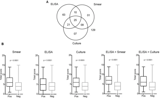
Diagnostic tests and clinical signs - ELISA, cultures, and smears were performed in all dogs. The positivity for each and the association of tests are described in Fig. 1A. Of the 265 dogs, 129 (48.68%) tested negative for all tests and were considered negative for CVL. Fifty-six dogs (21.13%) had positive ELISA and positive parasite culture results (n = 46) or/and positive parasite smear results (n = 35), and were considered positive for CVL (Fig. 1A). The next objective was to determine whether the total score value (range, 0-19), which combined all the signs, dif-fered according to the results of the diagnostic tests (Fig.
TABLE I
Baseline characteristics of sick dogs recruited in this study
CVL positive CVL negative All dogs
p value*
n = 56 n = 129 n = 265
Male n(%) 26 (46.43) 60 (46.51) 126 (47.55) 1.000**
Breed n(%)
Dalmatian 0 (0.00) 1 (0.77) 2 (0.75) 0.539***
Brazilian mastiff 0 (0.00) 1 (0.77) 3 (1.13)
German Shepherd 1 (1.79) 2 (1.55) 5 (1.89)
Pinscher 2 (3.57) 3 (2.33) 7 (2.64)
Pit bull 2 (3.57) 4 (3.10) 9 (3.40)
Poodle 2 (3.57) 9 (6.98) 14 (5.28)
Rottweiler 1 (1.79) 3 (2.33) 5 (1.89)
Yorkshire 0 (0.00) 0 (0.00) 2 (0.75)
Mongrel 46 (82.14) 94 (72.87) 201 (75.85)
Others 2 (3.57) 12 (9.30) 17 (6.42)
1B). The sign-based score was higher for the positive re-sults of all three individual diagnostic tests (ELISA, p < 0.001; parasite culture, p = 0.0021; and smear, p = 0.0003) and their combined results (ELISA and parasite culture, p < 0.0001; ELISA and parasite smear, p < 0.0001) than for the negative results of these tests (Fig. 1B).
A heat map analysis was performed for identifying clusters based on the signs of sick dogs (Fig. 2). Over-all, the presence of some signs was more frequent in sick dogs with CVL, as illustrated by lymph node en-largement, presence of ectoparasites, alterations in the bristles, and muzzle/ear lesions (Fig. 2). In addition, organ-related signs from the proposed score (systemic, cutaneous, and ocular groups) had a general trend for clustering. Clusters were observed between the ocular signs (keratoconjunctivitis and blepharitis) and between the cutaneous signs (skin lesion and muzzle/ear lesion, for example), demonstrating that these signs were char-acteristic of sick dogs from this endemic area (Fig. 2).
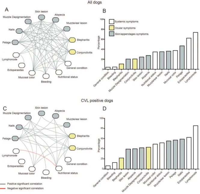
Networking the clinical signs according to CVL status - A higher complexity, measured by the network density (D = 0.346), was observed in all dogs when compared with the CVL-positive dogs (D = 0.165) (Fig. 3A, 3C, re-spectively); however, this could be attributed to the dif-ferent sample sizes. The majority of signs (except lymph node size and ectoparasite presence) had a high level of association in all dogs brought into the reference
veteri-nary hospital (Fig. 3A). The percentage of positivity for each sign for all dogs is illustrated in Fig. 3B. In the CVL-positive dogs, half of the signs (keratoconjunctivitis, mu-cosal colour, muzzle depigmentation, nutritional status, nails, bristles, and blepharitis) had the highest number of connections, suggesting their important role in the final clinical profile. The only significant negative correlation was between the lymph node size and nutritional status variables (Fig. 3C). The percentage of positivity for each sign in the CVL-positive dogs is demonstrated in Fig. 3D. The results showed that the overall positivity for the clini-cal signs was slightly more expressed in CVL-positive dogs than in all the sick dogs, and that the relative distri-butions of some signs differed between the two groups, as illustrated by serous or mucopurulent keratoconjunctivitis (42.86% and 20.65%, p = 0.001, Fig. 3D, B, respectively).
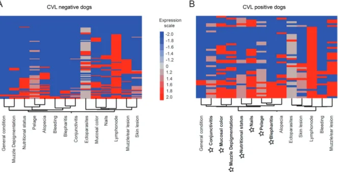
Heat map analyses according to the different signs were performed for CVL-negative and CVL-positive dogs (Fig. 4A-B, respectively). The results showed that the majority of signs were expressed more highly in the CVL-positive dogs than in the CVL-negative dogs. Moreover, the signs with the highest connectivity dem-onstrated in Fig. 3C also clustered in the CVL-positive heat map (highlighted in Fig. 4B) with smaller clusters between the following pairs of signs: mucosal colour and muzzle depigmentation; nutritional status and nails; and bristles and blepharitis (Fig. 4B).
TABLE II
Frequency of clinical signs in dogs with canine visceral leishmaniasis (CVL), ehrlichiosis, and without CVL
Clinical signs n(%)
1 2 3
p value 1 vs. 2
p value 1 vs. 3
p value 2 vs. 3 VL positive VL negative E. canis
n = 56 n = 129 n = 22
Apathy* 3 (5.36) 4 (3.10) 1 (4.54) 0.4341 1.000 0.5502
Nutritional status**
Thin 26 (46.43) 35 (27.13) 1 (4.54) 0.0023 < 0.0001 0.0274
Cachectic 3 (5.36) 0 (0.00) 0 (0.00)
Enlarged lymph nodes* 47 (83.93) 88 (68.22) 5 (22.73) 0.0308 < 0.0001 < 0.0001
Pale* 27 (48.21) 46 (35.66) 4 (18.18) 0.1404 0.0202 0.1425
Bleeding* 7 (12.50) 4 (3.10) 0 (0.00) 0.0194 0.1815 1.000
Bristles**
Regular 23 (41.07) 43 (33.33) 1 (4.54) 0.1100 0.0001 0.0035
Bad/opaque 9 (16.07) 11 (8.53) 0 (0.00)
Muzzle/ear lesion* 31 (55.36) 36 (27.91) 0 (0.00) 0.0005 <0.0001 0.0022
Onychogryphosis* 33 (58.93) 37 (28.68) 2 (9.09) 0.0001 <0.0001 0.0650
Skin lesion***
Presence 16 (28.57) 19 (14.73) 3 (13.64) 0.0162 0.0695 0.5727
Ulcer 6 (10.71) 6 (4.65) 0 (0.00)
Muzzle depigmentation* 23 (41.07) 17 (13.18) 0 (0.00) < 0.0001 0.0002 0.1352
Alopecia* 22 (39.29) 29 (22.48) 2 (9.09) 0.0308 0.0129 0.2511
Blepharitis* 12 (21.43) 8 (6.20) 0 (0.00) 0.0039 0.0159 0.6039
Keratoconjunctivitis**
Serous 21 (37.50) 12 (9.30) 1 (4.54) <0.0001 0.0009 0.4693
Mucopurulent 3 (5.36) 4 (3.10) 0 (0.00)
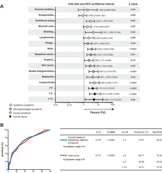
Association between clinical profile and CVL status - Ten of the 14 signs seemed to favour CVL, with special influences of onychogryphosis (long nails) (OR: 3.529; 95% CI: 1.832-6.796; p < 0.001), muzzle depigmentation
(OR: 4.651; 95% CI: 2.218-9.750; p < 0.001), and kerato-conjunctivitis (OR: 5.400; 95% CI: 2.549-11.441; p < 0.001) (Fig. 5A). Apathy (attitude), presence of ectoparasites, pale mucosal colour, and lymph node enlargement did not show any influence on CVL (p = 0.433, p = 0.609, p = 0.097, and p = 0.068, respectively; Fig. 5A). The cut-off values (≥ 6, ≥ 9, and ≥ 12) for the total clinical score (range, 0-19) established by using the ROC curve and C-statistics analy-ses (Fig. 5B) were also imputed in the regression model. The cut-off values ≥ 6 (OR: 4.151; 95% CI: 2.141-8.047; p < 0.001) and ≥ 9 (OR: 5.812; 95% CI: 2.399-14.083; p < 0.001) had almost similar performance in distinguishing between the CVL-positive and CVL-negative animals, and the cut-off value ≥ 12 had a lower precision (with a higher CI) than did the other described cut-off values (OR: 5.040; 95% CI: 1.213-20.20937; p = 0.026) (Fig. 5A).
The total clinical score (14 signs; range, 0-19) and a score based on the statistically different signs (10 signs; range, 0-14) shown in Fig. 5A had considerable power to distinguish between the positive and the CVL-negative dogs (Fig. 5B). The total score had an area un-der the curve (AUC) of 0.717 (p < 0.0001), and the cut-off value of ≥ 6 was the one with the best sensitivity for as -sociation (60.71%) and specificity (73.64%) to discrimi-nate the CVL-positive and CVL-negative dogs (Fig. 5B). With the higher cut-off values (≥ 9 and ≥ 12), the specificity increased (93.02% and 97.67%, respectively) despite the decrease in sensitivity (30.36% and 10.71%, respectively). Furthermore, the score based on
statisti-Fig. 1: canine visceral leishmaniasis (CVL) diagnostic tests and clinical signs. ELISA, smear, and culture tests for CVL were performed for all dogs brought to a reference veterinary hospital in the endemic area (A). Total clinical scores (range, 0-19) were higher in dogs with positive results on ELISA, culture, and smear tests and for the combinations of ELISA + culture or ELISA + smear tests (B). Data were compared using the Mann-Whitney test (B).
cally different signs had an AUC of 0.727 (p < 0.0001), and the cut-off value ≥ 3 had a sensitivity and specific -ity of 73.21% and 62.02%, respectively, to discriminate animals according to CVL status (Fig. 5B).
The score proposed herein was applied in dogs with eh-rlichiosis (but without CVL) in order to assess the ability of this score to test the specificity to CVL diagnosis. The clin-ical signs observed in the subjects with ehrlichiosis are de-scribed in Table II. With a cut-off value ≥ 3, the total score had an AUC of 0.9391 (p < 0.0001), and a sensitivity and specificity of 85.71% and 86.36%, respectively, to distin-guish the CVL-positive dogs from those with ehrlichiosis.
DISCUSSION
The serological and molecular diagnostic methods for CVL may be unavailable in endemic areas with lim-ited resources, and in such areas, the diagnosis may be based on only clinical signs. In the present study, dogs that tested positive for CVL with different diagnos-tic methods (ELISA, parasite smear, or culture) had a higher clinical score than did CVL-negative sick dogs. The cut-off value > 6 obtained from the clinical score proposed herein performed well in distinguishing CVL-positive dogs from CVL-negative sick dogs with 60.71% and 73.64% sensitivity and specificity, respectively.
Fig. 4: heat map analysis of the clinical signs of dogs according to canine visceral leishmaniasis (CVL) status. Two-way hierarchical cluster analysis (Ward’s method) was performed to identify patterns of associations between different signs for negative dogs (A) and CVL-positive dogs (B). The colours show the fold variation from the median values (log transformed) calculated for each sign. Highlighted in bold and with star symbols are those signs with the highest association observed in CVL-positive dogs in Fig. 3C.
Gouvêa et al. (2016), who conducted a study in the same area, found a cut-off value > 2 for a model based on the clinical score only (sensitivity = 75.3%; specificity = 65.9%) and a cut-off value > 9 for an association of IFAT + clinical score (sensitivity = 86.5%; specificity = 70%), thereby showing it was the best cut-off value for detect-ing the most infectious dogs and for disease control.
The clinical signs of CVL are important for diagno-sis, and the clinical score based on sick dogs’ clinical signs proposed herein was higher in subjects who tested positive for CVL on different diagnostic tests, suggest-ing it can help in diagnosis dependsuggest-ing on the cut-off value chosen. A group of clinical signs of Leishmania infection (keratoconjunctivitis, mucosal colour, muzzle depigmentation, nutritional status, nails, bristles, and blepharitis) was highly associated and clustered in the heat map results. Furthermore, the sole presence of ony-chogryphosis, muzzle depigmentation, or keratocon-junctivitis correlated highly with leishmaniasis. Manna et al. (2009) also created a severity clinical score that was associated with parasite load in CVL; however, this severity score was not compared with those of sick dogs without CVL to establish its diagnostic power. Other studies have demonstrated a model based on a scoring system combining the clinical signs and serological (IFAT) results as a tool to help in CVL control strategies (Proverbio et al. 2014, Gouvêa et al. 2016).
CVL diagnosis is still a challenge for veterinarians because of the lack of a perfect diagnostic test for this condition (Solano-Gallego et al. 2011). In the present study, CVL-positive dogs were diagnosed by both a positive serological test (ELISA) and a positive
parasito-logical test (parasite culture or smear) to confirm CVL diagnosis by using high-sensitivity and high-specificity methods, respectively. CVL diagnoses based only on an-ti-Leishmania antibody detection are not satisfactory as they do not discriminate between the disease and an as-ymptomatic condition; moreover, the antibodies cross-react with other pathogens (Sarkari et al. 2005, Zanette et al. 2014). For example, the conventional serological methods using crude antigen to detect antibodies against Leishmania spp. show cross-reactivity with Trypanoso-ma cruzi because both parasites share similar antigenic epitopes owing to their phylogenetic similarity (Zanette et al. 2014). Moreover, dogs infected with T. cruzi have not yet been detected in Teresina, PI. These aspects may have contributed to the different sensitivity and specific-ity results obtained in the studies that used serological methods for detecting CVL (de Arruda et al. 2016).
In both sick and CVL-positive dogs, lymph node en-largement was the most common sign. In several case series, lymphadenomegaly was reported as the most fre-quent clinical sign in dogs with CVL (Ciaramellaet al. 1997, Moreno & Alvar 2002, Amusategui et al. 2003, Mannaet al. 2009). However, lymph node enlargement is a frequent response to infectious agents, and it is not specific to CVL (Mylonakis et al. 2011, Rodríguez-Mo-rales et al. 2011, Salem & Farag 2014).
respectively. Intriguingly, the clinical spectrum of CVL may vary according to the phase of the disease, the dog’s immunity status, and previous specific therapies; thus, these features should be considered during CVL diagno-sis (Ciaramella et al. 1997, Manzillo et al. 2013).
In the network analysis, we observed that some clini-cal signs (keratoconjunctivitis, mucosal colour, muzzle depigmentation, nutritional status, nails, bristles, and blepharitis) were more associated with CVL positivity than were other signs. Moreover, these signs clustered
in the heat map analysis, indicating their important role in predicting CVL status. Ocular signs (i.e., blepharitis and keratoconjunctivitis), cutaneous signs (i.e., onycho-gryphosis and opaque bristles), and systemic signs (i.e., pale mucous membrane and weight loss) have been ex-tensively described in CVL clinical studies (Ciaramella et al. 1997, Amusategui et al. 2003, Manna et al. 2009, de Freitas et al. 2012, Manzillo et al. 2013). Cutaneous find-ings are very common in dogs with CVL, and the pres-ence of skin lesions (including ulcers), onychogryphosis,
alterations in the bristles, dermatitis, and alopecia should be carefully considered in sick dogs from endemic areas (Ciaramella et al. 1997, Ferrer 1999).
Among the 14 different clinical signs that comprise the clinical score proposed in this study, many could be used to differentiate CVL-positive dogs from other sick dogs, especially the presence of keratoconjunctivitis, ony-chogryphosis, and muzzle depigmentation in the CVL-positive dogs. Manciantiet al. (1988) proposed classifying dogs with CVL into asymptomatic, oligosymptomatic, and symptomatic cases, with the presence of onychog-ryphosis, keratoconjunctivitis, and cutaneous alterations being the clinical signs expected in symptomatic dogs. In-terestingly, chronic cutaneous changes and ocular lesions were also signs that characterised dogs with severe CVL (Ciaramella et al. 1997). In addition, the cut-off values for the clinical score proposed herein performed well (AUCs higher than 70%) in distinguishing CVL-positive dogs from other sick dogs (with different diseases). Higher cut-off values had better specificity for CVL diagnosis be-cause dogs with elevated clinical scores had more signs of this disease; although high cut-off values lacked sensitiv-ity, they could be used with good reliability for confirm-ing CVL in severely sick dogs in endemic areas.
In this study, 17 dogs had positive smear and/or cul-ture test results and negative ELISA results for CVL (Fig. 1A). Notably, when the clinical scores of the CVL-positive dogs (based on this study criteria) were com-pared to those with only positive smear and/or culture test results, the latter were found to have a lower score (p = 0.0057). This could probably be because these dogs were in the initial stages of infection and had not yet had a clinical response to leishmaniasis.
In this study, we had excluded CVL-positive dogs co-infected with E. canis and B. canis,which can pro-duce clinical findings similar to CVL (Ciaramella et al. 1997). Some studies have demonstrated that dogs with co-infection presented more severe clinico-pathological abnormalities and were frequently misdiagnosed in rou-tine veterinary practices (Andrade et al. 2014). Thus, we have applied our clinical model only in dogs with CVL and ehrlichiosis. When the clinical score was applied in dogs infected with E. canis, we observed a great dis-criminatory power to distinguish dogs with CVL from those with ehrlichiosis, suggesting that the ability of this score to detect CVL when E. canis infection was not pre-viously excluded. Dogs with other diseases requiring po-tential differential diagnosis for CVL were also included in the present study. Dogs with trypanosomiasis caused by T. cruzi have a general lymphadenopathy (Barret al. 1991), which is a usual clinical sign found in all dogs in this study. Furthermore, severe seborrhoea, focal or diffuse alopecia, pustules, and blemishes are frequent-ly observed in 12% to 23% of dogs with canine atopic dermatitis and could be found in the CVL-negative sick dogs (Hensel et al. 2015). Similarly, bacterial skin dis-eases, superficial and deep folliculitis, and keratinisa-tion disorders may also be confused with clinical signs of CVL and should be considered in the CVL-negative dogs (Cardoso et al. 2011) .
This study proposes a clinical sign-based score for CVL diagnosis that can help veterinarians reliably iden-tify dogs with CVL. Although there is no evidence sup-porting dog culling for CVL control, culling is still prac-ticed in many endemic areas; this makes determining a correct diagnosis of CVL even more important (Costa 2011). The clinical score proposed herein is a guide for veterinarians mainly from highly endemic areas with limited diagnostic resources. Moreover, this clinical score can help confirm the accuracy of some diagnostic test results (i.e., serological methods). Nevertheless, di-agnostic tests are still the best way to confirm CVL in sick dogs and should not be totally replaced by diagnosis based only on clinical signs.
Appropriate CVL diagnosis is a big challenge in en-demic areas with limited diagnostic resources. We report here that a score based on the clinical signs of sick dogs in a highly endemic region can help CVL diagnosis. Over-all, the clinical score proposed had a good performance in discriminating CVL-positive dogs from other sick dogs. Furthermore, some clinical signs were more cor-related with CVL positivity, particularly the ocular and cutaneous signs. The results of this study may help vet-erinarians correctly diagnose CVL in sick dogs; however, further studies with larger samples are needed to confirm and validate this clinical score because this study only examined dogs brought to the veterinary hospital or col-lected by the Zoonosis Control Center. The higher preva-lence of CVL among these dogs could have influenced the positive predictive values of the diagnostic tests.
ACKNOWLEDGEMENTS
To all employees of the veterinary hospital of the Federal University of Piauí, Teresina, Brazil; our team at FIOCRUZ-Bahia, Brazil, Jorge Tolentino, Adorielze Leite, Nataly Al-exandrino, and Andrezza Kariny, for help with logistics; and Global Science Editing, UK, for English language corrections.
REFERENCES
Alvar J, Vélez ID, Bern C, Herrero M, Desjeux P, Cano J, et al. Leish-maniasis worldwide and global estimates of its incidence. PLoS ONE. 2012; 7(5): e35671.
Amusategui I, Sainz A, Rodriguez F, Tesouro MA. Distribution and relationships between clinical and biopathological parameters in canine leishmaniasis. Eur J Epidemiol. 2003; 18(2): 147-56. Andrade GB, Barreto WT, Santos LL, Ribeiro LR, Macedo GC,
Sou-sa KC, et al. Pathology of dogs in Campo Grande, MS, Brazil naturally co-infected with Leishmania infantum and Ehrlichia canis. Rev Bras Parasitol Vet. 2014; 23: 509-15.
Badaró R, Reed SG, Barral A, Orge G, Jones TC. Evaluation of the mi-cro enzyme-linked immunosorbent assay (ELISA) for antibodies in American visceral leishmaniasis: antigen selection for detection of infection-specific responses. Am J Trop Med Hyg. 1986; 35(1): 72-8. Barr SC, Gossett KA, Klei TR. Clinical, clinicopathologic, and para-sitologic observations of trypanosomiasis in dogs infected with North American Trypanosoma cruzi isolates. Am J Vet Res. 1991; 52(6): 954-60.
Cardoso MJL, Machado LHA, Melussi M, Zamarian TP, Carnielli CM, Ferreira Jr JCM. Skin diseases in dogs: a review of 257 cas-es. Arch Vet Sci. 2011; 16(2): 66-74.
et al. A retrospective clinical study of canine leishmaniasis in 150 dogs naturally infected by Leishmania infantum. Vet Rec. 1997; 141(21): 539-43.
Costa CH. How effective is dog culling in controlling zoonotic vis-ceral leishmaniasis? A critical evaluation of the science, politics and ethics behind this public health policy. Rev Soc Bras Med Trop. 2011; 44(2): 232-42.
da Costa-Val AP, Cavalcanti RR, Gontijo NF, Michalik MS, Alexan-der B, Williams P, et al. Canine visceral leishmaniasis: relation-ships between clinical status, humoral immune response, haema-tology and Lutzomyia (Lutzomyia) longipalpis infectivity. Vet J. 2007; 174(3): 636-43.
Dantas-Torres F. The role of dogs as reservoirs of Leishmania parasites, with emphasis on Leishmania (Leishmania) infantum and Leish-mania (Viannia) braziliensis. Vet Parasitol. 2007; 149(3-4): 139-46. de Araújo VEM, Pinheiro LC, Almeida MCM, Menezes FC, Morais
MHF, Reis IA, et al. Relative risk of visceral leishmaniasis in Brazil: a spatial analysis in urban area. PLoS Negl Trop Dis. 2013; 7(11): e2540.
de Arruda MM, Figueiredo FB, Marcelino AP, Barbosa JR, Werneck GL, Noronha EF, et al. Sensitivity and specificity of parallel or serial serological testing for detection of canine Leishmania in-fection. Mem Inst Oswaldo Cruz. 2016; 111(3): 168-73.
de Castro MB, Machado RZ, de Aquino LP, Alessi AC, Costa MT. Experimental acute canine monocytic ehrlichiosis: clinicopatho-logical and immunopathoclinicopatho-logical findings. Vet Parasitol. 2004; 119(1): 73-86.
de Freitas JC, Lopes-Neto BE, de Abreu CR, Coura-Vital W, Braga SL, Reis AB, et al. Profile of anti-Leishmania antibodies related to clinical picture in canine visceral leishmaniasis. Res Vet Sci. 2012; 93(2): 705-9.
Ferrer L. Clinical aspects of canine leishmaniasis. In: R Killick-Ken-drick, editor. Canine leishmaniasis: an updat. Proceedings of a Canine Leishmaniasis Forum. Barcelona: Sitges; 1999. pp. 6-10. Gouvêa MV, Mendonça IL, Cruz MSP, Costa CHN, Braga JU,
Wer-neck GL. Predictive factors for Leishmania infantum infection in dogs examined at a veterinary teaching hospital in Teresina, state of Piauí, Brazil. Rev Soc Bras Med Trop. 2016; 49(1): 107-11. Hensel P, Santoro D, Favrot C, Hill P, Griffin C. Canine atopic derma-titis: detailed guidelines for diagnosis and allergen identification. BMC Vet Res. 2015; 11: 196.
Jefferies R, Ryan UM, Irwin PJ. PCR-RFLP for the detection and dif-ferentiation of the canine piroplasm species and its use with filter paper-based technologies. Vet Parasitol. 2007; 144(1-2): 20-7. Mancianti F, Gramiccia M, Gradoni L, Pieri S. Studies on canine
leishmaniasis control. 1. Evolution of infection of different clini-cal forms of canine leishmaniasis following antimonial treat-ment. Trans R Soc Trop Med Hyg. 1988; 82(4).
Manna L, Reale S, Vitale F, Gravino AE. Evidence for a relationship between Leishmania load and clinical manifestations. Res Vet Sci. 2009; 87(1).
Manzillo VF, Muccio TD, Cappiello S, Scalone A, Paparcone R, Fio-rentino E, et al. Prospective study on the incidence and progres-sion of clinical signs in naïve dogs naturally infected by Leishma-nia infantum. PLoS Negl Trop Dis. 2013; 7(5): e2225.
Moreno J, Alvar J. Canine leishmaniasis: epidemiological risk and the experimental model. Trends Parasitol. 2002; 18(9).
Mylonakis ME, Borjesson DL, Leontides L, Siarkou VI, Theodorou K, Koutinas AF. Cytologic patterns of lymphadenopathy in ca-nine monocytic ehrlichiosis. Vet Clin Pathol. 2011; 40(1): 78-83. Proverbio D, Spada E, de Giorgi GB, Perego R, Valena E. Relationship
between Leishmania IFAT titer and clinicopathological manifesta-tions (clinical score) in dogs. BioMed Res Int. 2014; 2014: 412808. Ribeiro RR, da Silva SM, Fulgêncio GO, Michalick MS, Frezard FJ.
Relationship between clinical and pathological signs and severity of canine leishmaniasis. Rev Bras Parasitol Vet. 2013; 22(3): 373-8. Rodríguez-Morales O, Ballinas-Verdugo MA, Alejandre-Aguilar R,
Reyes PA, Arce-Fonseca M. Trypanosoma cruzi connatal trans-mission in dogs with Chagas disease: experimental case report. Vector Borne Zoonotic Dis. 2011; 11(10): 1365-70.
Salem NY, Farag HS. Clinical, hematologic, and molecular findings in naturally occurring Babesia canis vogeli in Egyptian dogs. Vet Med Int. 2014; 2014: 270345.
Sarkari B, Michael C, Hommel M. A capture ELISA for the diagnosis of visceral leishmaniasis using a monoclonal antibody against a leishmanial urinary antigen. Iran Biomed J. 2005; 9: 117-22. Solano-Gallego L, Miró G, Koutinas A, Cardoso L, Pennisi MG,
Fer-rer L, et al. LeishVet guidelines for the practical management of canine leishmaniosis. Parasit Vectors. 2011; 4: 86.
Souza AP, Soto M, Costa JML, Boaventura VS, Oliveira CI, Cristal JR, et al. Towards a more precise serological diagnosis of human tegumentary leishmaniasis using Leishmania recombinant pro-teins. PLoS ONE. 2013; 8(6): e66110.
Tafuri WL, de Oliveira MR, Melo MN. Canine visceral leishmani-osis: a remarkable histopathological picture of one case reported from Brazil. Vet Parasitol. 2001; 96(3): 203-12.
Wen B, Rikihisa Y, Mott JM, Greene R, Kim HY, Zhi N, et al. Com-parison of nested PCR with immunofluorescent-antibody assay for detection of Ehrlichia canis infection in dogs treated with doxycycline. J Clin Microbiol. 1997; 35(7): 1852-5.