Bioeffects of albumin-encapsulated
microbubbles and real-time myocardial
contrast echocardiography in an
experimental canine model
1Laboratório de Investigação em Isquemia Miocárdica,
Unidade Clínica de Aterosclerose, 2Laboratório de Ecocardiografia, 3Laboratório de Patologia, 4Divisão Clínica, Instituto do Coração,
Faculdade de Medicina, Universidade de São Paulo, São Paulo, SP, Brasil
P.M.M. Dourado1,
J.M. Tsutsui2, J.M.T. Santos2,
V.D. Aiello3, W. Mathias Jr.2,
J.A.F. Ramires4 P.L. da Luz1
and A.C.P. Chagas1
Abstract
Myocardial contrast echocardiography has been used for assessing myocardial perfusion. Some concerns regarding its safety still remain, mainly regarding the induction of microvascular alterations. We sought to determine the bioeffects of microbubbles and real-time myocardial contrast echocardiography (RTMCE) in a closed-chest canine model. Eighteen mongrel dogs were randomly assigned to two groups. Nine were submitted to continuous intravenous infusion of perfluorocarbon-exposed sonicated dextrose albumin (PESDA) plus continuous imaging using power pulse inversion RTMCE for 180 min, associated with manually deflagrated high-mechanical index impulses. The control group consisted of 3 dogs submitted to continu-ous imaging using RTMCE without PESDA, 3 dogs received PESDA alone, and 3 dogs were sham-operated. Hemodynamics and cardiac rhythm were monitored continuously. Histological analysis was per-formed on cardiac and pulmonary tissues. No hemodynamic changes or cardiac arrhythmias were observed in any group. Normal left ventricular ejection fraction and myocardial perfusion were main-tained throughout the protocol. Frequency of mild and focal micro-hemorrhage areas in myocardial and pulmonary tissue was similar in PESDA plus RTMCE and control groups. The percentages of positive microscopical fields in the myocardium were 0.4 and 0.7% (P = NS) in the PESDA plus RTMCE and control groups, respectively, and in the lungs they were 2.1 and 1.1%, respectively (P = NS). In this canine model, myocardial perfusion imaging obtained with PESDA and RTMCE was safe, with no alteration in cardiac rhythm or left ventric-ular function. Mild and focal myocardial and pulmonary microhemor-rhages were observed in both groups, and may be attributed to surgical tissue manipulation.
Correspondence
P.M.M. Dourado
Laboratório de Investigação em Isquemia Miocárdica
Unidade Clínica de Aterosclerose Instituto do Coração, FM, USP Av. Dr. Enéas C. Aguiar, 44 05403-900 São Paulo, SP Brasil
Fax +55-11-3069-5447 E-mail: pmmdourado@terra.com.br
Publication supported by FAPESP.
Received June 17, 2005 Accepted March 3, 2006
Key words
•Microbubbles •Ultrasound •Bioeffects
•Myocardial contrast
Introduction
Recently, the assessment of myocardial perfusion by echocardiography following intravenous injection of ultrasound contrast agents has proved to be feasible (1). The development of low-mechanical index im-aging techniques that cause minimal micro-bubble destruction has permitted simulta-neous analysis of myocardial perfusion and function in real time (2). Real-time myocar-dial contrast echocardiography (RTMCE) increases the sensitivity of dobutamine stress testing for the detection of coronary artery disease (3,4) and can distinguish between stunned and infarcted myocardium after acute ischemia (5). However, some concerns re-garding the safety of this new imaging mo-dality remain, mainly because some animal studies have demonstrated damage to the endothelial system in the presence of microbubbles and ultrasound (6-9). Although investigations of the interaction of ultra-sound and contrast agents in vivo are scarce, some effects have been reported in the litera-ture (10) such as capillary damage in organs that contain air such as the lungs (7), damage to the microvasculature (11),and limited capillary ruptures in the heart (12). More-over, simultaneous exposure of isolated rab-bit hearts to ultrasound and a contrast agent has been found to result in transient depres-sion of left ventricular contractile function, an increase in coronary perfusion pressure and in lactate production, and in limited mechanical index-dependent capillary rup-ture (12). Nevertheless, the clinical impor-tance of these effects has not been estab-lished (13).
The bioeffects of the mechanical index during RTMCE imaging are particularly important since one of the current applica-tions of this technique is the determination of myocardial blood flow. The kinetics of microbubbles in the myocardium can be de-termined by applying a high-mechanical in-dex flash impulse to destroy the microbubbles
in the myocardium, followed by measure-ment of microbubble replenishmeasure-ment velocity and plateau intensity. The application of high-mechanical index pulse sequences may induce microvascular damage (14).
The aim of the present study was to deter-mine the effect of intravenous infusion of albumin-encapsulated microbubbles and RTMCE with intermittent application of high-mechanical index flash impulses on the cardiac and pulmonary microvasculature of dogs.
Material and Methods
Animal preparation
Eighteen mongrel dogs were anesthe-tized intravenously with 30 mg/kg sodium pentobarbital, intubated and mechanically ventilated with a respirator pump (model 670, Takaoka Medical, São Paulo, SP, Bra-zil). The anesthesia was supplemented with additional intravenous doses of sodium pen-tobarbital (2-3 mL) when necessary. A 7-Fr catheter was inserted into the left carotid artery and connected to a Biopac Systems device (model TSD-104, Biopac Systems, Santa Barbara, CA, USA) to record arterial pressure and heart rate. Another 7-Fr cath-eter was inserted into the femoral vein to infuse drugs and ultrasound contrast and a third catheter was placed into the jugular vein to record central venous pressure. He-modynamic parameters and electrocardio-graphic rhythm were monitored continu-ously. At the end of each experiment, all dogs received an additional dose of 30 mg/ kg sodium pentobarbital before euthanasia.
Myocardial contrast echocardiography
decafluorobutane gas with a 3:1 mixture of 5% dextrose and 5% human serum albumin and the mixture was sonicated for 75 s. The mean concentration of microbubbles pro-duced with this technique was 1.4 x 10⁹
micro-bubbles per mL and the mean size was 4.6 ± 0.2 µm (as reported in Ref. 2). RTMCE was obtained by continuous intravenous infu-sion of 0.2 mL/kg PESDA diluted in 80 mL of normal saline at a rate of 2-3 mL/min, injected through the femoral vein.
Echocardiographic images were acquired in a closed-chest model. RTMCE was per-formed using a commercially available ul-trasound system (HDI 5000, Philips Medi-cal Systems, Bothell, WA, USA) equipped with a 4-2 MHz broadband transducer. Im-aging was obtained by the power pulse inversion modality at a low-mechanical index (0.1). Specific instrumentation set-tings were low dynamic range, frame rate >25 Hz, and maximal line density. After adjustments, these parameters were constant for each experiment. Animals in the groups that received RTMCE were exposed to con-tinuous imaging using power pulse inver-sion RTMCE for 180 min. A manually trig-gered, transient, high-mechanical index (1.2) imaging flash was deflagrated at each 12 heartbeats to destroy microbubbles within the myocardium and to permit subsequent myocardial replenishment (15,16). Although the analysis of myocardial perfusion was performed using the apical four-, three-, and two-chamber views, continuous imaging with RTMCE was performed most of the time using the short-axis view at the level of papillary muscles. The images were stored on videotape and on an optical disk for off-line processing and analysis.
Study protocol
The protocol was reviewed and approved by the Institutional Animal Use Committee of the Heart Institute, University of São Paulo Medical School. Experiments were carried
out in accordance with the American Heart Association on Research Animal Use (17). The dogs were treated with standard ration and fasted overnight before the experiments. The effects of ultrasound-induced mi-crobubble destruction were evaluated in two groups. The PESDA plus RTMCE group consisted of 9 dogs weighing 17 ± 2 kg which received intravenous albumin-encap-sulated microbubbles, PESDA and RTMCE for 180 min, associated with manually defla-grated high-mechanical index flash impulses to destroy the microbubbles every 12 heart-beats. The control group consisted of 9 dogs weighing 17 ± 2 kg. In this group, 3 dogs received intravenous infusion of PESDA for 180 min and no ultrasound (PESDA alone), 3 dogs underwent RTMCE imaging for 180 min associated with manually deflagrated high-mechanical index flash impulses with-out infusion of PESDA (RTMCE alone), and 3 dogs were sham-operated and did not receive PESDA or ultrasound.
Left ventricular systolic function and myocardial perfusion were evaluated at base-line and at 90 and 180 min during the proto-col. Left ventricular ejection fraction was estimated by two-dimensional images (18). The presence of premature ventricular con-tractions (PVC) or any other cardiac ar-rhythmias was monitored continuously with a one-lead electrocardiogram connected to the echocardiographic system. Histological analysis of lung and heart samples was per-formed at the end of the protocol.
Histological analysis
lung (2.0 x 1.0 cm), one from the hilar region and the other sub-pleural, from the apex of the cranial lobe, were also examined. After fixation in 10% buffered formalin, the frag-ments were submitted to standard histologi-cal processing. Five-micrometer thick sec-tions were obtained, stained with hematoxy-lin-eosin and examined semi-quantitatively under the light microscope. The mean num-bers of 100X microscopical fields evaluated were 100 for the lung fragments and 120 for the hearts. We searched for areas of necro-sis, contraction bands, and hemorrhage. The percentage of “positive” microscopic fields relative to the total number of analyzed fields was determined.
Statistical analysis
Continuous and normally distributed data are reported as mean ± one standard devia-tion (SD) and qualitative data are reported as proportions. Comparisons of hemodynamic data and left ventricular ejection fraction at baseline and at 90 and 180 min of treatment were performed by repeated-measures anal-ysis of variance or the Friedman test, as appropriate. Inter- and intra-group compari-sons of continuous variables were performed by the unpaired and paired Student t-test, respectively. Comparison of proportions was performed by the exact Fisher test. A P value
<0.05 (two-sided) was considered to be sta-tistically significant.
Results
Intravenous infusion of PESDA micro-bubbles permitted the analysis of myocar-dial perfusion and left ventricular function in all dogs during the protocol. Two dogs were excluded, one from the PESDA plus RTMCE group and the other from the PESDA group because of hemodynamic instability before the beginning of microbubble infu-sion, which was attributed to the effects of anesthesia and hypovolemia. Baseline left ventricular ejection fraction was normal in all dogs with no difference between PESDA plus RTMCE and control groups (0.70 ± 0.03 vs 0.69 ± 0.04; P = 0.74). No changes in left ventricular ejection fraction were ob-served at 90 or 180 min of the protocol in the PESDA plus RTMCE group (P = 0.8) and in the control group (P = 0.56). Myocardial perfusion was normal in all animals.
At baseline, no differences were observed in heart rate (128.6 ± 23.8 vs 128.8 ± 23.9 bpm; P = 0.98), mean arterial blood pressure (93.1 ± 9.9 vs 91.3 ± 16.2 mmHg; P = 0.69) and central venous pressure (1.6 ± 1.5 vs 1.5 ± 2.0 cmH2O; P = 0.52) between PESDA
plus RTMCE and control groups. No statis-tically significant changes occurred in these parameters at 90 and 180 min of protocol in both groups.
PVC were observed in one dog of the control group that received RTMCE alone. In this case, PVC were observed immediately after high-mechanical index flash impulses. No PVC or other arrhythmias were observed in dogs that received PESDA plus RTMCE, PESDA alone, or were sham-operated.
Histopathological findings
No macroscopic abnormalities were ob-served in the heart or lungs of any animal. Table 1 shows the frequency of
microscopi-Table 1. Histological findings for myocardial and pulmonary tissues from dogs submit-ted to perfluorocarbon-exposed sonicasubmit-ted dextrose albumin (PESDA) and real-time myocardial contrast echocardiography (RTMCE).
Histological findings PESDA + RTMCE Control group (N = 8)
(N = 8)
PESDA RTMCE
Sham-alone alone operated
Myocardium
Focal hemorrhage 2 1 2 1
Contraction bands 0 0 0 1
Lung
Focal alveolar hemorrhage 3 0 1 2
Perivascular focal hemorrhage 2 0 0 2
Table 2. Mean percentages of 100X microscopical fields presenting microhemorrhage in myocardium and lung sections.
PESDA + RTMCE Control group (N = 8)
(N = 8)
PESDA RTMCE
Sham-alone alone operated
Myocardium 0.4% 1.3% 0.6% 0.6%
Lung 2.1% 0.0% 1.3% 1.7%
PESDA = perfluorocarbon-exposed sonicated dextrose albumin; RTMCE = real-time myocardial contrast echocardiography.
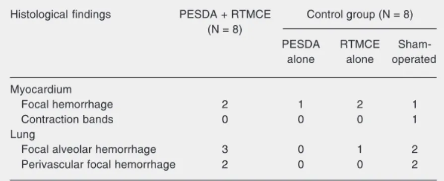
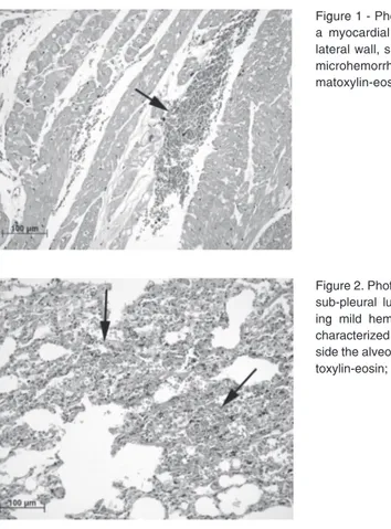
Figure 2. Photomicrography of a sub-pleural lung sample show-ing mild hemorrhage (arrows), characterized by erythrocytes in-side the alveolar spaces. Hema-toxylin-eosin; bar = 100 µm. Figure 1 - Photomicrography of a myocardial sample from the lateral wall, showing a focus of microhemorrhage (arrow). He-matoxylin-eosin; bar = 100 µm.
cal lesions obtained by histopathological analysis of the myocardium and lung samples. No significant difference was de-tected in animals from the PESDA plus RTMCE and control groups.
Mild and focal hemorrhage in the myo-cardium was similarly observed in both groups (Figure 1). Myocardium contraction-bands were observed in one sham-operated dog.
Table 2 shows the mean percentages of 100X microscopical fields presenting mi-crohemorrhages in the myocardium and lung samples. The mean percentages of positive microscopical fields in the myocardium were 0.4 and 0.7% in the PESDA plus RTMCE and control groups, respectively.
Focal and mild regions of alveolar hem-orrhage in pulmonary tissue were observed in the PESDA plus RTMCE and control groups (Figure 2). No significant difference was detected between groups when compar-ing the hilar and sub-pleural fragments of pulmonary tissue. The mean percentages of positive microscopical fields in the lungs were 2.1 and 1.1% in the PESDA plus RTMCE and control group, respectively.
Discussion
In recent years, myocardial contrast echo-cardiography has been demonstrated to be a feasible and accurate method for the evalua-tion of myocardial perfusion in many clini-cal settings (2-5). In addition, the combina-tion of intravenous contrast agents and RTMCE with application of high-mechani-cal index impulses for microbubble destruc-tion followed by myocardial replenishment has permitted the quantification of myocar-dial blood flow (19,20). However, some is-sues still need to be addressed before the widespread use of this technique in clinical practice. Previous studies have demonstrated adverse effects, including microvasculature damage, with contrast agents and ultrasound. Although these studies were restricted to
PESDA permitted simultaneous assessment of myocardial perfusion and function with a good safety profile, with no cardiac or pul-monary tissue necrosis as confirmed by pathological analysis. Although some lim-ited areas of focal microhemorrhage were present, they were detected at a similar rate in both the control and PESDA plus RTMCE groups. Pulmonary perivascular microhem-orrhage was observed in two dogs from the RTMCE protocol, and myocardial contrac-tion bands were detected in only one of the dogs from the control group (sham-oper-ated). These bands may correspond to recent ischemic myocardial necrosis. However, all these lesions represented minimal tissue in-volvement when compared to the total area, and could be attributed to the surgical ma-nipulation as well as the trauma resulting from mechanical ventilation.
The effects of microbubble cavitation by ultrasound have been already demonstrated in vitro and in vivo (12,21,22). The micro-bubbles used in our study (PESDA) consist of decafluorobutane gas surrounded by an albumin shell, with sufficient stability to cross the pulmonary capillary barrier and to reach the left cardiac cavities and coronary microcirculation. Porter and Xie (23) were the first to show the safety profile and utility of intravenously injected PESDA in 28 pa-tients, using intermittent harmonic triggered imaging. The authors demonstrated the pos-sibility of obtaining myocardial perfusion using a small bolus of PESDA at a concen-tration of 1.4 x 10⁹ microbubbles/mL - 0.005
mL/kg in dogs and 0.0025 mL/kg in humans - with no significant adverse effects. Other experimental studies have shown no change in cardiac output or in systolic and diastolic pressure in the left ventricle, with a minimal increase in mean systolic pulmonary pres-sure when using a 0.30 mL/kg dose of PESDA (24-26). Mathias Jr. et al. (27) stud-ied 68 patients who underwent dobutamine stress echocardiography using PESDA for enhancement of endocardial border
delinea-tion. None of the patients presented serious cardiac arrhythmia or important side effects and no significant changes were observed in blood pressure, respiratory frequency or oxy-gen saturation.
In the present study, we used the dose of 0.20 mL/kg PESDA diluted in 80 mL nor-mal saline, administered continuously for 180 min at a rate of 2-4 mL/min, which is approximately seven times higher than the dose recommended for humans. This high dose and long exposure did not result in any adverse effects in dogs. There is some evi-dence that the intravenous injection of con-trast agents is associated with induction of premature ventricular depolarizations when using high-mechanical index ultrasound. In general, the extension of bioeffects produced by contrast agents is proportional to the pres-sure amplitude (Mega Pascals) of expopres-sure. Lower frequencies (for example, 1 to 2 MHz) of exposure are more effective than higher frequencies. One study using intravenous contrast agents has reported an increased number of PVC when applying end-sys-tolic-triggered imaging at a mechanical in-dex of 1.5 (28). The induction of arrhyth-mias was related to both the dose of the contrast agent and the acoustic pressure stud-ied. In contrasty, Raisinghani et al. (29) reported no increase in the number of PVC in patients being imaged with triggered ul-trasound at a high-mechanical indexduring dipyridamole stress. In our study, we did not observe changes in mean arterial blood pres-sure, central venous prespres-sure, or heart rate during any of the protocols. PVC were ob-served in one dog from the control group which did not receive microbubbles. No ar-rhythmias were observed in the dogs which received PESDA plus RTMCE, even after high-mechanical index flash impulses.
Limitations
possibility of minor microvascular or endo-thelial damage, which could only be de-tected by electron microscopy. On the other hand, even if these lesions occurred, they did not cause significant acute tissue damage, as demonstrated by our results. Tissue sam-pling was considered adequate, since no macroscopical lesions were detected and because sections were taken from areas most prone to injury due to their topographical location.
The effects of RTMCE and other cur-rently used contrast agents such as lipid-encapsulated microbubbles were not tested
in the present study. Although the safety profile of commercially available lipid-en-capsulated microbubbles has already been demonstrated in humans (30), the bioeffect of such a prolonged exposure to ultrasound has not been previously reported. Further studies are still necessary to address this issue.
In this experimental model, we demon-strated that RTMCE permitted simultaneous evaluation of myocardial perfusion and func-tion, without significant myocardial or pul-monary lesion in the setting of prolonged infusion of PESDA.
References
1. Wei K, Jayaweera AR, Firoozan S, Linka A, Skyba DM, Kaul S. Quantification of myocardial blood flow with ultrasound-induced de-struction of microbubbles administered as a constant venous infu-sion. Circulation 1998; 97: 473-483.
2. Porter TR, Xie F, Silver M, Kricsfeld D, Oleary E. Real-time perfu-sion imaging with low mechanical index pulse inverperfu-sion Doppler imaging. J Am Coll Cardiol 2001; 37: 748-753.
3. Elhendy A, O’Leary EL, Xie F, McGrain AC, Anderson JR, Porter TR. Comparative accuracy of real-time myocardial contrast perfu-sion imaging and wall motion analysis during dobutamine stress echocardiography for the diagnosis of coronary artery disease. J Am Coll Cardiol 2004; 44: 2185-2191.
4. Tsutsui JM, Xie F, McGrain AC, Mahrous H, Hankins J, O’Leary EL, et al. Comparison of low-mechanical index pulse sequence schemes for detecting myocardial perfusion abnormalities during vasodilator stress echocardiography. Am J Cardiol 2005; 95: 565-570. 5. Dourado PM, Tsutsui JM, Mathias Jr W, Andrade JL, da Luz PL,
Chagas AC. Evaluation of stunned and infarcted canine myocardi-um by real time myocardial contrast echocardiography. Braz J Med Biol Res 2003; 36: 1501-1509.
6. Dalecki D, Keller BB, Carstensen EL, Neel DS, Palladino JL, Noordergraaf A. Thresholds for premature ventricular contractions in frog hearts exposed to lithotripter fields. Ultrasound Med Biol
1991; 17: 341-346.
7. Dalecki D, Child SZ, Raeman CH, Cox C, Penney DP, Carstensen EL. Age dependence of ultrasonically induced lung hemorrhage in mice. Ultrasound Med Biol 1997; 23: 767-776.
8. Porter TR, Xie F. Therapeutic ultrasound for gene delivery. Echocar-diography 2001; 18: 349-353.
9. Price RJ, Skyba DM, Kaul S, Skalak TC. Delivery of colloidal par-ticles and red blood cells to tissue through microvessel ruptures created by targeted microbubble destruction with ultrasound. Circu-lation 1998; 98: 1264-1267.
10. Chen S, Kroll MH, Shohet RV, Frenkel P, Mayer SA, Grayburn PA. Bioeffects of myocardial contrast microbubble destruction by echo-cardiography. Echocardiography 2002; 19: 495-500.
11. Unger EC, Hersh E, Vannan M, Matsunaga TO, McCreery T. Local
drug and gene delivery through microbubbles. Prog Cardiovasc Dis
2001; 44: 45-54.
12. Ay T, Havaux X, Van Camp G, Campanelli B, Gisellu G, Pasquet A, et al. Destruction of contrast microbubbles by ultrasound: effects on myocardial function, coronary perfusion pressure, and microvascu-lar integrity. Circulation 2001; 104: 461-466.
13. Miller DL, Gies RA. The influence of ultrasound frequency and gas-body composition on the contrast agent-mediated enhancement of vascular bioeffects in mouse intestine. Ultrasound Med Biol 2000; 26: 307-313.
14. MacRobbie AG, Raeman CH, Child SZ, Dalecki D. Thresholds for premature contractions in murine hearts exposed to pulsed ultra-sound. Ultrasound Med Biol 1997; 23: 761-765.
15. Becker H, Burns P. Handbook of contrast echocardiography: Left ventricular function and myocardial perfusion. New York: Springer-Verlag Publishers; 2000.
16. Pelberg RA, Wei K, Kamiyama N, Sklenar J, Bin J, Kaul S. Potential advantage of flash echocardiography for digital subtraction of B-mode images acquired during myocardial contrast echocardiogra-phy. J Am Soc Echocardiogr 1999; 12: 85-93.
17. Anonymous. Position of the American Heart Association on the use of research animals. A statement for health professionals from a task force appointed by the Board of Directors of the American Heart Association. Circ Res 1995; 57: 330-331.
18. Schiller NB, Shah PM, Crawford M, DeMaria A, Devereux R, Feigenbaum H, et al. Recommendations for quantitation of the left ventricle by two-dimensional echocardiography. American Society of Echocardiography Committee on Standards, Subcommittee on Quantitation of Two-Dimensional Echocardiograms. J Am Soc Echocardiogr 1989; 2: 358-367.
19. Peltier M, Vancraeynest D, Pasquet A, Ay T, Roelants V, D’hondt AM, et al. Assessment of the physiologic significance of coronary disease with dipyridamole real-time myocardial contrast echocardi-ography. Comparison with technetium-99m sestamibi single-photon emission computed tomography and quantitative coronary angiog-raphy. J Am Coll Cardiol 2004; 43: 257-264.
Quinones MA, et al. Identification of hibernating myocardium with quantitative intravenous myocardial contrast echocardiography: comparison with dobutamine echocardiography and thallium-201 scintigraphy. Circulation 2003; 107: 538-544.
21. Porter TR, Xie F. Ultrasound, microbubbles, and thrombolysis. Prog Cardiovasc Dis 2001; 44: 101-110.
22. Porter TR, Everbach C, Kricsfeld D, Xie F. Myocardial cavitational activity during continuous infusion and bolus intravenous injections of perfluorocarbon-containing microbubbles. J Am Soc Echocardiogr
2001; 14: 618-625.
23. Porter TR, Xie F. Transient myocardial contrast after initial exposure to diagnostic ultrasound pressures with minute doses of intrave-nously injected microbubbles. Demonstration and potential mechan-isms. Circulation 1995; 92: 2391-2395.
24. Porter TR, Xie F, Kricsfeld D, Armbruster RW. Improved myocardial contrast with second harmonic transient ultrasound response imag-ing in humans usimag-ing intravenous perfluorocarbon-exposed soni-cated dextrose albumin. J Am Coll Cardiol 1996; 27: 1497-1501. 25. Porter TR, Kricsfeld A, Deligonul U, Xie F. Detection of regional
perfusion abnormalities during adenosine stress echocardiography with intravenous perfluorocarbon-exposed sonicated dextrose
albu-min. Am Heart J 1996; 132: 41-47.
26. Porter TR, Xie F, Kricsfeld A, Deligonul U, Kilzer K, Kricsfeld D. Myocardial perfusion abnormalities during low-dose dobutamine af-ter coronary reperfusion can be demonstrated with intravenous perfluorocarbon-exposed sonicated dextrose albumin ultrasound contrast. Am Heart J 1996; 131: 1079-1087.
27. Mathias Jr W, Arruda AL, Osório A, Mattos E, Boloneti C, Schwerz V, et al. Improved endocardial border delineation during dobuta-mine-atropine echocardiography using perfluorocarbon containing microbubbles. J Am Coll Cardiol 1999; 33: 465A.
28. van Der Wouw PA, Brauns AC, Bailey SE, Powers JE, Wilde AA. Premature ventricular contractions during triggered imaging with ultrasound contrast. J Am Soc Echocardiogr 2000; 13: 288-294. 29. Raisinghani A, Wei KS, Crouse L, Villanueva F, Feigenbaum H,
Schiller NB, et al. Myocardial contrast echocardiography (MCE) with triggered ultrasound does not cause premature ventricular com-plexes: evidence from PB127 MCE studies. J Am Soc Echocardiogr
2003; 16: 1037-1042.