Evaluation of stunned and infarcted
canine myocardium by real time
myocardial contrast echocardiography
Instituto do Coração, Faculdade de Medicina, Universidade de São Paulo, São Paulo, SP, Brasil P.M.M. Dourado,
J.M. Tsutsui, W. Mathias Jr., J.L. Andrade, P.L. da Luz and A.C.P. Chagas
Abstract
Differentiation between stunned and infarcted myocardium in the setting of acute ischemia is challenging. Real time myocardial contrast echocardiography allows the simultaneous assessment of myocardial perfusion and function. In the present study we evaluated infarcted and stunned myocardium in an experimental model using real time myocardial contrast echocardiography. Sixteen dogs underwent 180 min of coronary occlusion followed by reperfusion (infarct model) and seven other dogs were submitted to 20 min of coronary occlusion followed by reperfusion (stunned model). Wall motion abnormality and perfusional myocardial defect areas were measured by planimetry. Risk and infarct areas were determined by tissue staining. In the infarct model, the wall motion abnormality area during coronary occlusion (5.52 ± 1.14 cm2) was larger than the perfusional myocardial defect area (3.71 ± 1.45 cm2; P < 0.001). Reperfusion resulted in maintenance of wall motion abnormality (5.45 ± 1.41 cm2; P = 0.43 versus occlu-sion) and reduction of perfusional myocardial defect (1.51 ± 1.29 cm2; P = 0.004 versus occlusion). Infarct size determined by contrast echocardiography correlated with tissue staining (r = 0.71; P = 0.002). In the stunned model, the wall motion abnormality area was 5.49 ± 0.68 cm2 during occlusion and remained 5.1 ± 0.63 cm2 after reperfu-sion (P = 0.07). Perfureperfu-sional defect area was 2.43 ± 0.79 cm2 during occlusion and was reduced to 0.2 ± 0.53 cm2 after reperfusion (P = 0.04). 2,3,5-Triphenyl tetrazolium chloride staining confirmed the absence of necrotic myocardium in all dogs in the stunned model. Real time myocardial contrast echocardiography is a noninvasive tech-nique capable of distinguishing between stunned and infarcted myo-cardium after acute ischemia.
Correspondence A.C.P. Chagas Unidade de Aterosclerose InCor, FM, USP
Av. Dr. Enéas Carvalho Aguiar, 44 05403-900 São Paulo, SP Brasil
Fax: +55-11-3069-5447 E-mail: antonio.chagas@incor.usp.br
Research supported by Instituto do Coração and Fundação Zerbini.
Publication supported by FAPESP.
Received February 6, 2003 Accepted August 19, 2003
Key words
•Myocardial perfusion •Contrast echocardiography •Myocardial stunning •Myocardial infarction
Introduction
After acute coronary occlusion, the resto-ration of epicardial blood flow results in a complex and variable myocardial reperfu-sion to the post-ischemic myocardium. Al-though flow to previously ischemic but
Myocardial contrast echocardiography is an evolving method that has the potential to assess myocardial perfusion in patients with acute coronary artery disease (2). The method has been validated for the assessment of risk area and infarct size during total coronary occlusion in experimental models (3-6). However, previous techniques using inter-mittent harmonic imaging have demonstrated limitations regarding the wall motion analy-sis and time required for image acquisition (2,5,7). Real time myocardial contrast echo-cardiography (RTMCE) is a recent technique that uses very low mechanical energy allow-ing the evaluation of microbubbles in the coronary circulation without their destruc-tion, and simultaneous assessment of myo-cardial perfusion and function (7-10).
The present study was undertaken to deter-mine by RTMCE the infarct size and stunned myocardium in an open-chest canine model of coronary occlusion and reperfusion, and their correlation with tissue staining.
Methods
Animal preparation
This study followed the guidelines of the “Position of the American Heart Associa-tion on Research Animal Use”. Twenty-three mongrel dogs were anesthetized with so-dium pentobarbital (30 mg/kgbody weight, intravenously), intubated and mechanically ventilated. The heart was exposed through left lateral thoracotomy and suspended in a pericardial cradle. The proximal left anterior descending coronary artery was dissected free from surrounding tissue, a transit-time flow probe (Transsonic Systems, Inc., Ithaca, NY, USA) connected to a digital flowmeter was placed around the vessel and, at the time of coronary occlusion, a non-traumatic oc-cluder was used. A 7F catheter was posi-tioned in the left carotid artery to monitor systemic arterial pressure (Biopac Systems, Goleta, CA, USA) and another catheter was
introduced into the jugular vein for drug infusion.
Study protocol
Sixteen dogs weighing 17.5 ± 3.6 kg were submitted to 180 min of left anterior descending coronary artery occlusion fol-lowed by 30 min of reperfusion (infarct mo-del). Baseline RTMCE was obtained when hemodynamic stability had been achieved. The left anterior descending coronary artery was then occluded and a flowmeter signal was used to confirm complete coronary oc-clusion. At 180 min of occlusion, RTMCE was performed to determine the perfusional myocardial defect (PMD) and wall motion abnormality (WMA) areas by planimetry. RTMCE was repeated 30 min after reperfu-sion in order to assess infarct area and re-sidual stunned myocardium. Hemodynamic data and epicardial left anterior descending coronary artery flow were determined at the same time as RTMCE acquisition.
In seven other dogs (mean weight = 20.5 ± 4.5 kg) the time of left anterior descending coronary artery occlusion was restricted to 20 min in order to cause ischemia without myocardial necrosis, followed by 30 min of reperfusion (stunned model). RTMCE, cor-onary flow measurements and hemodynam-ic data were obtained at baseline, at 20 min of coronary occlusion and at 30 min of reper-fusion.
Myocardial contrast echocardiography
these parameters were maintained constant for each experiment. Manually triggered, transient, high mechanical index imaging (flash) was deflagrated at peak contrast in-tensity to destroy microbubbles within the myocardium and allow subsequent myocar-dial replenishment (7,11).
The contrast agent used was perfluoro-carbon-exposed sonicated dextrose albumin (PESDA), that consisted of microbubbles containing decafluorobutane surrounded by an albumin shell, with a mean microbubble size of 3-5 µm, and a concentration of 109
microbubbles/ml(12). RTMCE was per-formed with continuous intravenous infu-sion of 0.1 ml/kg PESDA diluted in 80 ml of normal saline at a rate of 2-4 ml/min, in-jected through the jugular vein.
Left ventricular systolic function was visu-ally assessed and WMA area was determined by planimetry using a software built into the echocardiographic system. After the flash, we evaluated the myocardial perfusion and a single contrast-enhanced end-diastolic frame was used for the measurement of PMD area at the time of maximal myocardial replenishment. The myocardial thickness of the interventricu-lar septum and free wall segments was meas-ured in the anatomical specimens and com-pared to two-dimensional echocardiography in order to obtain the best correlation between the anatomical slice and the echocardiographic frame.
Risk area was defined by RTMCE as the PMD area during left anterior descending artery occlusion, and in nine dogs it was compared to the myocardium at risk as deter-mined by Evans blue staining. We consid-ered infarct size determined by RTMCE to be the residual PMD area after coronary reperfusion, and compared it to the anatomi-cal necrotic area determined by 2,3,5-triphenyl tetrazolium chloride (TTC) stain-ing (13). The stunned myocardium was de-fined as the area that remained with WMA but presented normal myocardial perfusion after reestablishment of coronary flow, with
the absence of infarction confirmed by tissue staining. In the infarct model, the stunned area was calculated as WMA minus PMD area after 30 min of coronary reperfu-sion.
The images were stored on videotape and an optical disk for off-line processing and analysis. All measurements were performed by two blinded observers and interobserver variability about the data for all dogs of the infarct model was determined.
Tissue staining
At the end of each experiment, the heart was arrested with intravenous potassium chloride and excised. The left anterior de-scending coronary artery was again occluded at the same site and cannulated immediately after occlusion. The ostium of the right cor-onary artery and the main left branch were also cannulated. Evans blue dye (1 mg/kg) was infused through the catheter positioned in the right coronary artery and left main branch and a 2% solution of TTC was simul-taneously injected into the left anterior de-scending coronary artery. Evans blue stained the regions not perfused by the left anterior descending coronary artery and was used to determine the risk area (unstained area). The heart was sectioned longitudinally and the slice corresponding to the echocardiographic view was incubated in TTC at 37ºC for 30 min. This technique stains viable myocardi-um brick red and does not stain necrotic tissue. Regions that failed to demonstrate brick red staining, appearing pale yellow, were considered to be infarcted myocardium (14-16). The stained slices were digitalized into an off-line computer. Risk areas, unstained by Evans blue, and necrotic, unstained by TTC, were measured by planimetry.
Statistical analysis
devia-measures analysis of variance or Friedman test, as appropriate. Correlations between the RTMCE and anatomical data, as well as interobserver variability, were determined by linear regression using Spearman’s rank statistic and agreement analysis. A P value <0.05 (two-sided) was considered to be sta-tistically significant.
Results
Technically adequate data for wall mo-tion and myocardial perfusion were obtained for all dogs. Hemodynamic data for the in-farct and stunned models are shown in Table 1. Left anterior descending coronary artery flow was reduced to almost zero during oc-clusion. During the first minute of reperfu-sion, we observed a period of reactive hype-remia with an increase of 2.3 times (50.8 ± 22.0 ml/min) the baseline left anterior de-scending coronary artery flow in the infarct model, and of 2.6 times (55.5 ± 25.6 ml/min) the baseline left anterior descending coro-nary artery flow in the stunned model. At the time of echocardiographic evaluation after reperfusion, left anterior descending coro-nary artery flow had returned to baseline levels.
Infarct model evaluation
At baseline, both left ventricular func-tion and perfusion were normal in all dogs. We observed WMA soon after coronary oc-clusion that persisted until reperfusion. Dur-ing coronary occlusion the WMA area (5.52 ± 1.14 cm2) was larger than the PMD area
(3.71 ± 1.45 cm2; P < 0.001). After coronary
reperfusion, there was maintenance of WMA area (5.52 ± 1.14 cm2 during occlusion and
5.45 ± 1.41 cm2 during reperfusion; P =
0.43) and a significant reduction of PMD area, from 3.71 ± 1.45 to 1.51 ± 1.29 cm2;
P = 0.004 (Figure 1).
During coronary occlusion, regions that failed to show contrast enhancement (risk
Figure 1. Wall motion abnormality (WMA) and perfusional myocardial defect (PMD) areas determined by real time myocardial contrast echocardiography at baseline, at 180 min of left anterior descend-ing coronary artery (LAD) occlusion and after 30 min of reperfusion in the infarct model. After reperfu-sion there was maintenance of WMA area and reduction of PMD area. *P < 0.05 for baseline vs
LAD occlusion; +P < 0.05 for LAD occlusion vs reperfusion (ANOVA).
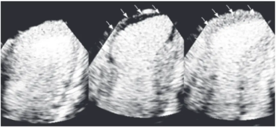
Figure 2. Representative example of real time myocardial contrast echocardiography in the infarct model showing normal myocardial perfusion at baseline (left) and the risk area during left anterior descending coronary artery occlusion (arrows) in the center. After 30 min of coronary reperfusion there was a clear reduction of perfusional defect size (arrows) and the infarcted area remained limited to the apical region (right).
tion (SD) and qualitative data as propor-tions. Within-group comparisons were per-formed by the paired Student t-test,
repeated-7
Area (cm
2) 6 5 4 3 2 1 0 -1
Baseline LAD occlusion Reperfusion
WMA PMD
*
* +
Table 1. Hemodynamic data for the experimental dogs submitted to infarct or stun-ning.
HR (bpm) MAP (mmHg) LAD flow (ml/min)
Infarct model
Baseline 135.4 ± 19.5 93.0 ± 19.2 21.6 ± 6.8
LAD occlusion 122.2 ± 31.5 91.7 ± 23.1 1.6 ± 2.3+
Reperfusion 116.5 ± 33.1* 88.6 ± 29.3 20.3 ± 8.2*
Stunned model
Baseline 135.0 ± 26.5 89.0 ± 14.3 22.1 ± 7.5
LAD occlusion 122.1 ± 21.0 84.3 ± 12.8 0.32 ± 0.34+
Reperfusion 119.1 ± 20.5* 80.6 ± 13.3 21.4 ± 8.9*
Data are reported as means ± SD. HR = heart rate; LAD = left anterior descending coronary artery; MAP = mean arterial blood pressure.
area) were well defined and generally trans-mural. In contrast, after reperfusion regions that remained with PMD (infarct area) were irregular in shape and frequently subendo-cardial (Figure 2).
In nine dogs of the infarct model with Evans blue staining, RTMCE consistently underestimated the risk area, which was smaller than that determined by tissue stain-ing (4.58 ± 1.30 vs 6.01 ± 0.91 cm2; P =
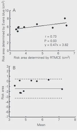
0.015). However, there was a good correla-tion between the RTMCE measurements and staining (r = 0.73; P = 0.03; Figure 3).
Infarct size determined by RTMCE was 1.51 ± 1.29 cm2 and necrotic area
deter-mined by TTC staining was 1.99 ± 1.23 cm2
(P = 0.29), with a strong linear correlation between these two measurements (necrotic area by TTC = 0.83 X infarct size by RTMCE + 0.73 cm2; r = 0.71; P = 0.002), as shown in
Figure 4.
After 30 min of coronary reperfusion, myocardial perfusion was reestablished in the left anterior descending coronary artery territory, except in the infarcted areas. How-ever, these regions continued to show im-paired contractility even with normal perfu-sion. The stunned myocardium area (myo-cardium with WMA and without PMD) de-termined by RTMCE was 4.00 ± 0.29 cm2.
Tissue staining confirmed the absence of infarction in these areas.
Stunned model evaluation
In the stunned model the WMA area was 5.49 ± 0.68 cm2 at 20 min of occlusion and
5.1 ± 0.63 cm2 after reperfusion (P = 0.07).
PMD area was 2.43 ± 0.79 cm2 during
coro-nary occlusion and was reduced to almost zero (0.2 ± 0.53 cm2) after reperfusion (P =
0.04) (Figure 5). TTC staining confirmed the absence of necrotic myocardium in all dogs of the stunned model.
Figure 6 demonstrates an example of nor-malization of perfusion with maintenance of WMA as observed in the stunned model.
Risk area determined by Evans blue (cm
2)
10
8
6
4
2
0
4 5 6 7
3
Risk area determined by RTMCE (cm2) r = 0.73 P = 0.03 y = 0.47x + 3.82
Risk area
3
1 0
-1 -2 -3
5 6 7 8
3
Mean -4
-5
-7 2
-6 A
B
Figure 3. Correlation between risk areas determined by Evans blue staining and by real time myocardial contrast echocardi-ography (RTMCE) (A) and a Bland-Altman plot showing the mean difference (solid line) and limits of agreement (dashed lines) between measurements of risk area by the two methods (B).
Necrotic area determined by TTC
(cm
2) 6
5
4
3
2
0 1
0 1 2 3 4 5
Infarct size determined by RTMCE (cm2)
Infarct size (cm
2) 5
3
1
-1
-3
-5
0 1 2 3 4 5
Mean A
B
Figure 4. Correlation between infarct size determined by real time myocardial contrast echo-cardiography (RTMCE) and ne-crotic area determined by 2,3,5-triphenyl tetrazolium chloride (TTC) staining (A) and a Bland-Altman plot showing the mean difference (solid line) and limits of agreement (dashed lines) be-tween infarct sizes determined by the two methods (B). r = 0.71
Interobserver variability
We observed excellent agreement be-tween the measurements of two observers for PMD area detected by RTMCE during coronary occlusion (r = 0.91; P < 0.001), with a mean difference of -0.20 (-1.35 to 0.95). Excellent agreement between two ob-servers was also obtained after reperfusion (r = 0.96; P < 0.001), with a mean difference of -0.03 (-0.40 to 0.34).
Discussion
Although some studies have demonstrated the usefulness of low-energy RTMCE to
detect coronary stenosis and perfusional de-fects in the setting of acute myocardial ische-mia (9,10,17,18), its value for the simulta-neous evaluation of myocardial function and perfusion in experimental models of infarc-tion has not been yet described. The present study showed for the first time the progres-sive changes that occur with myocardial func-tion and perfusion during coronary occlu-sion and after reperfuocclu-sion, distinguishing normally perfused, collateral-dependent and non-perfused myocardium. We demonstrated that RTMCE accurately measured the areas of stunned and infarcted myocardium and underestimated the risk area when compar-ing to tissue staincompar-ing.
The final infarct size is directly related to the risk area, which is also dependent on the duration of the ischemic period, collateral flow and efficacy of reperfusion (19,20). Therefore, its measurement is an important parameter for clinical decisions in the setting of acute myocardial infarction. In the pres-ent study we observed that left anterior de-scending coronary artery occlusion resulted in a larger area of myocardial dysfunction than of perfusion defect in both models. A possible explanation for this result is the presence of collateral flow from adjacent vascular beds allowing some degree of myo-cardial perfusion in the margins of regions supplied by the occluded artery and decreas-ing the size of the risk area determined by RTMCE. This result is consistent with previ-ous studies indicating that maturation of pre-existing collateral vessels, as opposed to de novo growth of vessels, constitutes an early
phase of collateral development in ischemic myocardium (21-23). Since we carried out the planimetry of perfusion defects by RTMCE only in the areas with total absence of myocardial contrast, regions with hypo-perfusion surrounded the central ischemic area were not included in the PMD measure-ment. Although collateral-derived myocar-dial blood flow may not be adequate to maintain normal myocardial systolic
func-Figure 5. Wall motion abnormal-ity (WMA) and perfusional myo-cardial defect (PMD) areas de-termined by real time myocar-dial contrast echocardiography at baseline, at 20 min of left an-terior descending coronary ar-tery (LAD) occlusion and after 30 min of reperfusion in the stunned model. After reperfu-sion there was maintenance of WMA area and reduction of PMD area to almost zero. *P < 0.05 for baseline vs LAD occlu-sion; +P < 0.05 for LAD occlu-sion vs reperfusion (ANOVA).
Figure 6. Representative example of real time myocardial contrast echocardiography in the stunned model showing normal myocardial perfusion at baseline (left) and risk area during left anterior descending coronary artery occlusion (arrows) in the center. After reperfusion (right) there was complete normalization of myocardial perfusion with maintenance of wall motion abnormality (arrows), demonstrating stunned myocardium.
7
Area (cm
2) 6 5 4 3
2
1 0 -1
Baseline LAD occlusion Reperfusion
WMA PMD
*
tion distal to an occluded vessel, it may be sufficient to maintain viability for prolonged periods of coronary occlusion. Therefore, we should consider that the area with a perfusional defect during coronary occlu-sion as determined by RTMCE reflects the real area at risk for future necrosis.
In areas that are not supplied by collateral flow, prolonged coronary occlusion produces myocardial necrosis, leading to irreversible damage at the microvascular level (24,25). If coronary reperfusion is achieved, either spon-taneously or by means of thrombolysis or angioplasty, a variable amount of myocardi-um can be salvaged. Previous studies have demonstrated that infarct-related artery pa-tency and myocardial perfusion are not nec-essarily concordant in humans (26,27). Even with epicardial coronary reflow, myocardial perfusion may not be achieved (the low-reflow or no-low-reflow phenomenon) because of microvascular disruption, plugging by debris or myocardial edema (14,28). Thus, the ability to identify the presence or ab-sence of myocardial perfusion by myocar-dial contrast echocardiography during the post-acute myocardial infarct period may offer important advantages over simple as-sessment of infarct-related artery patency during catheterization, and predicts left ven-tricular function and further remodeling (29). During the immediate period after acute infarction, the myocardium within the reper-fused bed contains a mixture of both ne-crotic and stunned viable tissue (24). In the present study we showed that after reperfu-sion the contractility remained altered, with maintenance of the WMA area determined by RTMCE as compared to the occlusion period. On the other hand, there was a sig-nificant reduction of PMD area with com-plete normalization of perfusion in the stunned model and maintenance of the per-fusion defect only in necrotic areas in the infarct model.
Therefore, RTMCE is a noninvasive method that allows simultaneous analysis of
contractility and perfusion and was capable of demonstrating stunned (normal perfusion and maintenance of WMA) and infarcted tissue (absence of perfusion and depressed contractility). In the present study only three dogs presented discrete underestimation of infarct size by myocardial contrast echocar-diography. One possible explanation for this result is that, because of the wave-front na-ture of myocardial damage following a pro-longed period of ischemia, a mismatch be-tween microvascular and myocyte necrosis can occur, resulting in areas of cellular death with relatively preserved perfusion. Irrevers-ibly injured myocardium is generally associ-ated with loss of microvascular integrity, which is proportional to the extension and severity of myocellular necrosis. In the poorly perfused areas, severe capillary damage and coagulation necrosis can be observed, but microvascular damage might also develop during reperfusion. Due to their ability to remain entirely within the intravascular space, microbubbles used as echocardiographic contrast agents are markers of blood flow and allow a noninvasive evaluation of mi-crovascular integrity. However, when we analyzed all the dogs of the infarct model, there was no statistical difference between the infarct size predicted by RTMCE and that determined by TTC staining, showing that RTMCE can accurately measure the necrotic area.
Limitations
ob-tained by RTMCE and anatomy represent an estimation of the true measurement of the spatial extent of risk area or infarcted myo-cardium. However, previous experimental studies have validated the good agreement between echocardiographic measurements and anatomic findings of risk area as well as infarct size using the methodology employed in this study (20,30). The planimetry of WMA included areas with any degree of segmental wall motion, such as those presenting akine-sia, hypokinesia or dyskinesia. We recog-nize that this approach does not consider qualitative changes in the segmental wall motion and could result in an underestima-tion of the improvement of wall mounderestima-tion after reperfusion. However, we noted that most segments presented akinesia soon after coro-nary occlusion that was maintained after reperfusion, without a significant change in the wall motion score index. In addition, we considered that a direct comparison between quantitative data of WMA and PMD was more suitable for the purpose of the study.
Another limitation is that visual assess-ment could lead to an overestimation of in-farct size by RTMCE, which is based on the evaluation of myocardial replenishment af-ter total destruction of microbubbles by a high emission power technique. Thus, bor-derline areas with some degree of residual perfusion could be interpreted as a contrast defect due to a late contrast replenishment.
In our study, infarct size was determined when there was full contrast replenishment of the normal areas but it is possible that borderline regions were included in the planimetry of contrast defect. Evaluation of myocardial blood flow by quantitative anal-ysis could distinguish between areas with very low reflow and areas with no reflow.
Clinical implications
Main and co-workers (31) recently dem-onstrated in a small number of patients that full-motion myocardial contrast echocardi-ography and the power pulse inversion tech-nique accurately predicted recovery of seg-mental function in patients with recent acute myocardial infarction.In the present study we demonstrate that RTMCE is a useful technique for visual assessment of stunned and infarcted myocardium. A rapid defini-tion of the efficacy of reperfusion therapy as well as the extent of its residual microvascu-lar damage has important implications for the treatment of acute myocardial infarction. It has been previously demonstrated that the presence of preserved microvascular flow in the post-reperfusion period is associated with a lower rate of fibrous scar and less ventric-ular remodeling (32-35). Thus the differen-tiation between stunned and infarcted myo-cardium by RTMCE has important clinical and prognostic implications.
References
1. Heyndrickx GR, Millard RW, McRitchie RJ, Maroko PR & Vatner SF (1975). Regional myocardial functional and electrophysiological al-terations after brief coronary artery occlusion in conscious dogs.
Journal of Clinical Investigation, 56: 978-985.
2. Kaul S (1997). Myocardial contrast echocardiography: 15 years of research and development. Circulation, 96: 3745-3760.
3. Armstrong WF, Mueller TM, Kinney EL, Tickner EG, Dillon JC & Feigenbaum H (1982). Assessment of myocardial perfusion abnor-malities with contrast-enhanced two-dimensional echocardiogra-phy. Circulation, 66: 166-173.
4. Tei C, Sakamaki T, Shah PM, Meerbaum S, Shimoura S, Kondo S & Corday E (1983). Myocardial contrast echocardiography: a reproduc-ible technique of myocardial opacification for identifying regional
perfusion deficits. Circulation, 67: 585-593.
5. Villanueva FS, Glasheen WP, Skelenar J & Kaul S (1993). Character-ization of spatial patterns of flow within the reperfused myocardium by myocardial contrast echocardiography; implications for determin-ing extent of myocardial salvage. Circulation, 88: 2596-2606. 6. White FC, Sanders M & Bloor CM (1978). Regional distribution of
myocardial blood flow after coronary occlusion and reperfusion in the conscious dog. American Journal of Cardiology, 42: 234-243. 7. Becker H & Burns P (2000). Handbook of Contrast
Echocardiogra-phy: Left Ventricular Function and Myocardial Perfusion. Springer-Verlag, New York.
ultrasound-induced destruction of microbubbles administered as a constant venous infusion. Circulation, 97: 473-483.
9. Porter TR, Xie F, Silver M, Kricsfield D & O’Leary E (2001). Real-time perfusion imaging with low mechanical index pulse inversion Dop-pler imaging. Journal of the American College of Cardiology, 37: 262-269.
10. Matsugata H, Peters B, Lafitte S, Strachan GM, Ohmori K & DeMaria AN (2001). Quantitative assessment of myocardial perfusion during graded coronary stenosis by real-time myocardial contrast echocar-diography. Journal of the American College of Cardiology, 37: 262-269.
11. Pelberg RA, Wei K, Kamiyama N, Skelenar J, Bin J & Kaul S (1999). Potential advantage of flash echocardiography for digital subtrac-tion of b-mode images acquired during myocardial contrast echo-cardiography. Journal of the American Society of Echocardiography, 12: 85-93.
12. Porter TR & Xie F (1995). Transient myocardial contrast following initial exposure to diagnostic ultrasound pressures with minute doses of intravenously injected microbubbles: demonstration and potential mechanisms. Circulation, 92: 2391-2395.
13. Jayaweera AR, Matthew TL, Skelenar J, Spotnitz WD, Watson DD & Kaul S (1990). Method for quantitation of myocardial perfusion during myocardial contrast echocardiography. Journal of the Ameri-can Society of Echocardiography, 3: 91-98.
14. Kloner R, Ganote CE & Jennings RB (1974). The no-reflow phenom-enon after temporary coronary occlusion in the dog. Journal of Clinical Investigation, 54: 1496-1508.
15. Fishbein MC, Meerbaum S, Rit J, Lado U, Kanmatsuse K, Mercier JC, Corday E & Ganz W (1981). Early phase acute myocardial infarct size quantification: validation of the triphenyl tetrazolium chloride tissue enzyme staining technique. American Heart Journal, 101: 593-600.
16. Chagas ACP (1992). Proteção miocárdica em isquemia e reperfusão: estudo experimental sobre a preservação miocárdica e a ocorrência de fibrilação ventricular na reperfusão, em cães. Doctoral thesis, Faculdade de Medicina, Universidade de São Paulo, São Paulo, SP, Brazil.
17. Lafitte S, Matsugata H, Peters B, Togni M, Strachan M, Kwan OL & DeMaria AN (2001). Comparative value of dobutamine and adeno-sine stress in the detection of coronary stenosis with myocardial contrast echocardiography. Circulation, 103: 2724-2730.
18. Lafitte S, Higashiyama A, Matsugata H, Peters B, Strachan M, Kwan OL & DeMaria AN (2002). Contrast echocardiography can assess risk area and infarct size during coronary occlusion and reperfusion: experimental validation. Journal of the American College of Cardiol-ogy, 39: 1546-1554.
19. Kemper AJ, O’Boyle JE, Cohen CA, Taylor A & Parisi AF (1984). Hydrogen peroxide contrast echocardiography; quantification in vivo
of myocardial risk area during coronary occlusion and of the necrotic area remaining after myocardial reperfusion. Circulation, 70: 309-317.
20. Kaul S, Glasheen W, Ruddy TD, Pandian NG, Weyman AE & Okada RD (1987). The importance of defining left ventricular area at risk in vivo during acute myocardial infarction: an experimental evaluation with myocardial contrast two-dimensional echocardiography. Circu-lation, 75: 1249-1260.
21. Kaul S, Pandian NG, Guerrero JL, Gillam LD, Okada RD & Weyman AE (1987). Effects of selectively altering collateral driving pressure on regional perfusion and function in occluded coronary bed in the dog. Circulation Research, 61: 77-85.
22. Schaper J, Borges M & Schaper W (1972). Ultrastructure of
is-chaemia-induced changes in precapillary anastomotic network in the heart. American Journal of Cardiology, 29: 851-859.
23. Kaul S, Glasheen WP, Oliner JD, Kelly P & Gascho JA (1991). Relation between antegrade blood flow through a coronary artery and the size of the perfusion bed it supplies: experimental and clinical implications. Journal of the American College of Cardiology, 17: 1403-1413.
24. Reimer KA & Jenning RB (1979). The wave front phenomenon of myocardial cell death. Laboratory Investigation, 40: 33-44. 25. Galiuto L, DeMaria AN, May-Newman K, Del Balzo U, Ohmori K,
Bhargava V, Flaim SF & Iliceto S (1998). Evaluation of dynamic changes in microvascular flow during ischaemia-reperfusion by myo-cardial contrast echocardiography. Journal of the American College of Cardiology, 32: 1096-1101.
26. Ito H, Tomooka T, Sakai N, Yu H, Higashino Y, Fujii K, Masuyama T, Kitabatake A & Minamino T (1992). Lack of myocardial perfusion immediately after successful thrombolysis: a predictor of poor re-covery of left ventricular function in anterior myocardial infarction.
Circulation, 85: 1699-1705.
27. Sabia PJ, Powers ER, Ragosta M, Sarembock IJ, Burwell LR & Kaul S (1992). An association between collateral blood flow and myocar-dial viability in patients with recent myocarmyocar-dial infarction. New England Journal of Medicine, 327: 1825-1831.
28. Johnson WB, Malone SA, Pantely GA, Anselone CG & Bristow JD (1988). No reflow and extent of infarction during maximal vasodila-tion in porcine heart. Circulation, 78: 462-472.
29. Lincoff AM & Topol EJ (1993). Illusion of reperfusion: does anyone achieve optimal reperfusion during acute myocardial infarction?
Circulation, 88: 1361-1374.
30. Grayburn PA, Erickson JM, Escobar J, Womack L & Velasco CE (1995). Peripheral intravenous myocardial contrast echocardiogra-phy using a 2% dodecafluoropentane emulsion: identification of myocardial risk area and infarct size in the canine model of is-chaemia. Journal of the American College of Cardiology, 26: 1340-1347.
31. Main ML, Magalski A, Chee NK, Coen MM, Skolnick DG & Good TH (2001). Full-motion pulse inversion power Doppler contrast echocar-diography differentiates stunning from necrosis and predicts recov-ery of left ventricular function after acute myocardial infarction.
Journal of the American College of Cardiology, 38: 1390-1394. 32. Agati L, Voci P, Bilotta F, Luongo R, Autore C, Penco M, Iacoboni C,
Fedele F & Dagianti A (1994). Influence of residual perfusion within the infarct zone on the natural history of left ventricular dysfunction after acute myocardial infarction: a myocardial contrast echocardio-graphic study. Journal of the American College of Cardiology, 24: 336-342.
33. Agati L, Voci P, Luongo R, Testa G, Mallus MT, Di Roma A, Fedele F & Dagianti A (1997). Combined use of dobutamine echocardiogra-phy and myocardial contrast echocardiograechocardiogra-phy to predict recovery of regional dysfunction after coronary revascularization in patients with recent myocardial infarction. European Heart Journal, 18: 771-779.
34. Ito H, Maruyama A, Iwakura K, Takiuchi S, Masuyama T, Hori M, Higashino Y, Fujii K & Minamino T (1996). Clinical implications of the “no-reflow” phenomenon: a predictor of complications and left ventricular remodeling in reperfused anterior wall myocardial infarc-tion. Circulation, 93: 223-228.