Thalidomide and pentoxifylline block
the renal effects of supernatants of
macrophages activated with
Crotalus
durissus cascavella
venom
Departamentos de 1Análises Clínicas e Toxicológicas, and 2Fisiologia e Farmacologia, Instituto de Biomedicina e
Unidade de Pesquisas Clínicas, Universidade Federal do Ceará, Fortaleza, CE, Brasil
3Departamento de Fisiologia, Universidade Estadual do Ceará,
Fortaleza, CE, Brasil A.M.C. Martins1,
A.C.L. Nobre2, A.C. Almeida2, G. Bezerra2, A.A.M. Lima2, M.C. Fonteles3 and H.S.A. Monteiro2
Abstract
Because thalidomide and pentoxifylline inhibit the synthesis and release of tumor necrosis factor-α (TNF-α), we determined the effect
of these drugs on the renal damage induced by supernatants of macrophages activated with Crotalus durissus cascavella venom in order to identify the role of TNF-α in the process. Rat peritoneal
macrophages were collected with RPMI medium and stimulated in vitro with C.d. cascavella venom (10 µg/ml) in the absence and presence of thalidomide (15 µM) or pentoxifylline (500 µM) for 1 h and washed and kept in culture for 2 h. Supernatant (1 ml) was tested on an isolated perfused rat kidney (N = 6 for each group). The first 30 min of each experiment were used as control. The supernatant was added to the perfusion system. All experiments lasted 120 min. The toxic effect of the preparation of venom-stimulated macrophages on renal parameters was determined. At 120 min, thalidomide (Thalid) and pentoxifylline (Ptx) inhibited (P < 0.05) the increase in perfusion pressure caused by the venom (control = 114.0 ± 1.3; venom = 137.1 ± 1.5; Thalid = 121.0 ± 2.5; Ptx = 121.4 ± 4.0 mmHg), renal vascular resistance (control = 4.5 ± 0.2; venom = 7.3 ± 0.6; Thalid = 4.5 ± 0.9; Ptx = 4.8 ± 0.6 mmHg/ml g-1 min-1), urinary flow (control = 0.23 ±
0.001; venom = 0.44 ± 0.01; Thalid = 0.22 ± 0.007; Ptx = 0.21 ± 0.009 ml g-1 min-1), glomerular filtration rate (control = 0.72 ± 0.06; venom
= 1.91 ± 0.11; Thalid = 0.75 ± 0.04; Ptx = 0.77 ± 0.05 ml g-1 min-1) and
the decrease in percent tubular sodium transport (control = 77.0 ± 0.9; venom = 73.9 ± 0.66; Thalid = 76.6 ± 1.1; Ptx = 81.8 ± 2.0%), percent tubular chloride transport (control = 77.1 ± 1.2; venom = 71.4 ± 1.1; Thalid = 77.6 ± 1.7; Ptx = 76.8 ± 1.2%), and percent tubular potassium transport (control = 72.7 ± 1.1; venom = 63.0 ± 1.1; Thalid = 72.6 ± 1.0; Ptx = 74.8 ± 1.0%), 30 min before and during the stimulation of macrophages with C.d. cascavella venom. These data suggest the participation of TNF-α in the renal effects induced by supernatant of
macrophages activated with C.d. cascavella venom.
Correspondence
H.S.A. Monteiro
Unidade de Pesquisas Clínicas Departamento de Fisiologia e Farmacologia
Faculdade de Medicina, UFC Caixa Postal 3229 60420-970 Fortaleza, CE Brasil
Fax: +55-85-281-5212 E-mail: martinsalice@hotmail.com
Received September 10, 2003 Accepted June 17, 2004
Key words
•Nephrotoxicity
•Crotalus durissus cascavella •Pentoxifylline
•Thalidomide
Introduction
Snakebitesare an important public health problem in Brazil. The genus Crotalus con-tains several species of snakes responsible for high morbidity and mortality rates (1). Crota-lus durissus cascavella is a snake usually found in the scrubland of the Brazilian Northeast (2). Crotalic venom causes neurotoxicity, systemic myotoxicity, edematogenic reactions, platelet aggregation, and acute renal failure. The most common complication observed in lethal snakebite victims in Brazil is acute renal fail-ure (3), a process that can occur even after specific antivenom treatment. The pathogen-esis of acute renal failure after snakebites ap-pears to be multifactorial (4). Some evidence suggests the possible existence of a direct nephrotoxic agent in the venom, but this does not exclude the release of mediators (5) or rhabdomyolysis as causative agents. Alterna-tively, these underlying causes can potentiate each other (6,7).
We demonstrated in isolated rat kidney that the venom of C.d. cascavella causes changes in renal function such as increase in perfusion pressure, urinary flow and percent sodium tubular transport (4). We have also reported that macrophages activated by C.d. cascavella venom release nephrotoxic media-tors (8).
The participation of tumor necrosis
factor-α (TNF-α) in renal injury has been recently
demonstrated (9-11). The protective effect of pentoxifylline against the damage induced by ischemia-reperfusion has been demonstrated in several experimental models (12,13).
The aim of this study was to investigate the action of thalidomide and pentoxifylline on the renal effects induced by supernatants of mac-rophages activated by C.d. cascavella venom.
Material and Methods
Macrophage cultures
Rat peritoneal macrophages were
col-lected with RPMI medium 4 days after an injection of 10 ml thioglycolate (3%, ip) and placed on plastic tissue culture dishes, as previously described (14). After incubation at 37ºC in a 5% CO2 atmosphere for 1.5 h,
the nonadherent cells were removed by wash-ing the dishes three times with RPMI medi-um. The cell pattern was determined by cell morphology analysis with a light microscope. The total cells (95% macrophages) were in-cubated at 37ºC in a 5% CO2 atmosphere for
2 h in fresh medium (control), and after this period in a medium containing 10 µg/ml C.d. cascavella venom. Thalidomide (15 µM) and pentoxifylline (500 µM) were added 30 min before the addition of venom and kept in the medium throughout the period of macro-phage stimulation with C.d. cascavella venom (10 µg/ml). The supernatant was dis-carded and, after additional washing, the cells were incubated for 1 h with 1 ml RPMI medium without venom or drugs. Cell-free incubation medium was obtained by cen-trifugation for 5 min and 1 ml of supernatant was adjusted to 1.3 x 10⁷ cells/ml and its
effects were tested on an isolated rat kidney. Six rats were used in each group of perfu-sion.Macrophage viability was determined by Trypan blue exclusion as described else-where (15). Macrophage viability ranged from 89 to 97%.
Isolated rat kidney
Adult Wistar rats of both sexes (240-280 g) were fasted with free access to water 24 h before each experiment. The animals were anesthetized with sodium pentobarbital (50 mg/kg body weight). The perfusion fluid was a modified Krebs-Henseleit solution of the following composition: 147 mM Na+, 5
mM K+, 2.5 mM Ca2+, 2 mM Mg2+, 110 mM
Cl-, 2.5 mM HCO
3-, 1 mM SO42-, and 1 mM
PO42-. Bovine serum albumin (BSA, 6 g/100
100 ml. BSA was previously dialyzed for 48 h at 4ºC in 1.5 liter of Krebs solution, and the solution was changedafter 24 h (16,17). The pH was adjusted to 7.4 and the perfusion system, based on Bowman’s technique (18), was modified (19) by the addition of an artificial lung to improve oxygenation (20) and of a 1.2-mm Millipore filter (21). Flow calibration and the resistance of the system were determined before each experiment. Perfusion pressure was determined at the tip of the stainless steel cannulae. The right renal artery was cannulated through the up-per mesenteric artery and the kidney was isolated (22-24) and submitted to uninter-rupted perfusion. After an equilibration pe-riod of 15 to 20 min, the experiments were carried out over a total period of time of 120 min. The supernatants of macrophages stim-ulated with C.d. cascavella venom plus phar-macological inhibitors were added 30 min after the beginning of the experiment. Perfu-sion pressure was measured at 5-min inter-vals. Urine and perfusate samples were col-lected every 10 min for the determination of sodium, chloride, potassium, and inulin lev-els and osmolality. Sodium and potassium concentrations were determined by flame photometry using a model 445 flame pho-tometer (Instrumentation Laboratory Inc., Lexington, MA, USA) and inulin levels were also determined (4,18). Chloride was deter-mined with a LabTest kit (LabTest Diagnós-tica, Lagoa Santa, MG, Brazil). The osmola-lity of the samples was measured with an Advanced Osmometer (Wescor 5100c, Needham Heights, MA, USA) at vapor pres-sure.
Drugs
C.d. cascavella venom was obtained from the regional ophiology laboratory of Forta-leza (LAROF-CE). Thalidomide was ob-tained from ICN Biomedical Inc., Aurora, OH, USA. Pentoxifylline, RPMI medium, albumin, inulin, and glucose were purchased
from Sigma, St. Louis, MO, USA. Thiogly-colate was obtained from Difco Laborato-ries, Detroit, MI, USA.
Statistical analysis
Data are reported as mean ± SEM and were analyzed statistically by analysis of variance (ANOVA) followed by the Bonfer-roni test. The level of significance was set at P < 0.05.
Results
Effect of tumor necrosis factor inhibitors
Previous results have shown that infu-sion of the supernatant of macrophages stim-ulated with C.d. cascavella venom (10 µg/ ml) after 30 min of internal control caused alterations of renal function parameters (8). In the present study, thalidomide and pen-toxifylline, TNF inhibitors, reversed all the renal changes promoted by the supernatant of macrophages stimulated with C.d. casca-vella venom.
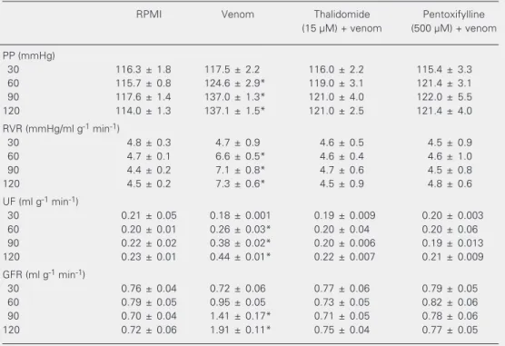
The data in Table 1 show that thalido-mide (15 µM) and pentoxifylline (500 µM) inhibited the increase in perfusion pressure, renal vascular resistance, urinary flow, and glomerular filtration rate induced by super-natants of macrophages stimulated with C.d. cascavella venom.
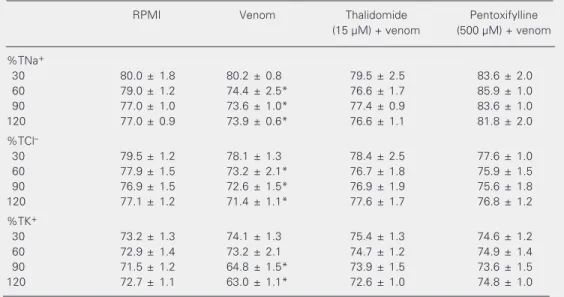
Treatment with thalidomide and pentoxi-fylline also inhibited the decrease in percent tubular sodium, chloride and potassium trans-port caused by supernatants of macrophages stimulated with C.d. cascavella venom as described in Table 2.
Discussion
Table 1. Thalidomide and pentoxifylline inhibit the renal effects of the supernatant of macrophages stimulat-ed with Crotalus durissus cascavella venom.
RPMI Venom Thalidomide Pentoxifylline
(15 µM) + venom (500 µM) + venom
PP (mmHg)
30 116.3 ± 1.8 117.5 ± 2.2 116.0 ± 2.2 115.4 ± 3.3
60 115.7 ± 0.8 124.6 ± 2.9* 119.0 ± 3.1 121.4 ± 3.1
90 117.6 ± 1.4 137.0 ± 1.3* 121.0 ± 4.0 122.0 ± 5.5
120 114.0 ± 1.3 137.1 ± 1.5* 121.0 ± 2.5 121.4 ± 4.0
RVR (mmHg/ml g-1 min-1)
30 4.8 ± 0.3 4.7 ± 0.9 4.6 ± 0.5 4.5 ± 0.9
60 4.7 ± 0.1 6.6 ± 0.5* 4.6 ± 0.4 4.6 ± 1.0
90 4.4 ± 0.2 7.1 ± 0.8* 4.7 ± 0.6 4.5 ± 0.8
120 4.5 ± 0.2 7.3 ± 0.6* 4.5 ± 0.9 4.8 ± 0.6
UF (ml g-1 min-1)
30 0.21 ± 0.05 0.18 ± 0.001 0.19 ± 0.009 0.20 ± 0.003
60 0.20 ± 0.01 0.26 ± 0.03* 0.20 ± 0.04 0.20 ± 0.06
90 0.22 ± 0.02 0.38 ± 0.02* 0.20 ± 0.006 0.19 ± 0.013
120 0.23 ± 0.01 0.44 ± 0.01* 0.22 ± 0.007 0.21 ± 0.009
GFR (ml g-1 min-1)
30 0.76 ± 0.04 0.72 ± 0.06 0.77 ± 0.06 0.79 ± 0.05
60 0.79 ± 0.05 0.95 ± 0.05 0.73 ± 0.05 0.82 ± 0.06
90 0.70 ± 0.04 1.41 ± 0.17* 0.71 ± 0.05 0.78 ± 0.06
120 0.72 ± 0.06 1.91 ± 0.11* 0.75 ± 0.04 0.77 ± 0.05
Macrophages were inoculated with 10 µg/ml venom in the presence or absence of 15 µM thalidomide or 500 µM pentoxifylline for 2 h at 37ºC. The supernatant was tested on the isolated rat kidney. Data are reported as means ± SEM for six kidney perfusions carried out for each set of conditions. PP = pentoxyfylline; RVR = renal vascular resistance; UF = urinary flow; GFR = glomerular filtration rate.
*P < 0.05 compared to other values in the same time group (ANOVA followed by the Bonferroni test).
alterations, immunologic reactions and di-rect nephrotoxicity. Some investigators have emphasized the importance of rhabdomy-olysis as a cause of acute renal failure after crotalid bites (7), while others have reported nephrotoxicity in the rat isolated kidney af-ter administration of crotalid venom (4,26). Perfusion of the isolated kidney has been extensively used as a model to study the vascular effect of biologically active sub-stances, preventing the interference of blood-borne cells, hormones and other factors with renal function transported by blood (16).
We have recently demonstrated the renal effect of the supernatant of macrophages acti-vated with C.d. cascavella venom (8). Acting as alarm cells, macrophages signal the pres-ence of foreign bodies by elaborating and releasing several substances, including
cyto-kines and arachidonic acid metabolites (27). An increase in serum TNF-α levels has
been reported to occur in mice injected with Bothrops asper and B. jararaca snake venom (28). It has been recently demonstrated that renal cells can produce and release TNF-α
(29) and that the kidney is highly sensitive to this cytokine. Hence, it is plausible to relate this protein to the renal damage associated with envenomation or other inflammatory stimuli, as reported by other investigators (9,10).
It has been observed that pentoxifylline can regulate calcium homeostasis (30) and thalidomide can affect the proliferation of endothelial cells and the expression of al-pha-beta 3 integrin on the surface of endo-thelial cells (31). Thalidomide inhibits
cytokine (29). Thalidomide and pentoxifyl-line blocked the synthesis and release of TNF-α from macrophages in culture after
the same period of incubation and concen-tration as used in our experiments (12). Us-ing a similar approach, it was demonstrated that thalidomide and pentoxifylline alone did not modify the functional parameters of the kidney (32).
In some situations, receptor-mediated events induced by TNF-α or Fas
(apoptosis-mediating surface antigen fas; CD95) may play a role in apoptosis in acute renal failure (33). Recently, it was demonstrated that pen-toxifylline may exert a protective effect
Table 2. Thalidomide and pentoxifylline inhibit the renal tubular effects of the supernatant of macrophages stimulated with Crotalus durissus cascavella venom.
RPMI Venom Thalidomide Pentoxifylline
(15 µM) + venom (500 µM) + venom
%TNa+
30 80.0 ± 1.8 80.2 ± 0.8 79.5 ± 2.5 83.6 ± 2.0
60 79.0 ± 1.2 74.4 ± 2.5* 76.6 ± 1.7 85.9 ± 1.0
90 77.0 ± 1.0 73.6 ± 1.0* 77.4 ± 0.9 83.6 ± 1.0
120 77.0 ± 0.9 73.9 ± 0.6* 76.6 ± 1.1 81.8 ± 2.0
%TCl
-30 79.5 ± 1.2 78.1 ± 1.3 78.4 ± 2.5 77.6 ± 1.0
60 77.9 ± 1.5 73.2 ± 2.1* 76.7 ± 1.8 75.9 ± 1.5
90 76.9 ± 1.5 72.6 ± 1.5* 76.9 ± 1.9 75.6 ± 1.8
120 77.1 ± 1.2 71.4 ± 1.1* 77.6 ± 1.7 76.8 ± 1.2
%TK+
30 73.2 ± 1.3 74.1 ± 1.3 75.4 ± 1.3 74.6 ± 1.2
60 72.9 ± 1.4 73.2 ± 2.1 74.7 ± 1.2 74.9 ± 1.4
90 71.5 ± 1.2 64.8 ± 1.5* 73.9 ± 1.5 73.6 ± 1.5
120 72.7 ± 1.1 63.0 ± 1.1* 72.6 ± 1.0 74.8 ± 1.0
Macrophages were inoculated with 10 µg/ml venom in the presence or absence of 15 µM thalidomide or 500 µM pentoxifylline for 2 h at 37ºC. The supernatant was tested on the isolated rat kidney. Data are reported as means ± SEM for six kidney perfusions carried out for each set of conditions. %TNa+ = percent tubular
sodium transport; %TCl- = percent tubular chloride transport; %TK+ = percent tubular potassium transport.
*P < 0.05 compared to other values in the same time group (ANOVA followed by the Bonferroni test).
against ischemic acute renal failure by inhib-iting TNF-α production in rabbits (34).
In the present study, thalidomide and pentoxifylline reversed all renal alterations induced by the supernatants of macrophages stimulated with C.d. cascavella venom. Our findings suggest that thalidomide and pen-toxifylline inhibit the release of renal cyto-toxic mediators by macrophages activated with C.d. cascavella venom, and indicate the need for investigation of the possible partici-pation of TNF-α in this process. The results
also support the need for investigations of the possible use of these drugs in the treat-ment of snakebites.
References
1. Santoro MC, Sousa-e-Silva MC, Gonçalves RL, Almeida-Santos SM, Cardoso DF, Laporta-Ferreira IL, Saiki M, Peres CA & Sano-Martins S (1999). Comparison of the biological activities in venoms from three subspecies of the South American rattlesnake (Crotalus duris-sus terrificus, C. durissus cascavella and C. durissus collilineatus).
ComparativeBiochemistry and Physiology. Part C: Pharmacology and Endocrinology, 122: 61-73.
3. Ribeiro LA, Albuquerque MJ, De Campos VA, Katz G, Takaoka NY, Lebrao ML & Jorge MT (1998). Deaths caused by venomous snakes in the State of São Paulo: evaluation of 43 cases from 1988 to 1993.
Revista da Associação Médica Brasileira, 44: 312-318.
4. Martins AM, Monteiro HS, Junior EO, Menezes DB & Fonteles MC (1998). Effects of Crotalus durissus cascavella venom in the iso-lated rat kidney. Toxicon, 36: 1441-1450.
5. Sitprija V & Chaiyabutr N (1999) Nephrotoxicity in snake envenoma-tion. Journal of Natural Toxins, 8: 271-277.
6. Nancy G, Ahlstrom MD, Luginbuhl MDW & Tisher MDC (1991). Acute anuric renal failure after pygmy rattlesnake bite. Southern Medical Journal, 84: 783-785.
7. Azevedo-Marques MM, Cupo P, Coimbra TM, Hering SE, Rossi MA & Laure CJ (1985). Myonecrosis, myoglobinuria and acute renal failure induced by South-American rattlesnake (Crotalus durissus terrificus) envenomation in Brazil. Toxicon, 23: 631-636.
8. Martins AMC, Lima AAM, Toyama MH, MarangoniS, Fonteles MC & Monteiro HSA (2003). Renal effects of supernatant from macro-phages activated by Crotalus durissus cascavella venom: The role of phospholipase A2 and cyclo-oxygenase. Pharmacology and Toxi-cology, 92: 14-20.
9. Ruiz-Ortega M, Ruperez M, Lorenzo O, Esteban V, Blanco J, Mezzano S & Egito J (2002). Angiotensin II regulates the synthesis of pro-inflammatory cytokines and chemokines in the kidney. Kid-ney International, 82 (Suppl): 12-22.
10. Kornhauser C, Dubey LA, Gary ME, Perez-Luque EL, Malacara JM & Vagas-Origen A (2002). Serum and urinary insulin-like growth factor-1 and tumor necrosis factor in neonates with and without acute renal failure. Pediatric Nephrology,17: 332-336.
11. Yoshida N, Ikemoto S, Narita K, Sugimura K, Vada S, Yasumoto R, Kishimoto T & Nakatani T (2002). Interleukin-6, tumor necrosis factor alpha and interleukin-1B in patients with renal cell carcinoma.
Brazilian Journal of Cancer, 86: 1396-1400.
12. Gimenez RT, Navidad Novalvos R, Martinez Onturbe P, Santamaria Solis LV & Castillo ORJL (1995). Liver damage due to normothermic ischemia and reperfusion in the rat. The effects of pentoxifylline pretreatment. Revista Española de Enfermedades Digestivas, 87: 509-515.
13. Vardareli E, Saricam T, Koken T, Degirmenci T, Aral E & Erenoglu E (1998). The effect of alpha-tocopherol and pentoxifylline on ische-mia-reperfusion induced liver injury in rats. Hepato-Gastroenterol-ogy, 45: 1505-1508.
14. Rocha MFG, Soares AM, Flores CA, Steiner TS, Lyerly DM, Guerrant RL, Ribeiro RA & Lima AAM (1998). Intestinal secretory factor released by macrophages stimulated with Clostridium difficile toxin A: role of interleukin 1b. Infection and Immunity, 66: 4910-4916. 15. Korzeniewski C & Callewaert DM (1983). An enzyme-release assay
for natural cytotoxicity. Journal of Immunological Methods, 64: 313-320.
16. Fonteles M, Cohen JJ, Black AJ & Wertheim SJ (1983). Support of renal kidney function by long-chain fatty acids derived from renal tissue. American Journal of Physiology, 244: 235-246.
17. Hanson RW & Ballard FS (1968). Citrate, pyruvate and lactate con-taminants of commercial serum albumin. Journal of Lipidid Re-search, 9: 667-668.
18. Bowman RH (1970). Gluconeogenesis in the isolated perfused rat kidney. Journal of Biological Chemistry, 245: 1604-1612.
19. Fonteles MC, Greenberg RN, Monteiro HSA, Currie MG & Forte LR (1998). Natriuretic and kaliuretic activities of guanylin and uroguany-lin in the isolated perfused rat kidney. American Journal of Physiolo-gy, 275 (Part 2): F191-F197.
20. Hamilton RL, Benny NM, Williams MC & Severin-Ghaus EMA (1974). Simple and inexpensive membrane “lung” for small organ perfu-sion. Journal of Lipid Research, 15: 182-186.
21. Pegg DE (1971). Vascular resistance of the isolated rabbit kidney.
Cryobiology, 8: 431-440.
22. Balhlmann J, Giebisch G & Ochwadt B (1967). Micropuncture study of isolated perfused rat kidney. American Journal of Physiology, 212: 77-82.
23. Nishiitsutji-Uwo GM, Ross BD & Krebs HA (1967). Metabolic activi-ties of the isolated perfused rat kidney. Biochemical Journal, 103: 852-862.
24. Ross BD (1978). The isolated perfused rat kidney. Clinical Science and Molecular Medicine, 55 (Suppl): 513-521.
25. Burdmann EA, Woronik V, Prado EBA, Abdulkader RC, Saldanha LB, Barreto OCO & Marcondes M (1993). Snakebite induced acute renal failure: an experimental model. American Journal of Tropical Medi-cine and Hygiene, 48: 82-88.
26. Monteiro HSA, Da Silva IMSC, Martins AMC & Fonteles MC (2001). Effects of Crotalus durissus terrificus venom and crotoxinon the isolated rat kidney. Brazilian Journal of Medical and Biological Re-search, 34: 1347-1352.
27. Laskin DL & Pendino KJ (1995). Macrophages and anti-inflamma-tory mediators in tissue injury. Annual Review of Pharmacology and Toxicology, 35: 655-677.
28. Petricevich VL, Teixeira CF, Tambourgi DV & Gutierrez JM (2000). Increments in serum cytokine and nitric oxide levels in mice in-jected with Bothrops asper and Bothrops jararaca snake venoms.
Toxicon, 38: 1253-1266.
29. Gormley SM, McBride WT, Armstromg MA, McClean E, MacGowan SW, Campalani G & McMurray TJ (2002). Plasma and urinary cy-tokine homeostasis and renal function during cardiac surgery with-out cardiopulmonary bypass cytokine. Cytokine, 17: 61-65. 30. Szamatowicz J, Laudanski P & Czygier M (2000). Pentoxifylline and
verapamil influence on the phagocytosis of peritoneal macrophages from women with endometriosis. Ginekologia Polska, 71: 1022-1025.
31. Gelati M, Corsini E, Frigerio S, Poll B, Broggi G, Croci D, Silvani A, Boiardi A & Salmaggi A (2003). Effects of thalidomide on param-eters involved in angiogenesis: an in vitro study. Neurooncology, 64: 193-201.
32. Nobre ACL (2003). Efeito da microcistina-LR em rim e intestino isolado e na secreção de insulina. Doctoral thesis, Faculdade de Medicina, Universidade Federal do Ceará, Fortaleza, CE, Brazil. 33. Lieberthal W, Koh JS & Levine JS (1998). Necrosis and apoptosis in
acute renal failure. Seminars in Nephrology, 18: 505-518.