Risk factors for alloimmunization by
patients with sickle cell disease
1Fundação Hemominas, and 2Departamento de Pediatria, Serviço de Hematologia,
Hospital das Clínicas, Universidade Federal de Minas Gerais, Belo Horizonte, MG, Brasil M. Murao1
and M.B. Viana2
Abstract
Blood transfusion in patients with sickle cell disease (SCD) is limited by the development of alloantibodies to erythrocytes. In the present study, the frequency and risk factors for alloimmuniza-tion were determined. Transfusion records and medical charts of 828 SCD patients who had been transfused and followed at the Belo Horizonte Blood Center, Belo Horizonte, MG, Brazil, were
retrospectively reviewed. Alloimmunization frequency was 9.9%
(95% CI: 7.9 to 11.9%) and 125 alloantibodies were detected, 79% of which belonged to the Rhesus and Kell systems. Female patients developed alloimmunization more frequently (P = 0.03). The median age of the alloimmunized group was 23.3 years, compared to 14.6 years for the non-alloimmunized group (P < 0.0001). Multivariate analyses were applied to the data for 608 hemoglobin (Hb) SS or SC patients whose number of transfusions was recorded accurately. Number of transfusions (P = 0.00006), older age (P = 0.056) and Hb SC (P = 0.02) showed independent statistical associations with alloimmunization. Hb SC patients older than 14 years faced a 2.8-fold higher (95% CI: 1.3 to 6.0) risk of alloimmunization than Hb SS patients. Female Hb SC patients had the highest risk of developing alloantibodies. In patients younger than 14 years, only the number of transfusions was significant. We conclude that an increased risk of alloimmunization was associated with older patients with Hb SC, specially females, even after adjustments were made for the number of transfusions received, the most significant variable. Correspondence
M.B. Viana
Departamento de Pediatria Faculdade de Medicina, UFMG Av. Alfredo Balena, 190 30130-100 Belo Horizonte, MG Brasil
Fax: +55-31-3248-9664 E-mail: vianamb@medicina.ufmg.br
M.B. Viana is the recipient of a CNPq fellowship.
Received October 29, 2003 Accepted December 9, 2004
Key words
•Sickle cell disease •Red cell alloimmunization •Blood cell transfusion •Transfusion reactions
Introduction
Sickle cell anemia is one of the most common hereditary diseases in Brazil (1). Although its true incidence is unknown, it is estimated that 2 to 8% of the Brazilian popu-lation harbor the sickle cell trait, according to the intensity of the migratory flow of the black population in each region. It is esti-mated that more than two million Brazilians carry the sickle cell trait and more than 8,000
present the homozygous hemoglobin (Hb) SS disease (1).
complications, the number of patients re-ceiving intermittent transfusion continues to increase (2-7).
One of the main complications of red blood cell (RBC) transfusion is the develop-ment of alloimmunization to RBC antigens, with a frequency ranging from 2.6 to 76% in several studies (3,5,8-17).
Alloimmunization may limit the availabil-ity of compatible blood for future transfu-sions and can contribute to perinatal morbid-ity due to hemolytic disease in the newborn (2,6,9,18). The probability of the occur-rence of alloimmunization is related to sev-eral factors such as age, number of transfu-sions, phenotypic differences between the donor and the receptor, and genetic factors related to the antigenic response (2,3,5,8-15,19-21).
The antigens most frequently involved include the Rhesus, Kell, Kidd, Duffy, Lewis, and MNS systems (3,8-15,20,22). The ob-jective of the present study was to determine the frequency of alloimmunization to RBC antigens in patients with SCD who had been transfused at a large Brazilian Blood Center located in Belo Horizonte, MG, Brazil, and the possible association between alloimmuniza-tion and the number of transfusions, age, gender, and Hb phenotype.
Patients and Methods
Patients
All 1,780 patients with SCD (Hb SS, Hb SC, Hb Sß+-thal, Hb SD) enrolled at the Blood Center of Belo Horizonte up to December 31st, 2000 were investigated. Deceased pa-tients, those who did not maintain periodic control in the Service, and those for whom accurate data about previous transfusions were not available before they were admitted to the Blood Center were excluded. Of the remaining 1,544 patients, 645 (41.8%) had not been submitted to blood transfusion. Screening tests for antibodies after last
trans-fusion were not performed in 71 of the 899 patients selected for the study who did not show up on recall, resulting in a loss of 7.9% patients. The group analyzed in the present study comprised 828 patients phenotypically distributed as follows: 81.8% with Hb SS or Sß0-thal, 15.9% with Hb SC, 2.2% with Hb S+ -thal, and 0.1% with Hb SD. There were 405 (48.9%) males and 423 (51.1%) females. Median age was 18.4 years (1.2 to 78.8 years). Median age on the day of antibody screening was 15.4 years (0.5 to 76.1 years).
The diagnosis of SCD was confirmed by standard Hb electrophoresis, isoelectric fo-cusing, determination of Hb A2 and Hb F, and family studies, when necessary.
Data collection
The records of all patients were reviewed. The number of transfusions was obtained from the transfusion history recorded in medical charts, including those before ad-mission, from information given by the pa-tient or relative, and from the Blood Center form for transfusion prescription. For 16 patients on a chronic transfusion regimen, only transfusions administered before the study cut-off date of April 31st, 2001 were considered. Information from patients or relatives regarding the number of transfu-sions received was considered unreliable in 208 cases (25.1%). It was thus possible to determine the number of blood transfusions for 620 (74.9%) patients. Of these, 297 were females and 150 were 14 years or older. For 76, obstetric history was not available in the medical records; for the remaining ones, 18 had never been pregnant and 56 had at least one pregnancy. The high percentage of miss-ing data precluded valid statistical tests for this variable.
Laboratory protocol
samples using the indirect human antiglobu-lin test (indirect Coombs) in low-ionic-strength solution (LISS) by the tube method up to July 1991 and thereafter by the column agglutina-tion method, employing phenotyped “O” RBC. For the column agglutination method, the gel centrifugation technique was used (DiaMed AG, Cressier sur Morat, Switzer-land). All patients were screened before any transfusion. Starting in 1995, every sickle cell patient registered or scheduled for a new appointment at Hemominas Foundation had his/her RBC immunophenotyped and serum screened for alloantibodies.
Antibody identification. Antibody speci-ficity was determined with a standard panel of red cells reacting to known antigens using column agglutination and gel centrifugation (ID-Diapanel and ID-DiaPanel-P, DiaMed AG). The indirect Coombs test using LISS as a potentiator at 37ºC, the enzymatic papain-treated RBC test at 37ºC and the indirect Coombs test at 4ºC were always included. A positive serum that became negative after incubation with 2-mercaptoethanol was con-sidered evidence for an IgM subtype. When necessary, the monospecific direct Coombs test, adsorption and elution of antibodies, and the indirect Coombs test with selected RBC (specific antigens) were also employed. Natu-ral antibodies were considered those of the IgM type and of low titer.
Statistical analysis
For univariate analysis of the association between alloimmunization and age at anti-body screening, gender, number of transfu-sions and type of hemoglobinopathy, the chi-square test was used for discrete variables and the Mann-Whitney test was used for continuous variables with non-Gaussian dis-tribution. Multivariate logistic regression anal-ysis was done with the MINITAB software, version 13.20 (State College, PA, USA). Alloimmunization (positive or negative) was the dependent variable and the remaining
explanatory variables were the covariables. The continuous variables age and number of transfusions, given their non-Gaussian dis-tribution, were included in the model after being transformed into ordinal variables, clas-sified into ranks. Only variables with P < 0.1 were kept in the final multivariate model.
The study was approved by the Ethics Research Committee of Hemominas (resolu-tion 057/2000).
Results
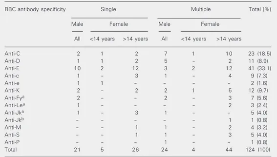
IgG alloantibodies were detected in 82 of the 828 patients who received transfusions (9.9%; 95% confidence interval, CI = 7.9 to 11.9%). Of these 82 patients, 29 tested positive before their first blood transfusion at Hemominas. A hundred and twenty-five IgG alloantibodies were detected. In one patient the antibody identification was not performed. Twenty-nine patients (35.4%) presented two or more alloantibodies; the presence of an autoantibody of the IgG class was also tected in 14 (17.1%). Cold antibodies de-tected in 3 patients were not included in the study. The specificities and frequencies of the alloantibodies are shown in Table 1. They did not differ significantly when patients who tested positive before first transfusion at Hemominas were compared with those who were negative. Of the 124 alloantibodies identified, 69.4% were directed against anti-gens of the Rhesus group and 9.7% were directed against antigens of the Kell group, corresponding to 79.1% of the alloantibodies detected.
Of the 82 alloimmunized patients, 37.8% were males and 62.2% females (P = 0.03); 78% were Hb SS or Sß0-thal, 20.7% Hb SC and 1.2% Hb Sß+-thal patients. The median age at antibody screening was 23.3 years in the alloimmunized group and 14.6 years in the non-alloimmunized group (P < 0.0001; Table 2).
of hemoglobinopathy, and number of trans-fusions, 620/828 patients whose number of transfusions was known were separated. The difference in the frequency of alloimmu-nization between this group and the group whose number of transfusions was unknown was not statistically significant (P = 0.24). The frequency of alloimmunization found in the group of patients with a known number of transfusion was 9.2% (57/620) with a 95% CI of 6.9 to 11.5%. Median age was 21.5 years for alloimmunized patients and 11.8 years for non-alloimmunized patients (P = 0.0001). Despite the higher frequency in females (11.1% as opposed to 7.4% for
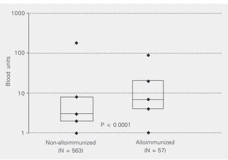
males), this difference was not statistically significant (P = 0.11). Median age was 25.3 years for the alloimmunized female patients and 15.1 years for the male patients. Alloim-munization was more frequent in Hb SC patients than in Hb SS patients (14 vs 8.1%; P = 0.048). Alloimmunized patients received a median number of 7 blood transfusions, as opposed to 3 transfusions for non-alloimmu-nized patients (P < 0.0001, Figure 1). Table 3 lists the number of transfusions by age group and alloimmunization.
After multivariate adjustment (N = 608, since 11 patients with Hb Sß+-thal and one with Hb SD were excluded from this
analy-Table 1. Specificity of single or multiple IgG alloantibodies in 82 patients with sickle cell disease.
RBC antibody specificity Single Multiple Total (%)
Male Female Male Female
All <14 years >14 years All <14 years >14 years
Anti-C 2 1 2 7 1 10 23 (18.5)
Anti-D 1 1 2 5 - 2 11 (8.9)
Anti-E 10 2 12 3 2 12 41 (33.1)
Anti-c 1 - 3 1 - 4 9 (7.3)
Anti-e 1 1 - - - - 2 (1.6)
Anti-K 2 - 2 2 1 5 12 (9.7)
Anti-Fya 2 - - 2 - 3 7 (5.6)
Anti-Lea 1 - - - - 2 3 (2.4)
Anti-Jka 1 - 3 1 - - 5 (4.0)
Anti-Jkb - - - - - 1 1 (0.8)
Anti-M - - 1 1 - 2 4 (3.2)
Anti-S - - 1 1 - 3 5 (4.0)
Anti-P - - - 1 - - 1 (0.8)
Total 21 5 26 24 4 44 124 (100)
In one patient, antibody identification was not performed. RBC = red blood cell.
Table 2. Distribution of 828 patients with sickle cell disease according to median age, hemoglobin (Hb) phenotype, and gender.
Patients Number Median age1 Type of Hb2 Gender3
(range)
SS* SC/Sß+-thal/SD Male Female
Non-alloimmunized 746 14.6 (0.5 to 76.1) 613 133 374 372
Alloimmunized 82 23.3 (2.2 to 48.3) 64 18 31 51
Total 828 18.4 (1.2 to 78.8) 677 151 405 423
*Included Sß0-thal. 1P < 0.0001 (Mann-Whitney test); 2P = 0.36 (chi-square test); 3P = 0.03 (chi-square
sis), gender was not statistically significant (P = 0.35). Explanatory variables that re-mained statistically significant for alloimmu-nization were: higher number of transfusions received (P = 0.00006), older age (P = 0.056) and SC compared with SS disease (P = 0.02).
Because obstetric history at the time of alloantibody detection was not available for almost 50% of females older than 14 years, separate analyses were performed for pa-tients under or above this cut-off point. For those below 14 years, only the number of transfusions remained statistically signifi-cant in multivariate analysis (P = 0.0001). For those above 14 years, number of trans-fusions (P = 0.04) and type of SCD (P = 0.008) remained significant in multivariate analysis. These Hb SC patients were at a 2.8-fold higher risk of alloimmunization than SS patients (95% CI = 1.3 to 6.0). The median numbers of blood transfusions for Hb SC and SS patients were 3 and 6, respectively (P < 0.0001). Male and female patients received the same median number of transfusions (5; P = 0.87). The frequency of alloimmuniza-tion for patients younger or older than 14 years according to sex and type of SCD is presented in Table 4. The frequency of alloimmunization was higher for Hb SS chil-dren (<14 years old), although not statisti-cally significant (P = 0.24); no significant difference was observed between males and females. Hb SS children, however, received more blood transfusion than SC children: 12% of SS children received more than 15 transfusions whereas no SC child received this number of transfusions. A double posi-tive gradient from males to females and from Hb SS to SC was observed for patients older than 14 years. The highest frequency was observed in Hb SC female patients (12/46 = 26.1%).
Discussion
Of the 1,544 patients initially studied, 899
Blood units
1000 ...
...
...
...
Non-alloimmunized (N = 563)
Alloimmunized (N = 57) 100
10
1
P < 0.0001
Figure 1. Number of transfusions received by alloimmunized and non-alloimmunized patients with sickle cell disease. Higher to lower, the points indicate maximum number, 75th percentile, median, 25th percentile, and minimum number of transfusions (logarith-mic scale).
Table 3. Number of blood transfusions by age group and red blood cell alloimmuni-zation among 620 patients with sickle cell disease.
Age group Number of Positive tests for Number of
(years) patients alloimmunization transfusions
N % Median Range
0 to 9.9 254 12 4.7 3 1-140
10 to 19.9 170 15 8.8 4 1-178
20 to 29.9 100 17 17.0 5 1-111
30 or more 96 13 13.5 5.5 1-110
Table 4. Frequency of alloimmunization for patients younger or older than 14 years according to gender and type of sickle cell disease.
Patients under 14 years Patients above
of age (N = 333) 14 years of age (N = 275)
Male Female Male Female
Hb SS patients 6.6% (11/167) 4.8% (6/125) 9% (9/100) 13.7% (14/102)
Hb SC patients 0% (0/22) 0% (0/19) 14.8% (4/27) 26.1% (12/46)
(58.2%) had received blood transfusions. This transfusion rate among SCD patients is similar to those reported in other studies (3,15,23).
The rate of alloimmunization observed in the present study was 9.9%. Other studies have reported frequencies ranging from 2.6 to 47% (3,9-13,15-17,19,20,24). Studies presenting lower rates of alloimmunization (7.8 (8) and 6.0% (14)) included patients who were predominantly in the pediatric group. In Brazil, rates of 12.9 (15), 20.8 (16), and 3.2% (24) have been reported. Again the lowest figure corresponds to a population of 83.9% of children aged less than 15 years. In the present study, 403/828 patients were less than 15 years old.
The relatively low rate found in the pres-ent study may have been influenced by sev-eral factors. Patients were not monitored for RBC antibodies after each transfusion and hence transitory antibodies were probably not detected. Antibody screening was per-formed employing the tube method up to July 1991 and then replaced by gel centrifugation. Hence, antibodies present at low titers were probably not detected during that period.
Another factor that could contribute to the low frequency found in the present study may be the high phenotypic compatibility between patients and blood donors. Further studies are necessary to confirm the influ-ence of this factor on the frequency of alloimmunization. In Brazil, only one study (15) compared RBC phenotype between blood donors and patients with SCD. A higher frequency of C antigen in donors was reported (66.7% compared with 46.0% in patients; P < 0.01), but no difference was detected for other RBC antigens, thus dem-onstrating high compatibility between do-nors and patients with SCD in the study.
The specificities of the antibodies found in the present study are similar to those found in other reports of alloimmunization in SCD. This is certainly due to the high immunoge-nicity of antigens C, D, E, and K (22), and
also to the fact that RBC matching was not routinely employed for all of these antigens, except for antigen D.
Of the 124 alloantibodies identified, 8.9% were anti-D (7 associated and 4 isolated), a figure higher than reported in most interna-tional studies (8,11). A Brazilian study (15) reported 20% anti-D antibody among a total of 15 alloantibodies detected. The compara-tively high frequency of anti-D antibodies found in the present study may be attributed to several factors. Since the obstetric history of the patients was not always available in their medical files, secondary alloimmuniza-tion related to pregnancy is to be considered. Many patients had been transfused before they were admitted to Hemominas Founda-tion and even after that, many were fused in small hospitals in which blood trans-fusion is not adequately supervised.
Although alloimmunization is more fre-quent in females, in the present multivariate analysis gender was not a risk factor for alloimmunization (P = 0.29), similarly to data reported by others (3,25). Some investiga-tors have reported a higher risk for females (19,21).
A significant association between Hb phenotype and alloimmunization was ob-served even after multivariate adjustment in patients older than14 years. Patients with Hb SC presented a risk of alloimmunization al-most three times as high as that for patients with Hb SS, an association hitherto unre-ported. In the Cooperative Study of Sickle Cell Disease (3) such association was not found, and a rather lower rate of alloimmu-nization was observed in patients with Hb SC phenotype. This was attributed to a lower number of transfusions received by that group, as also observed by others (20,21). In the present study, the association between Hb SC phenotype and alloimmunization was observed only for patients older than 14 years. For those below this cut-off point alloimmunization frequency was higher for SS children, although not statistically signifi-cant. Patients older than 14 years with Hb SC disease received significantly fewer blood transfusions than those with the SS type. The number of transfusions for male and female patients was closely similar. Two non-excluding hypotheses for explaining the double positive gradient shown in Table 4 for patients above 14 years of age are possible: 1) older female patients and especially those with Hb SC disease developed alloimmuniza-tion at a higher frequency because previous pregnancy and not blood transfusion was an induction factor. Menarche occurs signifi-cantly earlier in Hb SC than SS patients (27). Additionally, sexual activity, which is lower among SCD females compared with con-trols (28) may be higher for Hb SC than for
SS females because their clinical status is usually better; 2) it is conceivable that there is a span of many years before the first transfusion is needed for Hb SC male or female patients. Wayne et al. (2) suggested that alloimmunization in patients with thalas-semia may occur more frequently if the blood transfusion is administered intermit-tently and started after childhood. Since patients with Hb SC are faced with this situation, it is possible that a similar mechan-ism underlies the phenomenon observed in the present investigation.
Based on the present results and on litera-ture data (3,6,11-14,29), RBC should be matched for C, E and K antigens in transfu-sions administered to patients with hemoglo-binopathies. The relevant roles of patient age and number of administered transfusions in the risk of alloimmunization were confirmed. In contrast to the international literature, older patients with Hb SC disease - and especially females - were at a higher risk to develop RBC alloimmunization, requiring further studies for verification and explana-tion of the observed phenomenon. Cohort studies based on neonatal screening and careful prospective obstetric data would be very helpful.
Acknowledgments
We gratefully acknowledge the contribu-tions of ours colleagues at Hemominas Foun-dation and in the Hematology Division, Uni-versity Hospital, Federal UniUni-versity of Minas Gerais, Belo Horizonte, MG, Brazil.
References
1. Zago MA (2001). Anemia falciforme e doença falciforme. In: Manual
de Doenças mais Importantes, Por Razões Étnicas, na População Afro-Descendente. Secretary of Health Policy, Ministry of Health, Brasília, DF, Brazil.
2. Wayne AS, Kevy SV & Nathan DG (1993). Transfusion
manage-ment of sickle cell disease. Blood, 81: 1109-1123.
3. Rosse WF, Gallagher D, Kinney TR, Castro O, Dosik H, Moohr J,
Wang W & Levy OS (1990). Cooperative Study of Sickle Cell Disease. Transfusion and alloimmunization in sickle cell disease.
Blood, 76: 1431-1437.
The preoperative transfusion in Sickle Cell Disease Study Group.
New England Journal of Medicine, 333: 206-213.
5. Davies SC & Harewood MR (1997). Blood transfusion in sickle cell
disease. Blood Reviews, 11: 57-71.
6. Vichinsky EP (2001). Current issues with blood transfusion in sickle
cell disease. Seminars in Hematology, 38 (Suppl): 14-22.
7. Vichinsky EP, Luban NLC, Wright E, Olivieri N, Driscoll C, Pegelow CH, Adams RJ & Stroke Prevention Trail in Sickle Cell Anemia (2001). Prospective RBC phenotype matching in a
stroke-preven-tion trial in sickle cell anemia: a multicenter transfusion trial.
Transfu-sion, 41: 1086-1092.
8. Sarnaik S, Schornack J & Lusher JM (1986). The incidence of development of irregular red cell antibodies in patients with sickle
cell anemia. Transfusion, 26: 249-252.
9. Cox JV, Steane E, Cunningham G & Frenkel EP (1988). Risk of alloimmunization and delayed hemolytic transfusion reactions in
patients with sickle cell disease. Archives of Internal Medicine,
148: 2485-2489.
10. Luban NLC (1989). Variability in rates of alloimmunization in differ-ent groups of children with sickle cell disease: effect of ethnic
background. American Journal of Pediatric Hematology/Oncology,
11: 314-319.
11. Vichinsky EP, Earles APNP, Johnson RA, Hoag MS, Williams A & Lubin B (1990). Alloimmunization in sickle cell anemia and
transfu-sion of racially unmatched blood. New England Journal of
Medi-cine, 322: 1617-1621.
12. Tahhan HR, Holbrook CT, Braddy LR, Brewer LD & Christie JD (1994). Antigen-matched donor blood in the transfusion
manage-ment of patients with sickle cell disease. Transfusion, 34: 562-569.
13. Norol F, Nadjahi J, Bachir D, Desaint C, Bataille MG, Beaujean F, Bierling P, Bonin P, Galacteros F & Duedari N (1994). Transfusion
and alloimmunization in sickle cell anemia patients. Transfusion
Clinique et Biologique, 1: 27-34.
14. Hmida S, Mojaat N, Maamar M, Bejaoui M, Mediouni M & Boukef K (1994). Red cell alloantibodies in patients with
hemoglobinopa-thies. Nouvelle Revue Française d’Hématologie, 36: 363-366.
15. Moreira JG, Bordin JO, Kuroda A & Kerbauy J (1996). Red blood cell alloimmunization in sickle cell disease: the influence of racial and antigenic pattern differences between donors and recipients
in Brazil. American Journal of Hematology, 52: 197-200.
16. Fabron Junior A (2000). Estudo da significância clínica de aloanticorpos eritrocitários em pacientes com anemia falciforme. Doctoral thesis, Escola Paulista de Medicina, UNIFESP, São Paulo,
SP, Brazil.
17. Olujohungbe A, Hambleton I, Stephens L, Serjeant B & Serjeant G (2001). Red cell antibodies in patients with homozygous sickle cell disease: a comparison of patients in Jamaica and the United
Kingdom. British Journal of Haematology, 113: 661-665.
18. Fabron Junior A, Moreira JG & Bordin JO (1999). Delayed hemolyt-ic transfusion reaction presenting as a painful crisis in a patient with
sickle cell anemia. São Paulo Medical Journal, 117: 38-39.
19. Orlina AR, Unger PJ & Koshy M (1978). Post-transfusion
alloimmu-nization in patients with sickle cell disease. American Journal of
Hematology, 5: 101-106.
20. Oliva PG, Otero PB, Villalonga CG, Veranes MB, Aldama LO & Echevarria MG (1985). Aloimunización en pacientes con
enfer-medad por hemoglobina S politransfundidos. Sangre, 30:
406-410.
21. Reisner EG, Kostyu DD, Phillips G, Walker C & Dawson V (1987). Alloantibody responses in multiply transfused sickle cell patients.
Tissue Antigens, 30: 161-166.
22. Mollison PL, Engelfriet CP & Contreras M (1997). Blood
Transfu-sion in Clinical Medicine. 10th edn. Blackwell Science, Oxford, UK. 23. Thame JR, Hambleton IR & Searjeant GR (2001). RBC transfusion in sickle cell anemia (HbSS): experience from the Jamaican Cohort
Study. Transfusion, 41: 596-601.
24. Andrade SR, Brito GD, Calado NB, Marcos CPM, Serafim ESSM & Rodrigues S (1999). Red blood cell alloimmunization in patients
with sickle cell anemia. Revista Brasileira de Análises Clínicas, 31:
195-197.
25. American Association of Blood Banks (AABB) (1999). Technical
Manual. 13th edn. AABB, Bethesda, MD, USA.
26. Spanos T, Karageorga M, Ladis V, Peristeri J, Hatziliami A & Kattamis
C (1990). Red cell alloantibodies in patients with thalassaemia. Vox
Sanguinis, 58: 50-55.
27. Serjeant GR, Singhal A & Hambleton IR (2001). Sickle cell disease and age at menarche in Jamaican girls: observations from a cohort
study. Archives of Disease in Childhood, 85: 375-378.
28. Samuels-Reid JH, Scott RB & Brown WE (1984). Contraceptive
practices and reproductive patterns in sickle cell disease. Journal of
the National Medical Association, 76: 879-883.
29. Castro O, Sandler SG, Houston-Yu P & Rana S (2002). Predicting the effect of transfusing only phenotype-matched RBCs to patients
with sickle cell disease: theoretical and practical implications.