High pressure near infrared study of the
mutated light-harvesting complex LH2
1Department of Biology, University of Munich, Munich, Germany
2Department of Physics E13, Technical University Munich, Garching, Germany P. Braun1,
R. Gebhardt2, L. Kwa1 and W. Doster2
Abstract
The pressure sensitivities of the near infrared spectra of the light-harvesting (LH2) complex and a mutant complex with a simplified BChl-B850 binding pocket were compared. In the mutant an abrupt change in the spectral properties occurred at 250 MPa, which was not observed with the native sample. Increased disorder due to collapse of the chromophore pocket is suggested.
Correspondence
W. Doster
Technische Universität München Physikdepartment E13 D-85748 Garching Germany
Fax: +49-89-289-12-473 E-mail: [email protected]
Presented at the 3rd International Conference on High Pressure Bioscience and Biotechnology, Rio de Janeiro, RJ, Brazil, September 27-30, 2004.
Received January 21, 2005 Accepted May 12, 2005
Key words
•Light-harvesting complex •High pressure optical
spectroscopy
•Protein compressibility
•Antenna proteins •High pressure light
scattering
Introduction
The peripheral light-harvesting (LH2) complex from Rhodobacter sphaeroides (Rb. sphaeroides) constitutes an ideal model to investigate the effect of external stress on the stability of membrane proteins. The LH2 complex contains several pigment molecules, which constitute a series of intrinsic molecu-lar probes. These individual chromophores, therefore, are able to monitor localized struc-tural changes when examined by spectro-scopic methods as a function of external stresses such as applied hydrostatic pres-sure. Protein-cavities or voids, if they are empty in the interior of soluble proteins, may result in poor packing. The correlation be-tween protein stability and packing quality, however, is disputed (1). Internal voids can be induced in membrane embedded LH2 or related pigment-binding proteins by either the removal of a specific chromophore (2) or as a result of site-directed mutagenesis (3).
The peripheral antenna of purple
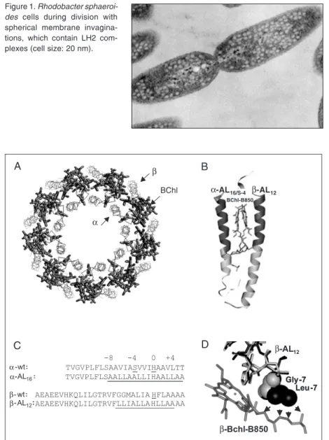
photo-synthetic bacteria is one of the best-charac-terized membrane proteins. These proteins, which are present in many photosynthetic bacteria, absorb solar photons and transfer the resulting excitonic energy to the so-called core antenna protein (or LH1) that surrounds the bacterial reaction centers. From the LH1, the energy is further transferred to the reac-tion centers, where it is transformed into potential chemical energy. This photosyn-thetic machinery is embedded in the thyla-koid membrane, in bacteria typically in the intra-cytoplasmatic membrane. In the case of Rb. sphaeroides, these membranes are spherical invaginations of the intra-cytoplas-matic membrane, which become vesicles upon cell disruption (Figure 1).
polypeptides called α and ß. The nine α -apo-proteins form a hollow cylinder with the nine ß-apo-proteins arranged radially out-side. One (or two) carotenoid (rhodopsin glucoside) and three bacteriochlorophyll a (BChl a) molecules are non-covalently at-tached to each α/ß pair. Most of the α -polypeptide/ß-polypeptide interactions in LH2 occur close to the membrane/water in-terfaces, between the C- and
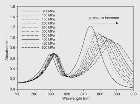
N-membrane-hugging termini, while the pigments (both BChl and carotenoid molecules) mediate most of the α-ßcontacts in the hydrophobic phase. In LH2, the 27 BChl molecules are organized into two discrete pools. Eighteen of the BChl molecules are sandwiched be-tween the α- and ß-apo-proteins and form a ring of overlapping BChl in close contact (see Figure 2A). These molecules absorb in the near-infrared at 850 nm and are known as the BChl-BChl-B850 molecules. The other nine BChl molecules lie toward the cyto-plasmic side of the membrane. These pig-ments exhibit spectroscopic properties close to monomeric BChls. They absorb in the near-infrared at 800 nm, and are denoted BChl-BChl-B800. Free in organic solvents, the Qy electronic transition of monomeric BChl a is located at 770 nm. The physico-chemical mechanisms underlying the shift of this transition to 800 or 850 nm when these molecules are bound to LH2 proteins have been extensively studied. For BChl-BChl-B800, the mechanism essentially de-pends on the interactions between the BChl molecules and their specific microenviron-ment (5), while for BChl-BChl-B850 it re-sults from a combination between BChl/ BChl interactions and interactions between each BChl and the surrounding protein (6-8). These interactions are very sensitive to localized structural (environmental) changes and therefore the positions of the Qy transi-tions constitute intrinsic molecular probes that are able to monitor the integrity of the LH2 structure. The electronic properties of LH2 yield information both about the struc-ture of the BChl binding sites within this protein (which in turns yields information about the tertiary structure of the polypep-tides in the vicinity of these molecules) and indirectly (although in a highly persuasive manner) about its quaternary structure. As a result, the LH2 family of proteins is ideal for the study of the stability of membrane-em-bedded hydrophobic α-helical membrane he-lices.
Figure 1. Rhodobacter sphaeroi-des cells during division with spherical membrane invagina-tions, which contain LH2 com-plexes (cell size: 20 nm).
Figure 2. A, Schematic view of the LH2 complex from the plane of the membrane. B, Elementary α and ß subunits with BChl-B850 (the mutated regions are shown in grey). C, Amino acid sequences of LH2 apoproteins and mutants. D, Structural alignment of residues at position-7 of wild-type LH2-ß (grey) and mutant LH2-ß (black). BChl = bacteriochloro-phyll; wt = wild type; AL = alanine mutant.
Previously, we have mutated both the α -and ß-apo-proteins of LH2 to produce com-plexes with substantially simplified BChl-B850 binding pockets (see Figure 2B,C). In spite of the extensive mutations, the spectral properties of these complexes remain un-changed (9,10). However, the mutations in-duced local packing defects around the BChl-B850 molecules(Figure 2D) and a signifi-cant reduction in thermal stability. Various antenna pigment-protein complexes from purple photosynthetic bacteria have been studied at high pressure in detergent, both at ambient temperature (2,11-13) and at cryo-genic temperatures (14). These studies have shown that increasing the pressure leads to a substantial red shift and broadening of the long wavelength Qyabsorption band both in LH1 and in LH2 in detergent. In the present study, the properties of LH2 are compared to the properties of the LH2 mutant complex in which the residues at the BChl/polypeptide interface have been systematically mutated (Figure 2B-D). This permits us to monitor the stability of model membrane proteins under external stress in the native lipid envi-ronment.
Material and Methods
Bacterial strains, plasmids, gene transfer, and growth conditions
The bacterial strains used in the present study were E. coli strain S17-1 (thi pro hsdR
-hsdM+ recA RP4-2 (Tc::mu Kan::Tn7), Rb.
sphaeroides strain DD13 (genomic deletion of both pucBA and pufBALMX; insertion of SmR and KanR genes, respectively) (15). The
mobilizable plasmids used were based on pRKCBC1 (TcR, a derivative of pRK415;
insertion of a 4.4-kb fragment encompass-ing pucBAC); briefly, this expression vector contains the pucBA genes as a 420-base pair KpnI-BamHI insert (15). Growth conditions for E. coli and Rb. sphaeroides were as described in Ref. 16. Conjugative transfer of
plasmid from E. coli S17-1 to Rb. sphaeroi-des was performed as described in Ref. 16.
Construction of the LH2 complex with the model BChl-BChl-B850 site was carried out as described in Refs. 9,10. Membranes were prepared from cells grown semi-aero-bically in the dark by disruption in a French pressure cell and subsequent centrifugation on a sucrose step gradient (10). Dynamic light scattering experiments on LH2 vesicles under pressure were performed using an ALV goniometer-correlator system (Fa. ALV-La-ser GmBH, Langen, Germany) and a fre-quency doubled NdYag laser (Coherent GmbH, 150 mW). The sample was trans-ferred to a specially designed four-window high-pressure cell manufactured by SITEC (Zürich, Switzerland). Optical absorption spectra were measured using the same cell, which was connected by optical fibers to a Perkin-Elmer UV/Vis Lambda II spectrom-eter (Überlingen, Germany).
Results
Dynamic light scattering experiments were performed with LH2 vesicles as func-tion of scattering angle at ambient pressure. The intensity correlation function was ana-lyzed in terms of two dynamic components: a fast component contributing 30% to the total scattering intensity and a slow fraction. From the resulting diffusion constants, we derive an average hydrodynamic radius us-ing the Stokes-Einstein relation:
D = kBT/(6 π ηRH) (Eq. 1)
where η is the viscosity of the solvent. RH of
aggre-Figure 3. Hydrodynamic radius (RH) of the LH2 vesicles at 20ºC versus pressure, corrected for solvent viscosity.
Pressure [MPa]
0 100 200 300 400 500
RH
[n
m
]
0 20 40 60 80 100
Figure 4. Near infrared absorption spectrum of wild-type LH2 vesicles.
gates by a factor of 60,000. The same experi-ments were performed at a 90º scattering angle in the pressure cell. The scattering intensity due to the two components remained approximately constant with pressure. How-ever, the radius of the large particles de-creased with pressure. Their effective com-pressibility, assuming the shape of the vesicle
to be approximately spherical, can be esti-mated from:
3 δRH/(δp RH) ≈δVves/(δp Vves) = -ßT
(Eq. 2)
where Vves is the volume of the vesicle and
ßT denotes the isothermal compressibility.
From the slope in Figure 3, correcting for the increase of the refractive index with pres-sure, we obtain ßT≈ 0.9 GPa-1. This value is
almost twice as high as the compressibility of water (0.56 GPa-1). The large apparent
compressibility suggests that the shape of the vesicular aggregates becomes more a-symmetric under pressure. The radius of the small particles varies only slightly with pres-sure, consistent with a compressibility domi-nated by the included water. The light scat-tering experiment demonstrated that the LH2 vesicles remain stable up to 500 MPa.
Figure 4 shows the near infrared spec-trum of wild-type LH2 (same scale for mu-tant and wild-type spectrum). The BChl-B850 band is stronger than the BChl-B800 band since 18 versus 9 BChl a molecules are contributing to the absorption. As shown previously for LH2 complexes in detergent, the application of pressure led to a signifi-cant red shift of the absorption maxima, which was more pronounced for BChl-B850 (11,14). The latter made excitonic contribu-tions reflecting direct interaccontribu-tions of BChl a dimers, in contrast to BChl-B800, in which the molecules are well separated and exci-tonic effects are negligible. The BChl-B800 pressure shift of 0.1 cm-1/MPa is typical for
Pressure [MPa]
0 100 200 300 400 500
d
/d
p
(cm
-1
MPa
)
-1.0 -0.9 -0.8 -0.7 -0.6 -0.5 -0.4 -0.3 -0.2
WT
Mut
/
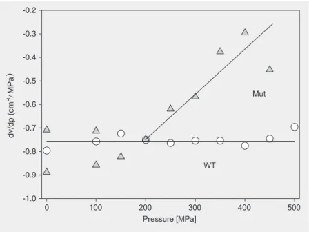
Figure 5. Near infrared absorption spectra of the mutant LH2 complex versus the indicated pressures.
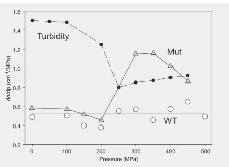
Figure 6. The pressure shift of the BChl-B850 line maximum (dν/dp) of wild-type (WT, circles) and mutant (Mut, triangles) LH2 from Gaussian fits in the frequency domain. at ambient pressure to about 19 nm at 500
MPa. Figure 5 shows the BChl-B850 spec-trum of the mutant complex. The mutant absorption band was red-shifted with in-creasing pressure similar to the red shift of this band in the wild type. The increase in line-width, however, was significantly more pronounced in the mutant BChl-B850 band. The line shapes of the absorption bands in the frequency domain were reasonably well approximated by a Gaussian distribution, indicating a dominance of inhomogeneous broadening. Figure 6 compares the pressure shifts of wild-type and mutant BChl-B850. At low pressures up to 200 MPa, the pres-sure shifts were identical within the experi-mental error, about -0.75 cm-1/MPa. This
suggests that the chromophore coupling in the BChl-B850 ring is similar for wild type and mutant. However, above 200 MPa, the negative pressure shift of the mutant de-creased, while it remained constant for the wild type. This result indicates that increas-ing the pressure beyond 200 MPa does not further enhance the chromophore interac-tions in the mutant. The change in the pres-sure sensitivity was quite abrupt, therefore suggesting a structural change. This is sup-ported by the pressure dependence of the bandwidth shown in Figure 7. The pressure sensitivity of the Gaussian width of the BChl-B850 band of wild-type LH2 remained ap-proximately constant with pressure. In con-trast, the Gaussian width of the mutant BChl-B850 band abruptly increased at 200 MPa, coinciding with the pressure at which the negative pressure sensitivity of the band de-creased. This indicates enhance energetic disorder, possibly related to loss of second-ary structure. Inspection of the BChl-B850 absorption spectrum of the mutant in Figure 5 suggests a transition from a reasonably well-defined state absorbing at 850 nm to-wards a more disordered structural state ab-sorbing near 875 nm. In the latter the chro-mophore-chromophore interactions have reached a limiting value.
wild-Figure 7. Pressure sensitivity of Gaussian line-width of BChl-B850 wild-type (WT, open circles) and mutant (Mut, triangles) and turbidity (filled circles) at 649 nm (OD -1) of the mutant sample.
type LH2. Interestingly, as shown in Figure 7, in the same pressure range we observe an abrupt decrease in the Raleigh scattering intensity for the mutant. This result implies that mutant-LH2 vesicles dissociate at least partially under pressure. This correlation is somewhat surprising since one expects that only the local properties of the protein struc-ture in the vicinity of the chromophore should affect the absorption spectra. Dynamic light scattering experiments performed with the mutant preparation indicate that the number of large vesicle (80 nm) was significantly enhanced compared to the native prepara-tion. That structural changes of the vesicles and of the membrane can modify the pres-sure sensitivity of BChl-B850 needs further investigation.
References
1. Cuff AL & Martin AC (2004). Analysis of void volumes in proteins and application to stability of the p53 tumour suppressor protein.
Journal of Molecular Biology, 344: 1199-1209.
2. Gall A, Ellervee A, Robert B et al. (2004). The effect of internal voids in membrane proteins: high-pressure study of two photochemical reaction centres from Rhodobacter sphaeroides. FEBS Letters, 560: 221-225.
3. Fyfe PK, Ridge JP, McAuley KE et al. (2000). Structural conse-quences of the replacement of glycine M203 with aspartic acid in the reaction center from Rhodobacter sphaeroides. Biochemistry, 39: 5953-5960.
4. McDermott G, Prince SM, Freer AA et al. (1995). Crystal structure of an integral membrane light-harvesting complex from photosynthetic bacteria. Nature, 374: 517-521.
5. Gall A, Fowler GJ, Hunter CN et al. (1997). Influence of the protein binding site on the absorption properties of the monomeric bacterio-chlorophyll in Rhodobacter sphaeroides LH2 complex. Biochemis-try, 36: 16282-16287.
6. Sauer K,Sauer K, Cogdell RJ et al. (1996). Structure-based calcula-tions of the optical spectra of the LH II bacteriochlorophyll-protein complex from Rhodopseudomonas acidophilia. Photochemistry and
Photobiology, 64: 564-576.
7. Braun P & Scherz A (1991). Polypeptides and bacteriochlorophyll organization in the light-harvesting complex B850 of Rhodobacter
sphaeroides R-26.1. Biochemistry, 30: 5177-5184.
8. Olsen JD, Sturgis JN, Westerhuis WH et al. (1997). Site-directed modification of the ligands to the bacteriochlorophylls of the light-harvesting LH1 and LH2 complexes of Rhodobacter sphaeroides.
Biochemistry, 36: 12625-12632.
9. Kwa LG, Garcia-Martin A, Vegh A et al. (2004). Hydrogen bonding in a model bacteriochlorophyll-binding site drives assembly of light
harvesting complex. Journal of Biological Chemistry, 279: 15067-15075.
10. Braun P, Olsen J, Strohmann B et al. (2002). Assembly of light-harvesting bacteriochlorophyll in a model transmembrane helix in its natural environment. Journal of Molecular Biology, 318: 1085-1095. 11. Gall A, Ellervee A, Sturgis JN et al. (2003). Membrane protein stability: high pressure effects on the structure and chromophore-binding properties of the light-harvesting complex LH2. Biochemis-try, 42: 13019-13026.
12. Gall A, Ellervee A, Bellissent-Funel MC et al. (2001). Effect of high pressure on the photochemical reaction center from Rhodobacter
sphaeroides R26.1. Biophysical Journal, 80: 1487-1497.
13. Sturgis JN, Gall A, Ellervee A et al. (1998). The effect of pressure on the bacteriochlorophyll a binding sites of the core antenna complex from Rhodospirillum rubrum. Biochemistry, 37: 14875-14880. 14. Reddy NRS, Wu HM, Jankowiak R et al. (1996). High pressure
studies of energy transfer and strongly coupled bacteriochlorophyll dimers in photosynthetic protein complexes. Photosynthesis
Re-search, 48: 277-289.
15. Jones MR, Fowler GJ, Gibson LC et al. (1992). Mutants of
Rhodo-bacter sphaeroides lacking one or more pigment-protein complexes
and complementation with reaction-centre, LH1 & LH2 genes.
Mo-lecular Microbiology, 6: 1173-1184.
16. Fowler GJ, Gardiner AT, MacKenzie RC et al. (1995). Heterologous expression of genes encoding bacterial light-harvesting complexes
in Rhodobacter sphaeroides. Journal of Biological Chemistry, 270:
23875-23882.
17. Zollfrank J, Friedrich J, Fidy J et al. (1991). Photochemical holes under pressure: compressibility and volume fluctuations in a protein.
Journal of Chemical Physics, 94: 8600-8603.
0.2 0.4 0.6 0.8 1.0 1.2 1.4 1.6
Mut
WT Turbidity
Pressure [MPa]
0 100 200 300 400 500
d/
d
p
(c
m
-1MPa
)