Standardizatio n o f a fluo rim e tric
assay fo r the de te rm inatio n o f
tissue angio te nsin-co nve rting
e nzym e activity in rats
1Laboratório de Genética e Cardiologia Molecular e Departamento de Clínica Médica/
LIM 13, Instituto do Coração, Faculdade de Medicina, Universidade de São Paulo, São Paulo, SP, Brasil
2Departamento de Bioquímica, Instituto de Biociências, Universidade Federal do
Rio Grande do Sul, Porto Alegre, RS, and Escola de Educação Física, Universidade de São Paulo, São Paulo, SP, Brasil
3Laboratório de Hipertensão, Instituto de Ciências Biológicas,
Universidade Federal de Minas Gerais, Belo Horizonte, MG, Brasil E.M. O liveira1,2,
R.A.S. Santos3 and
J.E. Krieger1
Abstract
The tripeptide Hip-His-Leu was used to standardize a fluorimetric method to measure tissue angiotensin-converting enzyme (ACE) ac-tivity in rats. The fluorescence of the o-phthaldialdehyde-His-Leu adduct was compared in the presence and absence of the homogenate (25 µl) to determine whether the homogenate from different tissues interfered with the fluorimetric determination of the His-Leu product. Only homogenates from lung and renal medulla and cortex showed significantly altered fluorescence intensity. To overcome this prob-lem, the homogenate from these tissues were diluted 10 times with assay buffer. The specificity of the assay was demonstrated by the inhibition of ACE activity with 3 µM enalaprilat (MK-422). There was a linear relationship between product formation and incubation time for up to 90 min for homogenates of renal cortex and medulla and liver, for up to 60 min for ventricles and adrenals and for up to 30 min for the aorta, lung and atrium homogenates. In addition, there was a linear relationship between product formation and the amount of protein in the homogenates within the following range: lung, 30-600 µg; renal cortex and medulla, 40-400 µg; atrium and ventricles, 20-200 µg; adrenal, 20-100 µg; aorta, 5-100 µg; liver, 5-25 µg. No peptidase activity against the His-Leu product (31 nmol), assayed in borate buffer (BB), was detected in the different homogenates except the liver homogenate, which was inhibited by 0.1 mM r -chloromercu-ribenzoic acid. ACE activity in BB was higher than in phosphate buffer (PB) due, at least in part, to a greater hydrolysis of the His-Leu product in PB. ACE activity of lung increased 20% when BB plus Triton was used. Enzyme activity was stable when the homogenates were stored at -20o
or -70o
C for at least 30 days. These results indicate a condition whereby ACE activity can be easily and efficiently assayed in rat tissue samples homogenized in BB using a fluorimetric method with Hip-His-Leu as a substrate.
Co rre spo nde nce
J.E. Krieger
Laboratório de Genética e Cardiologia Molecular Instituto do Coração, FM, USP Av. Dr. Eneas C. Aguiar, 44 05403-000 São Paulo, SP Brasil
Fax: + 55-11-3069-5022 E-mail: krieger@ incor.usp.br
Research supported by FINEP (No. 6693002300), FAPESP (No. 9546686), CNPq (No. 520696/95-6) and Fundação E.J. Zerbini. E.M. O liveira is the recipient of a CNPq doctoral fellowship (No. 140621/93-8).
Received February 24, 1999 Accepted March 9, 2000
Ke y wo rds
·Angiotensin-converting enzyme
Intro ductio n
Angiotensin-converting enzyme (ACE) is a dipeptidyl carboxypeptidase (EC 3.4.15. 1) that inactivates the vasodepressor com-pound bradykinin and activates the potent vasoconstrictor and growth-promoting sub-stance angiotensin II (Ang II) by removal of the carboxy-terminal dipeptide of angiotensin I (Ang I). The enzyme is present mainly in the lung (1) but also in plasma and other tissues (2,3). At these locations, ACE can influence the production of Ang II and may have important physiological contribution in the maintenance of cardiovascular homeo-stasis (4). Thus, measurements of ACE ac-tivity in tissues combined with molecular biology approaches can provide important information regarding the role of ACE in physiological and pathophysiological mech-anisms.
ACE activity has been determined by different methods (1,5-10). In general, meas-urements of ACE activity are based on the hydrolysis of synthetic tripeptides blocked at the amino terminus (6,7,10,11). Among the synthetic substrates, one of the most used has been hippuryl-L-histidyl-L-leucine (Hip-His-Leu). This tripeptide contains the termi-nal sequence of Ang I and its hydrolysis can be detected by the formation of hippuric acid (1) or by the formation of the dipeptide His-Leu (8,10). The fluorimetric detection of this dipeptide is a method of high sensitivity and is both a reproducible and simple assay for the determination of ACE activity.
In the present study ACE activity in dif-ferent rat tissues was determined using the fluorimetric assay described by Santos et al. (10) to determine ACE activity in rat serum. Our results indicate that borate buffer (BB) performs better than phosphate buffer (PB), which has been the buffer most frequently used to determine ACE activity (1,12). More-over, the hydrolysis of the His-Leu dipeptide was shown to be very high in tissues like liver, with a consequent underestimate of
ACE activity levels. The use of r
-chloro-mercuribenzoic acid (PCMB), a dipeptidase inhibitor, successfully resolves this prob-lem. Taken together, the present study ex-tends the initial work of Santos et al. (10) on the use of this method to determine tissue ACE activity in rat samples.
Mate rial and Me tho ds
Rat tissue s and se rum sam ple s
Normotensive male Wistar and SHR rats (200-250 g) were used and handled accord-ing to the NIH guidelines. ACE determina-tion was performed in serum, lung, kidney (cortex and medulla), heart ventricle and atrium, adrenal, liver and aorta. The animals were decapitated and blood was collected without anticoagulant, kept on ice, rapidly centrifuged at 3,000 rpm for 10 min and stored at -20o or -70oC. Tissue samples were
quickly harvested, rinsed, blotted and ho-mogenized in 0.4 M BB, pH 7.2, containing 0.34 M sucrose and 0.9 M NaCl, and frozen at -20o
or -70o
C (1 g tissue:10 ml buffer) or 0.1 M sodium PB, pH 7.2, containing 0.34 M sucrose and 0.3 M NaCl. Homogenates were centrifuged at 3,000 rpm for 10 min and the supernatants frozen at -20o or -70oC for
fluo-rimetric assay of ACE activity.
D e te rminatio n o f ACE activity
ACE activity was determined in rat tis-sues by the method described by Santos et al. (10) for the detection of ACE activity in plasma and serum. Briefly, serum (10 µl) and supernatants from homogenized tissues (20 µl) were incubated with 490 or 480 µl of assay buffer containing 5 mM Hip-His-Leu in 0.4 M sodium BB and 0.9 M NaCl, pH 8.3; sodium BB with 0.1% Triton X-100 with 0.9 M NaCl, pH 8.0, or 0.1 M sodium PB with 0.3 M NaCl, pH 8.0, for 15 or 30
min at 37oC. The reaction was stopped by the
product, His-Leu, was measured fluorimetric-ally at 365-nm excitation and 495-nm emis-sion with a fluoro-colorimeter (AMINCO)
as follows. One hundred µl of o
-phthaldial-dehyde (20 mg/ml) in methanol was added and after 10 min the solution was acidified with 200 µl 3 N HCl and centrifuged in a clinical centrifuge at 3,000 rpm for 10 min at room temperature. To correct for the intrin-sic fluorescence of the tissues and serum, time zero blanks (To) were prepared by add-ing tissue or serum after NaOH. All assays were performed in duplicate or triplicate.
D e mo nstratio n o f the validity o f the e
nzy-matic assay
Standard curves for His-Leu were ob-tained daily. There was a linear relationship between relative fluorescence and His-Leu concentration. Ten µl rat serum or 20 µl tissue homogenate (1 g tissue:10 ml buffer) did not modify the slope of the His-Leu standard curve, except for homogenates from lung, and renal medulla and cortex. To over-come this problem, homogenates from these tissues were diluted ten-fold with the assay buffer. When assays were carried out in sodium PB solution, His-Leu standards were prepared with the same buffer and assayed in parallel.
D e mo nstratio n o f the spe cificity o f the
e nzym atic assay
The specificity of the assay was deter-mined by the inhibition of ACE activity us-ing 3 µM enalaprilat (MK-422).
Line arity o f the assay with re gard to tim e
A 20-µl sample of homogenized tissue was incubated in assay buffer for up to 120 min (kidney medulla and cortex), 90 min (liver), 60 min (ventricle and adrenal) and 30 min (aorta, lung and atrium). The assay was carried out as described above.
Line arity o f the assay with re gard to pro te in
co nce ntratio n
The samples of homogenized tissues were tested within the following range: 30-600 µg for lung, 40-800 µg for renal medulla and cortex, 20-200 µg for ventricle, 20-400 for atrium, 20-200 µg for adrenal, 5-100 µg for aorta and 5-25 µg for liver. The homoge-nates were incubated for different periods of time. The samples were incubated in assay buffer as described above.
Me asure m e nt o f dipe ptidase activity
To determine the presence of dipeptidase activity in BB and PB 20 µl of tissue sample was incubated with 31 nmol His-Leu for 30 min. The relative fluorescence of the reac-tion mixture was compared with the value obtained at To as described above. In tissues where dipeptidase activity was detected, the samples were analyzed in the presence of the SH reagent PCMB (0.1 mM).
Pro te in de te rm inatio n
Protein was measured by the method of Bradford (13) using bovine serum albumin as a standard.
Statistical analysis
Data are reported as means ± SD. Statis-tical analysis was performed by the Student
t-test. P values of less than 0.05 were
consid-ered significant.
Re sults
intensity of the fluorescence signal, while the homogenate of other tissues did not. Diluting the homogenate of renal medulla, cortex and lung ten-fold with the assay buf-fer eliminated the interbuf-ference (Figure 1).
The specificity of the hydrolysis of Hip-His-Leu as a measure of ACE activity was demonstrated by greater than 98% inhibition of the enzymatic activity by 3 µM MK-422 (results not shown).
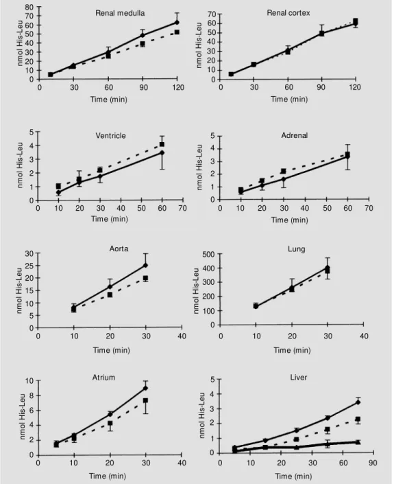
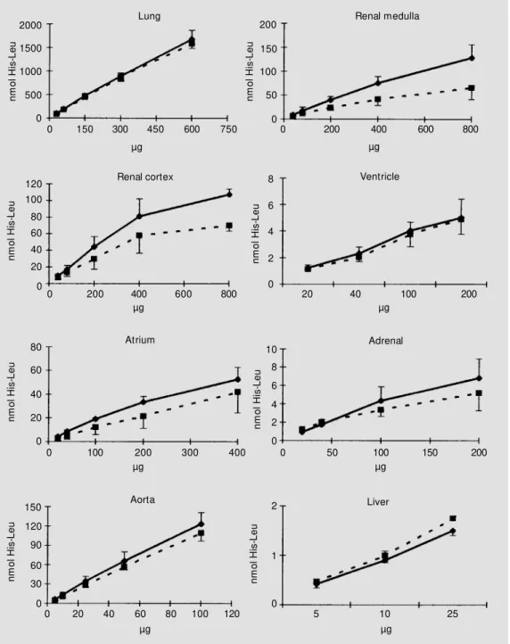
Production of the dipeptide His-Leu as a function of time of incubation is shown in Figure 2. The linearity of the assay as a function of time was maintained for up to 90 min for renal medulla and cortex and liver, up to 60 min for ventricle and adrenal, and up to 30 min for aorta, lung, and atrium. The linearity of the assay as a function of protein concentration (Figure 3) was maintained within the following range: lung, 30-600 µg; renal medulla and cortex, 40-400 µg; atrium and ventricle, 20-200 µg; adrenal, 20-100 µg; aorta, 5-100 µg; liver, 5-25 µg homoge-nates. Based on these results, 20 µl of the lung and aorta homogenates was incubated
for 15 min while the other tissues were incu-bated for 30 min to determine ACE activity. These results were obtained for both SHR and Wistar rats.
To investigate the possibility of nonspe-cific fluorescence development under the assay conditions, the relative fluorescence of the homogenate was evaluated under con-trol conditions (To) and after 1 h of incuba-tion of tissue homogenate samples from Wistar and SHR rats. This period of time was chosen because it was the maximum used for incubation. No difference between the two conditions was observed, indicating that the stability of the homogenates is main-tained under the assay conditions for up to 1 h (results not shown).
To determine whether dipeptidases in the homogenates can hydrolyze His-Leu, de-stroying the compound that gives the fluo-rescence to measure ACE activity, the ho-mogenates were incubated for 30 min with the dipeptide His-Leu. In 0.4 M BB dipepti-dase activity was found only in the liver (Table 1) and could be partially blocked by
F lu o re s c e n c e /n m o l H is -L e u 7 6 5 4 3 2 1 0 C o n tr o l R e n a l m e d u lla R e n a l m e d u lla ( d il 1 0 X ) R e n a l c o rt e x R e n a l c o rt e x ( d il 1 0 X ) L u n g L u n g ( d il 1 0 X ) V e n tr ic le A tr iu m A o rt a A d re n a l L iv e r Wistar SHR 8 * * * * * * Figure 1 - Influence of tissue
100 µM PCMB. This concentration of PCMB was chosen from a range tested (0-2 mM) because it produced the highest inhibition of dipeptidase activity without affecting ACE activity. Dipeptidase activity observed in so-dium BB and soso-dium PB is shown in Table 2. In the presence of sodium PB (Table 2), dipeptidase activity was detected in kidney (50%), ventricle (8%), serum (5%) and liver (65%). PCMB (100 µM) in the incubation
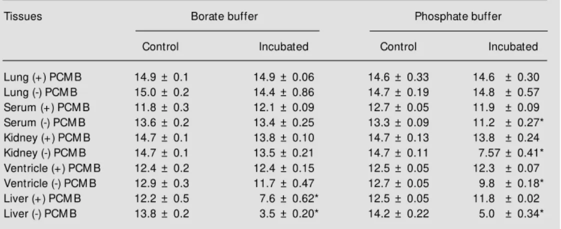
medium abolished His-Leu hydrolysis in these tissues. In contrast, in BB (Table 2) the dipeptidase activity, which was detected only in the liver (75% of His-Leu was hydro-lyzed), could be partially (38%) blocked by PCMB. In addition to the difference in dipeptidase activity, it is interesting to note that the specific activity of ACE determined using BB was consistently higher than with PB (Table 3).
n m o l H is -L e u 80 70 60 50 40 30 20 0 10 0
0 30 60 90 120 0 30 60 90 120
Time (min) Time (min) Renal cortex Renal medulla n m o l H is -L e u 5 4 3 2 0 1
0 10 20 30 70
Time (min) Ventricle
40 50 60
n m o l H is -L e u Time (min) n m o l H is -L e u 30 25 20 15 0 10
0 10 20 30
Time (min) Aorta 40 5 n m o l H is -L e u 10 8 6 4 0 2
0 10 20 30 40
n m o l H is -L e u Atrium Time (min) n m o l H is -L e u 70 60 50 40 30 20 10 5 4 3 1 0
0 10 20 30 70
Adrenal
40 50 60
n m o l H is -L e u 500 400 300 200 100
0 10 20 30
Time (min) Lung
40 0
0 10 20 30 60 90
5 4 3 2 0 1 Liver Time (min)
Figure 2 - Effect of incubation time on the formation of the His-Leu product by different tissue homogenates. His-Leu product formation w as detected as de-scribed in M aterial and M eth-ods. Data are reported as the mean ± SD of three independ-ent experim independ-ent s f or SHR (squares) and Wistar (lozenges) rat s. The ef f ect of 100 µM PCM B treatment on the activity of liver homogenate is indicated by the triangles.
The effect of release of the enzyme from the membranes was tested using the deter-gent Triton X-100. The ACE activity of lung homogenate increased 20% after treatment with 0.1% Triton X-100 (P<0.05) while no differences were detected in the other tissues (results not shown).
The effect of different times and tem-peratures of storage on ACE activity in
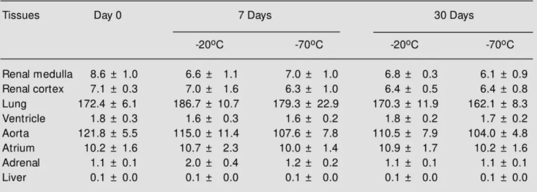
tis-sue homogenates was tested (Table 4). To this end, the homogenates were stored at -20o or -70oC for 7 and 30 days. The enzyme
activity was stable when stored at both tem-peratures for up to 30 days.
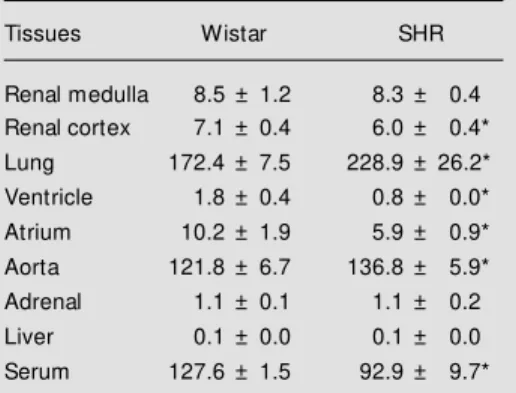
Finally, ACE activity was compared a-mong the different tissues from SHR and Wistar rats from our colony. ACE activity was lower in renal cortex, ventricle, atrium
Figure 3 - Effect of protein con-centration in the assay medium on the formation of the His-Leu product by different tissue ho-mogenates. The assay w as car-ried out for 15 min (lung, and renal cortex and medulla) or 30 min (other tissues) as described in M aterial and M ethods. Data are reported as the mean ± SD of three independent experi-ments for SHR (squares) and Wistar (lozenges) rats.
µg µg n m o l H is -L e u 2000
0 150 0 200 400 600 800
µg µg Renal medulla Lung µg Renal cortex µg µg Atrium Aorta n m o l H is -L e u Ventricle µg Adrenal Liver 1500 1000 500 0
300 450 600 750
120 100 80 60 40 n m o l H is -L e u 80 60 40 20 0 n m o l H is -L e u 150 90 60 30 0 n m o l H is -L e u 200 150 100 50 0 n m o l H is -L e u 8 6 4 2 0 n m o l H is -L e u 10 8 6 4 2 0 n m o l H is -L e u 2 1 0
20 40 100 200
0 50 100 150 200
5 10 25
0 200 400 600 800
100 200 300 400
0 20 40 60 80 100 120
20
0
0
Table 1 - M easurement of dipeptidase activity.
Tissue homogenates from SHR and Wistar rats w ere incubated for 30 min w ith 31 nmol His-Leu. Data are reported as the relative fluorescence of the incubated product and the values obtained at time zero (control). Data indicate the mean ± SD of three independent experiments. Only liver homogenate presented dipepti-dase activity. The effect of 100 µM r-chloromercuribenzoic acid (PCM B) on the dipeptidase activity is show n. * P<0.01 compared to incubated (-) PCM B (Student t-test).
Tissues Wistar SHR
Control Incubated Control Incubated
Renal medulla 5.7 ± 0.09 5.8 ± 0.04 5.7 ± 0.09 5.7 ± 0.06
Renal cortex 5.8 ± 0.08 5.7 ± 0.14 5.7 ± 0.05 5.6 ± 0.10
Lung 5.7 ± 0.01 5.8 ± 0.04 5.7 ± 0.07 5.8 ± 0.05
Ventricle 5.5 ± 0.05 5.3 ± 0.09 5.5 ± 0.07 5.2 ± 0.05
Aorta 5.7 ± 0.02 5.7 ± 0.02 5.9 ± 0.01 5.9 ± 0.02
Atrium 5.5 ± 0.05 5.3 ± 0.03 5.8 ± 0.05 5.6 ± 0.03
Adrenal 5.5 ± 0.04 5.5 ± 0.03 5.7 ± 0.03 5.5 ± 0.09
Liver (-) PCM B 5.8 ± 0.05 2.0 ± 0.09 5.8 ± 0.05 2.3 ± 0.03
Liver (+) PCM B 5.8 ± 0.05 3.8 ± 0.12* 5.8 ± 0.05 3.8 ± 0.12*
Table 2 - Effect of buffer and r-chloromercuribenzoic acid (PCM B) on the dipeptidase activity of tissue homogenates from Wistar rats.
The homogenates (20 µl) and serum (10 µl) w ere incubated for 30 min w ith 62 nmol His-Leu in the presence and absence of 100 µM PCM B. Data indicate the relative fluorescence of the incubated product and the values obtained at time zero (control). Data represent the mean ± SD of three independent experiments. * P<0.01 compared to control (Student t-test).
Tissues Borate buffer Phosphate buffer
Control Incubated Control Incubated
Lung (+) PCM B 14.9 ± 0.1 14.9 ± 0.06 14.6 ± 0.33 14.6 ± 0.30
Lung (-) PCM B 15.0 ± 0.2 14.4 ± 0.86 14.7 ± 0.19 14.8 ± 0.57
Serum (+) PCM B 11.8 ± 0.3 12.1 ± 0.09 12.7 ± 0.05 11.9 ± 0.09
Serum (-) PCM B 13.6 ± 0.2 13.4 ± 0.25 13.3 ± 0.09 11.2 ± 0.27*
Kidney (+) PCM B 14.7 ± 0.1 13.8 ± 0.10 14.7 ± 0.13 13.8 ± 0.24
Kidney (-) PCM B 14.7 ± 0.1 13.5 ± 0.21 14.7 ± 0.11 7.57 ± 0.41*
Ventricle (+) PCM B 12.4 ± 0.2 12.4 ± 0.15 12.5 ± 0.05 12.3 ± 0.07
Ventricle (-) PCM B 12.9 ± 0.3 11.7 ± 0.47 12.7 ± 0.05 9.8 ± 0.18*
Liver (+) PCM B 12.2 ± 0.5 7.6 ± 0.62* 12.5 ± 0.05 11.8 ± 0.02
Liver (-) PCM B 13.8 ± 0.2 3.5 ± 0.20* 14.2 ± 0.22 5.0 ± 0.34*
and serum, similar in renal medulla, adrenal and liver and higher in lung and aorta when Wistar tissues were compared to equivalent SHR tissues (Table 5).
D iscussio n
The study of tissue ACE is relevant be-cause several lines of evidence indicate that
it may be associated with the development of cardiac and vascular hypertrophy (14). How-ever, depending on the buffers used, the various methods for determination of ACE activity usually yield different results.
Hip-His-Leu or the natural substrate, Ang I. Ac-cording to Santos et al. (10), the use of
sodium BB instead of PB increased the Vmax
and reduced the Km of the enzyme when
measured in rat serum. Santos et al. (10) also observed lower dipeptidase activity against the His-Leu substrate when BB was used.
The Vmax of this enzyme was reduced and the
Km increased. These results indicate a
pro-tection of the His-Leu dipeptide used to meas-ure ACE activity by the buffer, with reduced hydrolysis by the dipeptidases. Hydrolysis of His-Leu leads to the underestimation of ACE activity due to the lower fluorescence of the free amino acids leucine and histidine (10). In the present study, we extended these
observations so as to optimize the conditions of the assay to determine ACE activity in several rat tissues.
Considering the differences in the pepti-dase content of tissues, it was necessary to investigate their possible interference with the detection of the His-Leu product. The interference was produced by lung and kid-ney medulla and cortex homogenates and was successfully resolved by diluting the homogenate 10 times with the assay buffer. The specificity of Hip-His-Leu hydroly-sis by ACE activity was demonstrated in the presence of the ACE inhibitor enalaprilat. This compound essentially abolished ACE activity at 3-µM concentration.
The linearity of the reaction was also evaluated as a function of time of incubation and protein concentration. Linearity was obtained for all tissues using 20 µl of the homogenate and a time of incubation of 30 min. Lung and aorta required incubation times of 15 min. This is in accordance with previous reports showing a high ACE activ-ity in lung and aorta (15,16).
Homogenate stability was maintained up to 1 h under the assay conditions when using BB.
Dipeptidase activity varied according to the tissue homogenates and the buffer used. The high dipeptidase activity observed in the Table 3 - Effect of borate and phosphate buffer on
the ACE activity of Wistar rat tissue homogenates.
The enzyme activity w as assayed as described in M aterial and M ethods. Results are reported as nmol His-Leu min-1 mg protein-1 or nmol His-Leu
min-1 ml-1. Data represent the mean ± SD of three
independent experiments. * P<0.01 compared to borate buffer (Student t-test).
Tissues Borate buffer Phosphate buffer
Lung 141.3 ± 4.8 28.6 ± 2.1*
Serum 131.0 ± 4.0 28.0 ± 2.3*
Kidney 8.6 ± 1.7 1.20 ± 0.18*
Ventricle 1.5 ± 0.12 0.16 ± 0.013* Liver 0.15 ± 0.017 0.023 ± 0.0054*
Table 4 - Stability of ACE activity in tissue homogenates stored at -20o or -70oC for 7 or 30 days.
Analysis w as performed on fresh and stored tissue homogenates. The enzyme activity w as assayed as described in M aterial and M ethods. Results are expressed as nmol His-Leu min-1 mg protein-1. Data
represent the mean ± SD of three independent experiments.
Tissues Day 0 7 Days 30 Days
-20oC -70oC -20oC -70oC
Renal medulla 8.6 ± 1.0 6.6 ± 1.1 7.0 ± 1.0 6.8 ± 0.3 6.1 ± 0.9
Renal cortex 7.1 ± 0.3 7.0 ± 1.6 6.3 ± 1.0 6.4 ± 0.5 6.4 ± 0.8
Lung 172.4 ± 6.1 186.7 ± 10.7 179.3 ± 22.9 170.3 ± 11.9 162.1 ± 8.3
Ventricle 1.8 ± 0.3 1.6 ± 0.3 1.6 ± 0.2 1.8 ± 0.2 1.7 ± 0.2
Aorta 121.8 ± 5.5 115.0 ± 11.4 107.6 ± 7.8 110.5 ± 7.9 104.0 ± 4.8
Atrium 10.2 ± 1.6 10.7 ± 2.3 10.0 ± 1.4 10.9 ± 1.7 10.2 ± 1.6
Adrenal 1.1 ± 0.1 2.0 ± 0.4 1.2 ± 0.2 1.1 ± 0.1 1.1 ± 0.1
liver was partially inhibited by 100 µM PCMB. Interestingly, in both SHR and Wistar rats, dipeptidase activity measured with BB was essentially absent in all tissue homoge-nates, except liver. In contrast, PB displayed dipeptidase activity in several other tissue homogenates. As was observed for serum, BB successfully minimized the interference of dipeptidase activity in the assay for rat tissues (10). The mechanism of the inhibito-ry effect of high ionic strength (NaCl) com-bined with sodium BB on the His-Leu dipep-tidase activity is not known.
The possibility of an enhancement of ACE activity produced by the detergent Tri-ton X-100 was also investigated. It is thought that the detergent can increase activity by displacing the enzyme from the membranes (3). In our protocol only the lung homoge-nate showed a significant increase of ACE activity, indicating no need for such treat-ment before the assay for the other tissues.
Although the objective of this study was not to compare ACE activity among rat strains, we observed that ACE activity var-ied when different tissues were compared between SHR and Wistar rats from the colony maintained at our facility (Heart Institute/ USP). These results are in general agreement with findings from other laboratories. How-ever, the discrepancies reported (15) may be explained, at least in part, by different assay conditions and genetic variability in the strains of the animals studied.
In summary, the results presented here indicate that the use of BB for the
fluorimet-ric detection of the His-Leu dipeptide is a simple, sensitive and reliable method for the determination of ACE activity in rat tissue samples. Although the sensitivity of this as-say for the determination of ACE activity in subcellular fractions was not investigated, the data obtained with the homogenates sug-gest that this method may be also applicable to subcellular fraction analysis. Precise ACE activity measurements combined with mo-lecular biology approaches will allow a bet-ter understanding of the regulatory mechan-isms involved in the control of circulating and tissue ACE activity. It is important to understand how the renin-angiotensin sys-tem is regulated under different physiologi-cal and pathologiphysiologi-cal conditions.
Table 5 - Determination of the ACE activity in several tissues of Wistar and SHR rats.
The enzyme activity w as assayed as described in M aterial and M ethods. Results are reported as nmol His-Leu min-1 mg protein-1 or nmol His-Leu
min-1 ml-1. Data represent the mean ± SD of three
independent experiments. * P<0.05 compared to Wistar rat tissue (Student t-test).
Tissues Wistar SHR
Renal medulla 8.5 ± 1.2 8.3 ± 0.4 Renal cortex 7.1 ± 0.4 6.0 ± 0.4*
Lung 172.4 ± 7.5 228.9 ± 26.2*
Ventricle 1.8 ± 0.4 0.8 ± 0.0*
Atrium 10.2 ± 1.9 5.9 ± 0.9*
Aorta 121.8 ± 6.7 136.8 ± 5.9*
Adrenal 1.1 ± 0.1 1.1 ± 0.2
Liver 0.1 ± 0.0 0.1 ± 0.0
Serum 127.6 ± 1.5 92.9 ± 9.7*
Re fe re nce s
1. Cushman DW & Cheung HS (1971). Spec-trophotometric assay and properties of the angiotensin-converting enzyme of rab-bit lung. Biochemical Pharmacology, 20: 1637-1648.
2. Yang HYT, Erdös EG & Levin Y (1972). Characterization of a dipeptide hydrolase (kininase II: angiotensin I converting en-zyme). Journal of Pharmacology and
Ex-perimental Therapeutics, 117: 291-300. 3. Welsch C, Grima M , Giesen EM , Helw ing
JJ, Barthelmebs M , Coquard C & Imbs JL (1989). Assay of tissue angiotensin con-verting enzyme. Journal of Cardiovascular Pharmacology, 14 (Suppl 4): S26-S31. 4. Dzau VJ (1988). Circulating versus local
renin-angiotensin system in cardiovascular homeo-stasis. Circulation,77 (Suppl I): I-4-I-13.
5. Piquilloud Y, Reinharz A & Roth M (1970). Studies on the angiotensin converting en-zyme w ith different substrates. Biochimi-ca et BiophysiBiochimi-ca Acta, 206: 136-142. 6. Bakhle YS (1974). Converting enzyme: in
enzyme and the regulation of vasoactive peptides. Annual Review of Biochemis-try, 45: 73-93.
8. Friedland J & Silverstein E (1976). A sen-sitive fluorimetric assay for serum angio-t ensin-converangio-t ing enzym e. Am erican Journal of Clinical Pathology, 66: 416-424. 9. Ondetti M A & Cushman DW (1982). En-zymes of the renin-angiotensin system and their inhibitors. Annual Review of Bio-chemistry, 51: 283-308.
10. Santos RAS, Krieger EM & Greene LJ (1985). An improved fluorometric assay of rat serum and plasma converting en-zyme. Hypertension, 7: 244-252. 11. Soffer RL (1981). Angiotensin-converting
enzyme. In: Soffer RL (Editor), Biochemi-cal Regulation of Blood Pressure. John
Wiley & Sons, New York.
12. Unger T, Schull B, Rascher W, Lang RE & Ganten D (1982). Selective activation of the converting enzyme inhibitor M K 421 and comparison of its active diacid form w ith captopril in different tissues of the rat. Biochemical Pharmacology, 31: 3063-3070.
13. Bradford M M (1976). A rapid and sensi-tive method for the quantitation of micro-gram quantities of protein utilizing the principle of protein-dye binding. Analytical Biochemistry, 72: 248-254.
14. Shunkert H, Dzau VJ, Tang SS, Hirsh AT, Apstein CS & Lorell BH (1990). Increased rat cardiac angiotensin converting enzyme activity and mRNA expression in pressure overload left ventricular hypertrophy:
ef-fects on coronary resistance, contractility, and relaxation. Journal of Clinical Investi-gation, 86: 1913-1920.
15. Grima M , Welsch C, Giesen-Crouse EM , Coquard C, Barthelmebs M & Imbs JL (1990). Age-related variations in tissue an-giotensin converting enzyme activities: comparison betw een spontaneously hy-pertensive and Wistar-Kyoto rats. Journal of Hypertension, 8: 697-702.