Repositório Institucional da Universidade de Brasília
repositorio.unb.br
Este artigo está licenciado sob uma licença Creative Commons Atribuição-NãoComercial 4.0 Internacional.
Você tem direito de:
Compartilhar — copiar e redistribuir o material em qualquer suporte ou formato. Adaptar — remixar, transformar, e criar a partir do material.
De acordo com os termos seguintes:
Atribuição — Você deve dar o crédito apropriado, prover um link para a licença e indicar se
mudanças foram feitas. Você deve fazê-lo em qualquer circunstância razoável, mas de
maneira alguma que sugira ao licenciante a apoiar você ou o seu uso Não Comercial — Você não pode usar o material para fins comerciais.
Sem restrições adicionais — Você não pode aplicar termos jurídicos ou medidas de caráter tecnológico que restrinjam legalmente outros de fazerem algo que a licença permita.
This article is licensed under a Creative Commons Attribution-NonCommercial 4.0 International License.
You are free to:
Share — copy and redistribute the material in any medium or format. Adapt — remix, transform, and build upon the material.
Under the following terms:
Attribution — You must give appropriate credit, provide a link to the license, and indicate if
changes were made. You may do so in any reasonable manner, but not in any way that
suggests the licensor endorses you or your use.
NonCommercial — You may not use the material for commercial purposes.
No additional restrictions — You may not apply legal terms or technological measures that legally restrict others from doing anything the license permits.
173
NANDROLONE INCREASES ANGIOTENSIN-I CONVERTING
ENZYME ACTIVITY IN RATS TENDONS
NANDROLONA AUMENTA A ATIVIDADE DA ENZIMA CONVERSORA DE ANGIOTENSINA
EM TENDÕES DE RATOS
LA NANDROLONA AUMENTA LA ACTIVIDAD DE LA ENZIMA DE CONVERSIÓN
ANGIOTENSINA EN TENDONES DE RATONES
Rita de Cassia Marqueti1
(Physiotherapist)
Nara Yumi Hashimoto2
(Physical Education) João Luiz Quaglioti Durigan1
(Physiotherapist) Lívia Larissa Batista e Silva1
(Physiotherapist)
Jeeser Alves de Almeida4
(Physical Education) Maria da Glória da Silva1
(Laboratory Technician) Edilamar Menezes de Oliveira2
(Biochemistry)
Heloisa Sobreiro Selistre de Araújo3
(Biochemistry)
1. Universidade de Brasília - UnB. Brasília, DF, Brasil.
2. Universidade de São Paulo - USP. São Paulo, SP, Brasil.
3. Universidade Federal de São Carlos - UFSCAR. São Carlos, SP, Brasil. 4. Universidade Federal de Mato Grosso do Sul - UFMS, MT, Brasil.
Correspondência:
Jeeser Alves de Almeida. Universidade Federal de Mato Grosso do Sul, Cidade Universitária, Campo Grande, Mato Grosso do Sul, Brasil. 79070-900.
ABSTRACT
Introduction: The renin-angiotensin system (RAS) has been associated with several biological processes of the human body, regulating, among others blood pressure and water and electrolytes balance. Moreover, RAS also regulates connective tissue growth. Recently, studies have shown that the use of nandrolone modifies the angiotensin-I converting enzyme (ACE) activity and increases collagen deposition in the heart. Objective: The aim of study was to evaluate the Angiotensin-I converting enzyme (ACE) activity in the superficial flexor tendon (SFT) and in serum after load exercise in combination with anabolic androgenic steroid (AAS) administration after training session and six weeks of detraining. Methods: Forty-eight Wistar rats were used into two groups (G1 and G2) subdivided into four subgroups: Sedentary (S); trained (T); AAS-treated (Deca-Durabolin, 5mg/kg, twice a week) sedentary rats (AAS) and AAS-treated and trained animals (AAST). Trained groups performed jumps in water: four series of 10 jumps each, followed by a 30 sec interval between the series, for seven weeks. Results: Training increased ACE activity in the SFT compared to the control group (p <0.05). Both AAS and AAST groups presented higher ACE activity levels (p < 0.05). The AAST increased the ACE activity only compared to the trained animals. Only the AAST group presented significant higher levels of ACE in the serum. In the G2 group, all experimental groups presented decreased ACE activity in the serum and in the tendon, as compared to the control group. Conclusion: This study indicates that AAS administration and its combination with exercise increased ACE activity of tendons. AAS abuse could compromise tendon adaptation causing maladaptive remodeling.
Keywords: steroids, renin-angiotensin system, exercise.
RESUMO
Introdução: O sistema renina-angiotensina (SRA) tem sido associado a importantes processos biológicos do corpo humano, regulando, entre outros processos, a pressão arterial e balanço hidroeletrolítico. Além disso, o SRA também regula o crescimento do tecido conjuntivo. Recentemente, foi demonstrado que a utilização de nandrolona modifica a atividade da enzima conversora de angiotensina (ECA) e aumenta a deposição de colágeno no coração. Objetivo: O objetivo do estudo foi avaliar a atividade de ECA no tendão flexor superficial (TFS) e no soro após exer-cício de força com administração de esteroides anabólicos androgênicos (EAA) durante sete semanas e após seis semanas de destreinamento. Métodos: Quarenta e oito ratos da linhagem Wistar foram divididos em dois grupos (G1 e G2) e subdivididos em quatro subgrupos: Sedentários (S); treinados (T); sedentários com EAA (EAAS) (Deca-Durabolin - 5mg/kg, duas vezes por semana) e treinados com administração de EAA (EAAT). Os grupos treinados realizaram saltos na água: quatro séries de 10 saltos cada, com intervalo de 30 seg entre as séries. Resultados: O treinamento aumentou a atividade de ECA no TFS em comparação ao controle (p<0,05). Os grupos tratados com EAA apresentaram maiores níveis de ECA (p<0,05). O grupo EAA-T mostrou atividade de ECA mais elevada quando comparada ao grupo T. Além disso, o grupo EAA-T apresentou maiores níveis de ECA no soro. No grupo G2, todos os subgrupos diminuíram a atividade de ECA tanto no soro quanto no tendão. Conclusão: Este estudo indica que a administração de EAA e sua combinação com o exercício aumenta a atividade de ECA nos tendões. O uso abusivo de EAA pode comprometer a adaptação tendínea no qual pode provocar remodelamento mal adaptativas.
Palavras-chave: esteroides, sistema renina-angiotensina, exercício.
RESUMEN
Introducción: El sistema renina-angiotensina (RAS) ha sido asociado con varios procesos biológicos del cuerpo humano, entre ellos, regular la presión arterial y el contenido de electrolitos. Además, el RAS también regula el te-jido conectivo. Recientemente, estudios han demostrado que el uso de nandrolona modifica la actividad de ACE e incrementa la deposición de colágeno en el corazón. Objetivo: En este modo, el objetivo del estudio fue evaluar la actividad de la enzima de conversión angiotensina (ACE) en el tendón flexor superficial (TFS) y en el suero después del ejercicio de resistencia en combinación con la administración de esteroides anabólico-androgénicos (AAS) después de la sesión de entrenamiento, y seis semanas de desentrenamiento. Métodos: Cuarenta y ocho ratones Wistar fueron divididos en dos grupos (G1 y G2) y subdivididos en cuatro grupos: sedentarios (S); entrenados (T); ratas sedentarias
ARTIGO ORIGINAL
ORIGINAL ARTICLE
174
Artigo recebido em 07/12/2014 aprovado em 21/01/2015. DOI: http://dx.doi.org/10.1590/1517-869220152103143667
INTRODUCTION
The renin angiotensin system (RAS) is classically the system that plays an important role in hemodynamic maintenance of human body1. The RAS regulates blood pressure, water and electrolyte
balan-ce. Among other functions, the RAS also regulates connective tissue cell growth and the metabolic behavior of loose and dense connective tissue and sites of tissue repair2.
From a pathological point of view, RAS activation results in va-soconstriction, cardiac hypertrophy and even fibrosis. In relation to the connective tissue, the RAS is involved the process of myocardial infarction and fibrosis in chronic liver disease3. Interestingly, several
studies have shown that RAS is involved in bone metabolism4. Thus,
strategies have been used to decrease the process of bone disorders through RAS inhibition5. However, little is known about RAS on tendon
tissue remodeling.
Tendon fibrosis is associated with skeletal muscle disuse and muscle and tendon damage6. Angiotensin II (AngII) has been shown to induce
collagen type I gene expression by activation of transforming growth factor-β1 (TGF-β1) signaling pathways and these effects were blocked by the AT1 receptor antagonist losartan7. TGF-β is widely regarded as
universal mediators of fibrogenesis in diverse tissues, including tendon8.
It is clearly demonstrated that AngII mediates atrial fibrosis by provoking mitogen activated protein kinases (MAPKs), and then stimulating en-dogenous synthesis of transforming growth factor-β1 (TGF-β1) and connective tissue growth factor (CTGF)9.
Also in this sense, the use of androgenic anabolic steroids (AAS) cause cardiac hypertrophy with interstitial fibrosis, which is associa-ted with a local RAS activation10. Normally, the use of AAS has been
associated with increased physical performance, wherein athletes and non-athletes use to increase the strength and muscle mass. Un-controlled use of AAS causes changes in biomechanical properties of tendons, reducing tendon flexibility, which increases the risk of tendon rupture11. Previous studies have also demonstrated that
ad-ministration of AAS inhibits the activity of matrix metalloproteinases (MMPs) and collagen gene expression, possibly impairing tendon12,13,
and cardiac remodeling14.
Recently, were investigated15 the effects of nandrolone on the ACE
activity, cardiac cytokines profile and other biomarkers. It was observed that the use of nandrolone increased the ACE activity, reduced the cytokines anti-inflammatory and increased of matrix type I collagen deposition, demonstrating that the use of AAS may cause cardiac hypertrophy associated to arterial hypertension.
Based on these previous studies, we raised the hypothesis that AAS-treatment would increase ACE activity similarly in the serum and tendon of rats and the association of exercise training and
ASS-tre-tratadas con AAS (Deca-Durabolin - 5 mg / kg dos veces a la semana) (AAS) y animales entrenados y tratados con AAS (AAST). Los grupos entrenados realizaron saltos en el agua: cuatro series de 10 saltos cada uno, con 30 segundos de intervalo entre las series, durante siete semanas. Resultados: El entrenamiento aumentó la actividad de ECA en TFS en comparación con el control (p <0,05). Los grupos AAS y AAST mostraron mayores niveles de ACE (p <0,05). El grupo AAST mostró alta actividad de ECA en comparación con el grupo T. Además, el AAST mostró niveles más altos de ACE en el suero. En G2, todos los grupos disminuyeron la la actividad ACE tanto en el suero como en el tendón si comparados con el grupo control. Conclusión: Este estudio indica que la administración de AAS y su combinación con el ejercicio aumenta la actividad de ECA en los tendones. El uso abusivo de AAS puede comprometer la adaptación del tendón, lo que puede causar remodelaciones mal adaptativas.
Palabras clave: esteroides, sistema renina-angiotensina, ejercicio.
atment could enhance this effect. However, the elevated ACE levels would return to basal values after 6 weeks without AAS administration. Therefore, the aim of present study was to evaluate the ACE activity in superficial flexor tendon (SFT) and in the serum after load exerci-se in combination with AAS administration. We also evaluated theexerci-se variables 6 weeks after the experimental groups had stopped both training and AAS treatment. This is the first study to investigate the effects of tendon ACE activity, bringing a new viewpoint for the role of this enzyme in tendons.
METHODS
Forty eight male rats (Wistar, Rattus norvegicus albinus; ~ 200g) were housed at room temperature with access to food and water ad libitum. All animal procedures were performed in accordance with the U.S.A. National Research Council’s Guide for the Care and Use of Laboratory Animals. The experimental procedures of this study were approved by the institutional Ethics Committee in Animal Research of the University (Protocol no. 004/2006).
Rats were used into two groups (G1, and G2 and) subdivided into four subgroups (six animals/subgroup): sedentary without AAS admin-istration (S); sedentary with AAS adminadmin-istration (AAS); trained (T); and trained with AAS administration (AAST).
Animals in the sedentary groups were not submitted to any type of physical activity. Animals in trained groups were exposed to jump training in a plastic tube 25 cm in diameter filled to a height of 30 cm, with water at constant temperature of 30 ± 2ºC. After a pre-training week, the animals initiated the experimental training protocol, which consisted of seven weeks of training with five training days (one ses-sion per day) per week. After seven weeks, G1 group was euthanized and G2 stopped both training and AAS treatment. G2 animals were euthanized after six weeks (S6, T6, AAS6, AAST6) of detraining.
Rats received 5 mg/kg of body mass (supraphysiological dose) of deca-durabolin (Nandrolone Decanoate; Organon do Brasil, São Paulo, Brazil) administered subcutaneously in their backs, twice a week. This dosage is comparable to the dosage frequently used by athletes. The experimental groups with no AAS treatment (S and T) received the vehicle only (Peanut oil with Benzyl alcohol). The treatment started in the first training week, and continued for seven weeks.
Training protocol
175
breathing. The overload was attached to the animal’s chest by means of a vest fitted to its body. The numbers of sets (2–4) and repetitions (5–10) were adjusted daily and gradually increased. All sessions were performed in the afternoon after 4h pm. After the pre-training week, animals were exposed to the experimental training protocol, which consisted of jumps in water, with the overload adjusted according to the animal’s body weight, as previously described12. Briefly, the training
protocol consisted of a first training week, in which the animal per-formed four sets of 10 jumps with a rest period of 30s between sets and overload at 50% of body weight. In the next six weeks, the training protocol consisted of the same number of sets, jumps, and resting intervals, but with increased overload (5% increase per week), so that in the last week it was 80% of body weight. An observer was present during all the training sessions. All animals were weighed three times/ week. The depth of the water column and the overload constituted barriers to avoid the rats to rest on their tails.
The G1 animals were euthanized immediately after the last training session. The G2 animals were euthanized after six weeks of detraining and no AAS treatment, respectively. Blood samples were obtained in ice without anticoagulant, centrifuged (10 min, 3000 x g at 4ºC) and serum was stored at 20ºC. The SFT tendon was immediately dissected from both posterior paws, frozen in liquid nitrogen and stored at -80ºC. The prompt responses on ECM remodeling of the SFT to the demands of jump training and the treatment with AAS in our previous studies have motivated the choice of this tendon in the current study.
Thesamples of serum were diluted in 0.1 M Tris-HCl buffer, pH 7.0, containing 50 mM NaCl, centrifuged (10 min., 3000 x g at 4ºC) and frozen at -20ºC. Tendon samples were homogenized and incubated in 0.5 ml of extraction buffer (10 mM Cacodylic Acid pH 5.0; 0.15 M NaCl; 1 µM ZnCl2, 20 mM CaCl2, 1.5 mM NaN3; 0.01% Triton X-100 [v/v]), at 4ºC for 24 hours. After this period the solution was centrifuged (10 min, 13.000 x g at 4ºC). The protein content of the samples was measured by BCA protein assay kit (Pierce, Rockford, IL), according to manufacturer’s instruction, using bovine serum albumin as standard.
For the assay5 μL serum extracts or 10 μL of homogenized tendon were incubated with the substrate Abz-FRK(Dnp)P-OH (Abz= ortho-aminobenzoic acid; Dnp = dinitrophenyl) in 0.1 M Tris-HCl buffer, pH 7,0, with 50 mM NaCl for 30 min at 37oC and centrifuged at 1000 x g
for 10 min. The assays were performed at 37ºC in 0.1 M Tris-HCl buffer, pH 7.0, containing 50 mM NaCl and 10 μM ZnCl2.
The enzymatic activity was continuously monitored with a Hitachi F-2000 (Tokyo, Japan) fluorometer by measuring fluorescence at λex = 320 nm and λem = 420 nm16. The slope was converted into μmol substrate
hydrolyzed/minute on the basis of a calibration curve obtained after complete hydrolysis of each peptide.For the determination of the ki-netic parameters, the enzyme concentration was chosen to hydrolyze less than 5% of the substrate present per unit time in order to obtain the initial rate. To correct for the inner filter effect we used an adjusting equation determined experimentally for 0.1 to 100 μM Abz-FR-OH, used as standard for fluorescence measurements15. The ACE activity
was expressed in uF.min-1.mL-1.mg-1 of protein.
Statistical analysis
All data were presented as mean ± standard error of the mean (SEM). All variables show normal distribution and homoscedasticity (Kolmogorov-Smirnov and Levene’s tests, respectively). Thus, two way ANOVA (training x AAS) followed by Tukey’s multiple paired analysis was used for comparisons among treatments. Differences were considered significant when p < .05. All data were analyses by using the Statistica 7.0 software package (Stat. Soft. Tusa Inc., OK, USA).
RESULTS
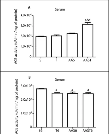
Neither jumping protocol nor AAS treatment altered ACE activity in the serum (figure 1A p >0.05). Only the association of AAS and training presented significant higher levels of ACE (p=0.01). However, six weeks after experimental treatments had stopped, the levels of ACE activity decreased in the T6, AAS6 and AAST6 groups compared to the control group (figure 1B; p=0.02; p=0.01; p = .01, respectively).
Training increased the ACE activity in SFT compared to the control group (figure 2A, p=0.01). Interestingly, both AAS and AAST groups presented higher values of ACE activity levels when compared to the control (figure 2A; p = 0.01; p =0.01; respectively). Six weeks after these experimental groups had stopped both training and AAS treatment, experimental groups decreased ACE activity compared to the control (figure 2B; p=0.01).
Figure 1. Angiotensin-converting enzyme (ACE) activity in the serum of sedentary animals (S), trained animals (T), sedentary with AAS administration (AAS) and trained with AAS administration (AAST). (A). ACE activity obtained immediately after the last training session. (B). ACE activity after 6 weeks of detraining and no AAS treatment.
DISCUSSION
This is the first study to investigate the effects of mechanical loa-ding resulting from jumping exercise combined with AAS on the ACE activity of the SFT. The results demonstrated that training increased the ACE activity. Likewise, the AAS administration had increased the ACE activity in both AAS and AAST groups. After stopping exercise and AAS administration, it was observed a reduction in ACE activity in both tendon and serum compared to the control group. These findings have clinical relevance since they could be associated to harmful effect on ECM remodeling and could indicate further risk factors to tendons dysfunction in AAS users on sports field.
Values were expressed as means ± SEM (p < .05). a, significant difference vs. S group; b, significant difference
vs. T group; c, significant difference vs. AAS group.
Serum
Serum
a a a A
B 4.0x10⁴
3.0x10⁴
2.0x10⁴
1.0x10⁴
0
abc
S T AAS AAST
S6 T6 AAS6 AAST6
A
CE ac
tivit
y (uF/min/mg of pr
ot
eín)
A
CE ac
tivit
y (uF/min/mg of pr
ot
eín)
3.0x10⁴
2.0x10⁴
1.0x10⁴
176
ECM remodeling is determined by an active and dynamic pro-cess and depends on the adequate balance between synthesis and degradation of their components16. Studies showed that RAS
cascade is involved in connective tissue cell growth and ECM re-modeling2, mainly in the heart following myocardial infarction17,18.
These enzymes cascade, including ACE expression has been detec-ted in human cardiac tissues and there is a correlation between the levels of ACE and collagen type I mRNA19. Although, there is
a lack of evidence that ACE activity is expressed by tendons, we demonstrated here the involvement of the local renin-angiotensin system on tendon tissue.
Interestingly, our results demonstrated that seven weeks of trai-ning increased the ACE activity, contrary to other reports20,21.
Proba-bly, these discrepancies could be attributed to variations amongst the studies, such as training models and type of tissue evaluation. Concerning training models, these authors20,21 have used a
swim-ming training exercise, whereas the presented study performed jumping exercise protocol. In addition, we assessed a tendon tissue differently from other studies, which analyzed cardiac muscles20,21,
lung and kidney20. Further investigations are needed to test these
discrepancies results.
It has been widely accepted that RAS cascade plays a pivotal role on the ECM remodeling, cell growth and fibrosis22. Rocha and
colle-agues reported20 an increase in the ACE activity on the left ventricle
after swimming training and AAS administration in rats. This exercise
training associated with AAS causes loss of the beneficial effects of left ventricle function induced by exercise training and maladaptive remodeling. Further deterioration of cardiac performance was detec-ted accompanying with a local activation of the RAS consistent with the hypertrophic response. Nevertheless, there is a lack of information about the ACE activity on tendons and this relation with training and or AAS administration.
The main approach of our study was the increase of ACE ac-tivity of tendon in the AAS and AAST groups. In serum, only AAS combined with training have increased the ACE activity. These results are very intriguing comparing previous studies using the same experimental model. We have shown that AAS treatment in association with load exercise reduced total concentration of matrix metallopeptidase 2 (MMP-2: enzyme responsible for ECM remode-ling and collagen degradation) and the percentage of the active mostly in SFT. The SFT responded more promptly to AAS treatment and this might be related to a higher cell density in this tendon12.
In addition, the biomechanical properties of SFT also was impaired by AAS treatment or AAS plus training, showing reduced tendon capacity to accommodate the initial tensional load, decreased ca-pacity to resist tension and reduced deformability, contributing to the high risk of tendon rupture during training in AAS consumers11.
We might presume that AAS treatment in association with high load exercise induce harmful effects in SFT consider the increase of ACE activity together with abolishment of MMP-2 activity and biomechanical changes.
In line with this, our data indicate that the increase of ACE activity could be associated with ECM remodeling, once the ACE modula-tes the TGF-β, an important mediator of fibrous tissue formation in repairing tissue. Also, ACE increase the CTGF synthesis, which is a potent stimulator of type I collagen synthesis and fibrosis23. It is
possible to suggest that these results could be associated to tendon maladaptive remodeling, once we have previously demonstrated that AAS alters the biomechanical properties of tendons, reducing tendon flexibility, led to greater stiffness and would increase the risk of tendon rupture in AAS abusers11. Recently, we also showed
that AAS and ASS associated to training decreased the expression of key genes involved in tendon ECM remodeling13. Clinically,
the-se findings are important in sports field, since AAS administration and their combination with exercise demonstrated harmful effects in relation to tendon ECM remodeling. The effect on tendons is likely a reflection of the delayed anabolic response of this tissue as compared to the highly vascularized and androgen-responsive associated skeletal muscle.
It is interesting to note that ACE activity has reduced in both tendon and serum after 6 weeks of detraining and upon cessation of AAS treat-ment in all experitreat-mental groups. The detraining induces a decrease in the synthetic activities of the tenocytes and the interruption of sudden build-up of MMPs activity24, which is in agreement with the reduction
of ACE activity after interruption of training. In addition, the literature has reported that many of the side effects are reversible with cessation of AAS special with reproductive/endocrine system25, however there is
a lack of studies to clarify whether cessation of AAS could be reversible in the tendon tissue.
Nevertheless, the reversibility of ACE activity levels after 6 weeks without AAS administration can be associated with a reversible of side effects caused by these drugs. Further researches in order to assess the MEC remodeling, such as MMPs activity and tissue morphology after six weeks of detraining and cessation AAS administration period will be performed.
Figure 2. Angiotensin-converting enzyme (ACE) activity in SFT of sedentary animals (S), trained animals (T), sedentary with AAS administration (AAS) and trained with AAS administration (AAST). (A). ACE activity obtained immediately after the last training session. (B). ACE activity after 6 weeks of detraining and no AAS treatment.
Values were expressed as means ± SEM (p<.05). a, significant difference vs. S group; b, significant diffe-rence vs.T group.
SFT
SFT ab
a
a ab A
B
S T AAS AAST
S6 T6 AAS6 AAST6
A
CE ac
tivit
y (uF/min/mg of pr
ot
eín)
A
CE ac
tivit
y (uF/min/mg of pr
ot
eín)
80
60
40
20
0
20
15
10
5
177
CONCLUSION
In conclusion, this study indicated that AAS administration and their combination with exercise increased ACE activity, suggesting the activation of the local renin-angiotensin system on tendon tissue. This effect can be reversed with interruption of AAS treatment. Taking together these results with previous studies it is possible to speculate
that ACE activity is associated to harmful effect of AAS administration on tendon remodeling, and this fact has a clinical importance related to greater risk of tendon rupture.
All authors have declared there is not any potential conflict of interests concerning this article.
REFERENCES
1. Nishimura H. Physiological evolution of the renin-angiotensin system. Jpn Heart J. 1978;19(5):806-22.
2. Rush JW, Aultman CD. Vascular biology of angiotensin and the impact of physical activity. Appl Physiol Nutr Metab. 2008;33(1):162-72.
3. Felmeden DC, Lip GY. The renin-angiotensin-aldosterone system and fibrinolysis. J Renin Angiotensin Aldosterone Syst. 2000;1(3):240-4.
4. Haznedaroğlu IC, Tuncer S, Gürsoy M. A local renin-angiotensin system in the bone marrow. Med Hypotheses. 1996;46(6):507-10.
5. Gebru Y, Diao TY, Pan H, Mukwaya E, Zhang Y. Potential of RAS inhibition to improve metabolic bone disorders. Biomed Res Int. 2013;2013:932691.
6. Kjaer M. Role of extracellular matrix in adaptation of tendon and skeletal muscle to mechanical loading. Physiol Rev. 2004;84(2):649-98.
7. Heinemeier K, Langberg H, Olesen JL, Kjaer M. Role of TGF-beta1 in relation to exercise-induced type I collagen synthesis in human tendinous tissue. J Appl Physiol (1985). 2003;95(6):2390-7.
8. Wang Q, Usinger W, Nichols B, Gray J, Xu L, Seeley TW, et al. Cooperative interaction of CTGF and TGF-β in animal models of fibrotic disease. Fibrogenesis Tissue Repair. 2011;4(1):4.
9. Shokri S, Hemadi M, Bayat G, Bahmanzadeh M, Jafari-Anarkooli I, Mashkani B. Combination of running exercise and high dose of anabolic androgenic steroid, nandrolone decanoate, increases protamine deficiency and DNA damage in rat spermatozoa. Andrologia. 2014;46(2):184-90.
10. Ma TK, Kam KK, Yan BP, Lam YY. Renin-angiotensin-aldosterone system blockade for cardiovascular diseases: current status. Br J Pharmacol. 2010;160(6):1273-92.
11. Marqueti RC, Prestes J, Wang CC, Ramos OH, Perez SE, Nakagaki WR, et al. Biomechanical responses of different rat tendons to nandrolone decanoate and load exercise. Scand J Med Sci Sports. 2011;21(6):e91-9.
12. Marqueti RC, Prestes J, Paschoal M, Ramos OH, Perez SE, Carvalho HF, et al. Matrix metallopeptidase 2 activity in tendon regions: effects of mechanical loading exercise associated to anabolic-androgenic steroids. Eur J Appl Physiol. 2008;104(6):1087-93.
13. Marqueti RC, Heinemeier KM, Durigan JL, de Andrade Perez SE, Schjerling P, et al. Gene expression in distinct regions of rat tendons in response to jump training combined with anabolic androgenic steroid
14. Marqueti RC, Micocci KC, Leite RD, Selistre-de-Araujo HS. Nandrolone inhibits MMP-2 in the left ventricle of rats. Int J Sports Med. 2012;33(3):181-5.
15. Franquni JV, do Nascimento AM, de Lima EM, Brasil GA, Heringer OA, Cassaro KO, et al. Nandrolone dec-anoate determines cardiac remodeling and injury by an imbalance in cardiac inflammatory cytokines and ACE activity, blunting of the Bezold-Jarisch reflex, resulting in the development of hypertension. Steroids. 2013;78(3):379-85.
16. Janssens S, Lijnen HR. What has been learned about the cardiovascular effects of matrix metallopro-teinases from mouse models? Cardiovasc Res. 2006;69(3):585-94.
17. Sun Y, Weber KT. RAS and connective tissue in the heart. Int J Biochem Cell Biol. 2003;35(6):919-31.
18. Kanayama G, Barry S, Hudson JI, Pope HG Jr. Body image and attitudes toward male roles in anabolic-androgenic steroid users. Am J Psychiatry. 2006;163(4):697-703.
19. Hafizi S, Wharton J, Morgan K, Allen SP, Chester AH, Catravas JD, et al. Expression of functional an-giotensin-converting enzyme and AT1 receptors in cultured human cardiac fibroblasts. Circulation. 1998;98(23):2553-9.
20. Rocha FL, Carmo EC, Roque FR, Hashimoto NY, Rossoni LV, Frimm C, et al. Anabolic steroids induce cardiac renin-angiotensin system and impair the beneficial effects of aerobic training in rats. Am J Physiol Heart Circ Physiol. 2007;293(6):H3575-83.
21. Barauna VG, Magalhaes FC, Krieger JE, Oliveira EM. AT1 receptor participates in the cardiac hypertrophy induced by resistance training in rats. Am J Physiol Regul Integr Comp Physiol. 2008;295(2):R381-7.
22. Zannad F, Rossignol P, Iraqi W. Extracellular matrix fibrotic markers in heart failure. Heart Fail Rev. 2010;15(4):319-29.
23. Evans NA, Bowrey DJ, Newman GR. Ultrastructural analysis of ruptured tendon from anabolic steroid users. Injury. 1998;29(10):769-73.
24. Frizziero A, Fini M, Salamanna F, Veicsteinas A, Maffulli N, Marini M. Effect of training and sudden detraining on the patellar tendon and its enthesis in rats. BMC Musculoskelet Disord. 2011;12:20.