ORIGINAL ARTICLE
p27
KIP1expression in gastric cancer: Differential pathways in the
histological subtypes associated with
Helicobacter pylori
infection
MARKÊNIA KÉLIA SANTOS ALVES, VALESKA PORTELA LIMA, ÂNGELA ROSA ANDRÉ,
MÁRCIA VALÉRIA PITOMBEIRA FERREIRA, MARCOS AURÉLIO PESSOA BARROS &
SILVIA HELENA BAREM RABENHORST
Department of Pathology and Forensic Medicine, Section of Microbiology, Federal University in Ceará, Fortaleza, Brazil
Abstract
Objective.Decreases in p27KIP1and C-MYC expression have been associated withHelicobacter pyloriinfection. Furthermore,
C-MYC seems to be a transcriptional repressor of p27KIP1. Therefore, in a series of gastric adenocarcinomas we studied the
association of p27KIP1expression withH. pylorigenotype (vacA,cagA,cagE andvirB11) and the involvement of C-MYC in this
process.Material and methods.Expression of p27KIP1and C-MYC was determined by immunohistochemistry in 84 gastric
adenocarcinoma samples andH. pyloriinfection and genotype were determined by polymerase chain reaction.Results.Most
p27KIP1-negative cases (94.0%) were
H. pylori-positive and 44.8% were C-MYC-positive. In the diffuse gastric cancer subtype,
p27-negative-C-MYC-positive was the most frequent combination (cluster II), and was associated with the more pathogenic
H. pyloristrains. Although an association with p27KIP1andH. pyloristrain was found in the intestinal gastric cancer subtype,
negativity for p27KIP1and C-MYC markers was the most frequent cluster, followed by cluster II, and both were present,
independent of theH. pylorigenotype.Conclusions.Reduced expression of p27KIP1was closely linked toH. pyloriinfection,
and was dependent on the more pathogenic strains. Moreover, intestinal and diffuse subtypes showed distinct carcinogenic pathways influenced byH. pyloristrains. These data add insight to the differential influence and relevance ofH. pylorigenotype
in gastric cancer development.
Key Words:C-MYC, gastric cancer, Helicobacter pylori genotypes, histological subtypes, p27KIP1
Introduction
Gastric cancer is one of the most frequent malignan-cies worldwide and one of the leading causes of cancer mortality in Brazil [1,2]. Adenocarcinoma, the most common type, has been classified by Laurèn [3], according to its clinical and histological features, into two main types, diffuse and intestinal, with dis-tinct carcinogenetic pathways. Intestinal gastric ade-nocarcinoma, which is well differentiated, is the most frequent type and has a more favorable prognosis. Its etiology depends on environmental factors. Diffuse-type adenocarcinoma is poorly differentiated, has a poor prognosis, and its relationship with environmen-tal factors is controversial [4–6].
Helicobacter pylori infection is the major
environ-mental factor that contributes to the development of human gastric cancer [7,8]. The ability ofH. pylorito
promote gastric carcinogenesis is related to the host’s
genetic susceptibility and to specific bacterial viru-lence factors [9,10]. H. pylori is highly prevalent in
human populations and has remarkable genetic diver-sity [11,12]. This diverdiver-sity has been found to affect the bacterial virulence and consequently the disease out-come [13–15]. Two well-established virulence factors
ofH. pylori are vacuolating cytotoxin A (VacA) and
the cytotoxin-associated gene A (CagA) [16,17]. The VacA protein has potential to influence gastric
epi-thelial cell cycling and epiepi-thelial cell-signal transduc-tion. The vacA gene is present in all strains and
Correspondence: Markênia Kélia Santos Alves, MS, 3485, Osvaldo Cruz Street, Dionísio Torres, Fortaleza 60125151, Ceará State, Brazil. E-mail: [email protected]
(Received 27 September 2009; accepted 29 November 2009)
comprises two variable regions: the signal sequence (s1ands2) and the middle region (m1andm2), which
combine mosaicism. Strains bearings1andm1alleles
have long been noted as being more virulent than
s2m2strains [5,18,19]. ThecagA gene is considered a
marker for the presence of thecagpathogenicity island
(cag-PAI). It is located within the right portion of the cag-PAI and induces intense inflammatory responses
and alterations of the gastric epithelium [20,21].
cag-PAI also includes the cagE and virB11genes,
located in the right and left regions of cag-PAI,
respectively. ThevirB11 gene encodes the
homony-mous protein, which has a key role due to its structural position in the formation of the type IV secretion system, through which CagA is injected into host cells, and because it exhibits adenosine triphosphate synthase activity [16,22,23].cagE is also considered to
play a role in constructing the type IV secretion system and some authors consider it to be a marker of the integrity of cag-PAI, like the cagA gene
[15,24,25].
Despite the association ofH. pylori infection with
the development and progression of gastric cancer, the precise molecular mechanisms responsible for the promotion of gastric cancer byH. pyloriremain poorly
understood. One of the proposed mechanisms is modulation of gastric epithelial cell-cycle kinetics by alterations of cell-cycle regulators, like p27KIP1, p53, and p21WAF1/CIP1 [26
–29]. Although some
studies have already described an association between
H. pyloriand p53 mutation [30–32], knowledge of the
relationship betweenH. pyloriand p27KIP1expression
is still limited. p27KIP1 is a member of the Cip/Kip family of cyclin-dependent kinase inhibitors (CDKI). It binds to a wide variety of cyclin/CDK complexes, including CDK2 and -4, inhibiting kinase activity and blocking the G1/S transition necessary for cell-cycle progression [33–36]. In addition, p27
KIP1 has been implicated in the regulation of apoptosis and cell differentiation, and in the response to infl
am-matory stimuli [37,38]. The p27KIP1protein level is mainly regulated through degradation by ubiquitin-dependent proteolysis [38,39]. However, some studies have demonstrated a decreased expression of p27KIP1 due to transcriptional repression by C-MYC, a helix–loop–helix zipper transcriptional
factor [40,41]. The presence of the C-MYC protein has also been shown to block the nuclear transport of p27KIP1from the cytoplasm, mediated by increase in synthesis of cyclins D1 and D2 [40,42].
In gastric cancer, decreased p27KIP1protein expres-sion is an indicator of poor prognosis, and is associ-ated with more aggressive characteristics and tumor proliferation [28,35,43]. The association between
H. pylori and reduced p27KIP1 expression has been
indicated by somein vitrostudies and others involving
eradication of this microorganism [39,44,45]. How-ever, only a few papers have analyzed in vivo the
association between H. pylori infection and the
expression of the p27KIP1 protein and they were restricted to gastritis and intestinal metaplasia [27,44]. Moreover, there are noin vivostudies which
relate H. pylorigenotypes with p27KIP1and C-MYC
expression in gastric cancer. Thus we examined, in a series of gastric adenocarcinomas, the relationship between the H. pylori genotype and p27KIP1 protein
expression and also the involvement of C-MYC in this process. Since the intestinal and diffuse tumors are different entities, the data were also analyzed consid-ering these histological subtypes.
Material and methods
Clinical specimens
The study was approved by the Ethics Committee of the Hospital Complex of the Federal University of Ceará and all subjects signed an informed consent form before inclusion. Samples from 84 patients with gastric adenocarcinoma who had undergone gastrec-tomy were collected from two hospitals in Ceará State, Brazil: Walter Cantídeo Hospital at the Federal University of Ceará and Santa Casa de Misericórdia Hospital, both located in Fortaleza, the state capital. The histological classification was made according
to the Laurèn classification [3] by the team’s
pathologists.
DNA extraction
Genomic DNA was extracted from frozen tumor tissue using the cetyltrimethyl ammonium bromide technique, adapted from the method of Foster and Twell [46]. DNA extraction was done only in frag-ments that showed>80% tumor cells, and the quality
was assessed by 1% agarose gel electrophoresis with ethidium bromide staining. Also, the amount of DNA was determined using a NanoDrop 3300
fluorospectrometer.
Detection of H. pylori and the presence of vacA, cagA, cagE, and virB11 genes
TheH. pyloriinfection was detected by amplification
of theureC gene using primers for polymerase chain
alleles vacA, and the cagA, cagE, and virB11 genes
was identified using primer sets shown in the pub-lished literature. These are shown in Table I. PCR mixtures, for amplification ofcagE andvirB11 genes,
were prepared in a volume of 20ml using MasterMix
(Taq DNA Polymerase, dNTPs and MgCl2) accord-ing to the manufacturer’s instructions (Promega,
Madison, WI), with addition of 0.8% Tween 20, 0.3 mM (virB11) or 1 mM (cagE) of each primer
and 1ml of the DNA sample.
The cagA, vacA s1/s2, and vacA m1 genes were
amplified in a 25-ml volume containing 2.5ml of 10
PCR buffer (Invitrogen, Cergy Pontoise, France), 1% Tween 20, 1.5 mM MgCl2 (Invitrogen), 200 mM (each) of dNTPs (Invitrogen), 1 U of Platinum Taq
polymerase (Invitrogen), 0.4 mM (ureC, cagA, vacA s1/s2, and vacA m1) or 0.3 mM (vacAm2) for each
primer and 1ml ofH. pyloriDNA.
The PCR products were analyzed by 1% agarose gel electrophoresis with ethidium bromide staining. The size of the amplification product was used to
confirm the identity of the PCR product. The sample
was consideredH. pylori-positive when anureC
frag-ment of 294 bp was amplified. For confirmation of the
specificity of reaction, PCR products from ureC
gene were cloned with a TOPO TA Cloning kit
(Invitrogen, Carlsbad, CA) and sequenced using the ABI PRISMBigDye Terminator v.3.0
cycle-sequencing kit (Applied Biosystems, Foster City, CA)
and ABI PRISM 3100 DNA Sequencer
(Applied Biosystems). vacA, cagA, cage, and virB11
genes were considered positive when a specific
fragment was detected (Table I). DNAse-free water was used as a negative control. DNA preservation was also confirmed by amplification of different genes by other approaches under study in the laboratory. Ran-dom samples were re-analyzed for confirmation of the
results.
Immunohistochemistry
Immunostaining was performed according to a pre-viously described protocol of Hsu et al. [49]. For antigen retrieval, deparaffinized sections were
pre-treated by heating in a microwave oven in 10 mM citrate buffer, pH 6.0, for 20 min. After cooling, sections were then immersed in 3% hydrogen perox-ide for 10 min to block endogenous peroxidase activ-ity. Sections were then incubated in a moist chamber overnight at 4C with primary antibody. The primary
antibodies used were p27KIP1 (clone SX53G8; dilution 1:50; DakoCytomation; Glostrup, Zealand, Denmark) and C-MYC (clone 9E11; dilution 1:100; Novocastra; Newcastle upon Tyne, UK). After rins-ing with phosphate-buffered saline (PBS), the slides were incubated with secondary antibody followed by streptavidin–biotin–peroxidase complex (LSAB+
sys-tem; DakoCytomation), both for 30 min at room temperature with a PBS wash between each step. The reaction was revealed with diaminobenzidine–
H2O2 (DAB+ system; DakoCytomation), counter-stained with Harry’s hematoxylin and mounted. A confirmed case of nuclear C-MYC-positive and
Table I. PCR primer sets used for genotypingH. pylori.
Gene Primer sequence Reference
Annealing temperature (C)
Size (bp) of PCR product
ureC F: -5¢AAGCTTTTAGGGGTGTTAGGGGTTT3¢; R: -5¢AAGCTTACTTTCTAACACTAACGC3¢
47 55 294
vacA
s1/s2 F: -5¢ATGGAAATACAACAAACACAC3¢; R: -5¢CTGCTTGAATGCGCCAAAC3¢
48 55 259/286
m1 F: -5¢GGTCAAAATGCGGTCATGG3¢; R: -5¢CCATTGGTACCTGTAGAAAC3¢
55 290
m2 F: -5¢GGAGCCCCAGGAAACATTG3¢; R: -5¢CATAACTAGCGCCTTGCAC3¢
52 192
cagA 48
F: -5¢ATAATGCTAAATTAGACAACTTGAGCGA3¢; R: -5¢TTAGAATAATCAACAAACATAACGCCAT3¢
56 297
cagE F: -5¢TTGAAAACTTCAAGGATAGGATAGAGC3¢; R: -5¢GCCTAGCGTAATATCACCATTACCC3¢
15 50 509
virB11 F: -5¢TTAAATCCTCTAAGGCATGCTAC3¢; R: -5¢GATATAAGTCGTTTTACCGCTTC3¢
15 49 491
p27-positive human breast carcinoma was used as a positive control. Controls of primary antibody spec-ificity included omission or substitution of primary antiserum by normal bovine serum.
Immunostaining analyses
The immunohistochemical evaluation was performed by two experienced analysts independently using direct light microscopy. Any conflicting results were
jointly considered to give a consensual determination. Protein expression was quantified by manual count-ing of at least 1000 tumor cells in 10 differentfields at
a magnification of400. The labeling index (LI) was
expressed as the percentage of tumor-positive cells for nuclear or cytoplasmic staining in each sample [50]. Only cases with scores‡5% were considered positive.
Also, the pattern of staining (focal, multifocal, or diffuse) was observed. In the diffuse pattern, the staining of tumor cells occurs uniformly, distributed throughout the whole sample. In the focal staining pattern, staining is restricted to the same region and there can be one tofive staining foci. Finally, in the
multifocal pattern, more thanfive foci are observed,
distributed non-uniformly.
Statistical analyses
The analyses were carried out using the statistical software SPSS version 15.0 (SPSS Inc., Chicago, IL). Statistically significant differences were evaluated
by the chi-square test or Fisher’s exact test.
Correla-tions between p27KIP1 expression and the H. pylori genotype group were analyzed by Spearman’s rank
correlation coefficient. The results were considered
statistically significant when P-values were<0.05.
Results
Of the 84 analyzed cases, 59 were males and 25 were females. The median age was 64.5 years (range 23–90 years). The classification of the gastric tumors
was as follows: 2.4% (2/84) were stage IA, 10.7% (9/84) were IB, 23.8% (20/84) were II, 19.0% (16/84) were IIIA, 10.7% (9/84) were IIIB, and 33.3% (28/84) were IV.
Detection of p27KIP1, H. pylori infection, and vacA, cagA, cagE, and virB11 genes
Of the analyzed cases, p27KIP1 expression was detected in only 20.2% (17/84) andH. pyloriinfection
was present in 95.2% (80/84). All p27KIP1-positive cases were H. pylori-positive, with a diffuse stain
pattern observed in 76.5% of the cases (13/17), focal in 17.6% (3/17) and multifocal in 5.9% (1/17). The LI ranged from 5.6% to 41%. Three cases with exclusive cytoplasmic p27KIP1staining and two cases with concomitant nuclear and cytoplasmic p27KIP1 staining were also H. pylori-positive. On the other
hand, among the 79.8% (67/84) of p27KIP1-negative cases, 94.0% (63/67) were H. pylori-positive. All H. pylori-negative cases were also p27KIP1-negative.
TheH. pylori vacAs1m1combination was the most
frequently observed (72.5%; 58/80), withs1m2,s2m1,
and s2m2 being detected in 15.0% (12/80), 5.0%
(4/80) and 7.5% (6/80) of the cases, respectively. The H. pylori cag-PAI genes studied had similar
frequencies: 65.0% (52/80) for cagA, 53.8% for cagE (43/80), and 61.2% (49/80) for thevirB11 gene.
Relationship between H. pylori vacA, cagA, cagE, and virB11 genes and p27KIP1detection
To investigate the relationship between H. pylori cagA, cagE, virB11, and vacA genes and p27KIP1
expression in the pathogenesis of gastric cancer, the cases were divided into groups according to the pres-ence of the studied H. pylori genes. The division
criteria utilized were cag-PAI integrity and presence
of the vacA alleles, as described in Table II. This
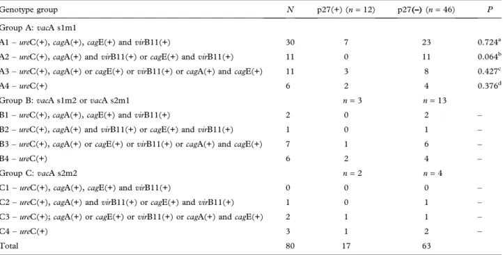
Table also shows that the highest frequency of lack of p27KIP1expression (76.7%; 23/30) occurred in cases withH. pylorisubgroup A1 strain (vacA s1m1,cagA, cagE, and virB11), followed by subgroup A2 strains
(vacA s1m1 and virB11- as left-side marker of cag
-PAI and cagE or cagA- as right-side marker of cag
-PAI). Statistical analysis was carried out only in the A group due to insufficient numbers of cases in the B
and C groups. Although the A1 subgroup had the highest number of p27KIP1-negative cases, a repre-sentative number of p27KIP1-positive cases were observed in this subgroup. In subgroup A2, only p27KIP1-negative cases were found. However, this was not statistically significant (P = 0.064). It is interesting to note that in group B (vacA s1m2 or
s2m1) and group C (vacA s2m2) the majority of
p27KIP1-negative cases were in strains with only one or nocag-PAI gene.
Association of p27KIP1negativity with the A1 sub-group was observed only when the 63 p27KIP1 -negative cases were analyzed. From this analysis, there was a statistical difference between the A1 subgroup and the A2 and A3 subgroups (P = 0.006
and P= 0.022, respectively). Also, Spearman’s rank
p27KIP1-negative cases of the A1 subgroup with the A2 (rS = –0.349; P = 0.005) and A3 subgroups
(rS=–0.289;P=0.022). Additionally, as the A group
was the most representative, the 46 p27KIP1-negative cases belonging to this group were analyzed separately (Figure 1) and there was also a statistical difference between the A1 and A2 (P <0.001) and between
the A1 and A3 (P = 0.004) subgroups. Spearman’s
rank correlation showed a negative correlation between p27KIP1-negative cases of the A1 subgroup with the A2 (rS = –0.561; P < 0.001) and A3
subgroups (rS= –0.459; P= 0.001).
Relationship between H. pylori genotype group and p27KIP1detection and histological type
No statistical difference between intestinal and diffuse histological subtypes related to p27KIP1 expression was observed. However, when the cases were distrib-uted according to the H. pylori genotype groups, a
statistical difference between these histological sub-types in the A1 subgroup was found (P = 0.038) (Table III). In the A group, a tendency towards reduction of p27KIP1 negativity could be observed with loss of studiedcag-PAI genes in both histological
types (Table III). This tendency was not observed in cases from the B group. Most of the p27KIP1-positive cases (13/17) were found in the intestinal subtype, but without a statistical difference (P= 0.39).
Among the intestinal subtype (Table III), the majority of the p27KIP1-positive cases were observed in the A1 subgroup (53.8%; 7/13), besides a high number of p27KIP1-negative cases in this subgroup. Six of these p27KIP1-positive cases showed a predom-inant high LI and diffuse staining pattern. Two cases had exclusive cytoplasmic p27 staining and two cases had concomitant nuclear and cytoplasmic p27 stain-ing, all belonging to the A group. On the other hand, in the diffuse subtype tumors, despite the small num-ber of cases, the p27KIP1-positive cases were present
Table II. Comparison between the results of p27 nuclear expression inH. pylorigenotype groups in the 80 cases of gastric adenocarcinoma
analyzed.
Genotype group N p27(+) (n=12) p27(–)(n=46) P
Group A:vacA s1m1
A1–ureC(+),cagA(+),cagE(+) andvirB11(+) 30 7 23 0.724a
A2–ureC(+),cagA(+) andvirB11(+) orcagE(+) andvirB11(+) 11 0 11 0.064b A3–ureC(+),cagA(+) orcagE(+) orvirB11(+) orcagA(+) andcagE(+) 11 3 8 0.427c
A4–ureC(+) 6 2 4 0.376d
Group B:vacA s1m2 orvacA s2m1 n=3 n=13
B1–ureC(+),cagA(+),cagE(+) andvirB11(+) 2 0 2 –
B2–ureC(+),cagA(+) andvirB11(+) orcagE(+) andvirB11(+) 1 0 1 – B3–ureC(+),cagA(+) orcagE(+) orvirB11(+) orcagA(+) andcagE(+) 7 1 6 –
B4–ureC(+) 6 2 4 –
Group C:vacA s2m2 n=2 n=4
C1–ureC(+),cagA(+),cagE(+) andvirB11(+) 0 0 0 –
C2–ureC(+),cagA(+) andvirB11(+) orcagE(+) andvirB11(+) 1 0 1 – C3–ureC(+);cagA(+) orcagE(+) orvirB11(+) orcagA(+) andcagE(+) 2 1 1 –
C4–ureC(+) 3 1 2 –
Total 80 17 63
aA1 versus other groups. bA2 versus other groups. cA3 versus other groups. dA4 versus other groups.
A1 0 20 40 60
Percentage of negative p27 cases
80 100
A2 p=0.004
p<0.001
A3 A4
p27(-)
Figure 1. Comparison of 46 p27KIP1-negative cases among
in the less pathogenic A subgroups, with statistical significance (P = 0.045) for the A3 subgroup.
Although all cases from the subgroups A1 and A2 were p27-negative (13/28), considering both sub-groups there was no statistical significance when
they were compared to the other groups
(P = 0.067). Only one case in this group had focal cytoplasmic staining with low LI.
Figure 2 shows the distribution of the 46 p27KIP1 -negative cases according to theH. pyloriA group and
histological subtypes. From this Figure it is possible to observe that, in both intestinal and diffuse subtypes, there was a decrease in p27KIP1 negativity according to the less pathogenic group. The intestinal subtype tumors showed statistical significance between
sub-group A1 versus subsub-groups A2 and A3 (P=0.010 and
P=0.025, respectively). Also, in the intestinal tumors,
Spearman’s rank correlation test showed that the
presence of p27KIP1-negative cases in the A1 sub-group was inversely correlated with the A2 (rS =
–0.501; P = 0.004) and A3 subgroups (rS= –0.459;
P=0.009). In the diffuse subtype tumors, a statistical
significance was only observed between subgroup
A1 versus subgroup A2 (P=0.022) and Spearman’s
rank correlation test showed that the presence of p27KIP1-negative cases in the A1 subgroup was inversely correlated with the A2 subgroup (rS=–0.707;P =0.003).
Relationship between p27KIP1and C-MYC expression and association with H. pylori genotype group
To verify the involvement of C-MYC with p27KIP1 negativity, both markers were analyzed together. C-MYC positivity was detected in 40.5% (34/84) of the cases, and 85.3% (29/34) of these were p27-negative. Considering the p27KIP1-negative cases, 44.8% (30/67) were C-MYC-positive, 14 of which were intestinal and 16 diffuse subtype. Among the p27KIP1 cytoplasmic staining cases, only one was C-MYC-positive and it belonged to the diffuse
Table III. Comparison of p27 nuclear positivity among theH. pylorigenotype groups according to the histological subtypes.
Genotype group
Intestinal (I)
P
Diffuse (D)
P
ID
p27(+) p27(–) p27(+) p27(–) P
Group A
A1 7 13 0.188a 0 10e 0.107a 0.038*
A2 0 8 0.076b 0 3e 0.618b
–
A3 1 7 0.375c 2 1 0.045c 0.152
A4 1 3 1.000d 1 1 0.134d 1.000
Subtotal 9 31 – 3 15 – –
Group B
B1 0 1 – 0 1 – –
B2 0 0 – 0 1 – –
B3 0 5 – 1 1 – –
B4 2 2 – 0 2 – –
Subtotal 2 8 – 1 5 – –
Group C
C1 0 0 – 0 0 – –
C2 0 0 – 0 1 – –
C3 1 0 – 0 1 – –
C4 1 0 – 0 2 – –
Subtotal 2 0 – 0 4 – –
Total;n(%) 13 (25) 39 (75) – 4 (14.3) 24 (85.7) – –
aA1 versus other groups. bA2 versus other groups. cA3 versus other groups. dA4 versus other groups.
eA1+A2 versus other groups (P =0.067).
subtype. In histological subtypes, no statistical differ-ence was observed between p27KIP1 and C-MYC expression.
There was no statistical difference between C-MYC expression and H. pylori genotype group.
However, when this association was examined con-sidering the histological subtypes, there was a signif-icant difference in C-MYC expression between the intestinal and diffuse tumors in theH. pylorisubgroup
A1 (P = 0.039), with a predominance of C-MYC negativity in the intestinal subtype.
For the combined C-MYC and p27KIP1 analysis, considering the H. pylori genotype groups, the data
were divided into four clusters (I–IV), according to
the expression of these genes. This analysis showed that these clusters had different frequencies according to the histological subtype (Figure 3). In diffuse
tumors, the most frequent cluster was the p27-negative-C-MYC-positive (cluster II). It was pre-dominant in H. pylori strains A1 and A2, with a
significantly higher frequency (P=0.002) when
com-pared to p27-negative-C-MYC-negative (IV). On the other hand, in the intestinal tumors, the cluster II had similar frequency among all subgroups, and there was a statistically significant difference between intestinal
and diffuse tumors, related to the presence of this cluster, in the H. pylori A1 subgroup (P = 0.018). Additonally, in diffuse tumors, positivity for both markers (I) was more frequent in the A3 and A4 subgroups, especially in relation to the A3 subgroup (P=0.055). The opposite was observed in the
intes-tinal tumors, where positivity for both markers was restricted to the A1H. pylorigenotype. Negativity for
both markers (IV) was not so frequent in the diffuse
p=0.025 p=0.022
A1
A2
A3
A4
Intestinal 0
10 20 30
Percentage of negative p27 cases
40 50 60 70 80 90 100
Diffuse p=0.010
Figure 2. Comparison of 46 p27-negative cases amongH. pylorigenotype groups A1, A2, A3, and A4 according to histological type.
0
Intestinal
A1 A2 A3 A4
Diffuse
A1 A2 A3 A4
10 20 30 40 40
Percentage of p27 and c-MYC clusters
60 70 80 90 100
**
b
p27(+)C-MYC(+) (I)
p27(–)C-MYC(+) (II)
p27(+)C-MYC(–) (III)
p27(–)C-MYC(–) (IV)
Figure 3. Percentage of clusters I (n=4), II (n=21), III (n=8), and IV (n=25) according to theH. pylorigenotype subgroup and histological classification in 40 intestinal tumors and 18 diffuse tumors of group A (n=58). **P=0.002 (cluster II versus cluster IV),P=0.018 (histological subtype versus cluster II).bP
tumors. Conversely, it was the most frequent cluster in the intestinal tumors, followed by cluster II, which was present regardless of the H. pylorigenotype. In
addition, the p27-positive-C-MYC-negative cases (III) were only observed in the intestinal tumors.
Discussion
The association ofH. pyloriinfection with the
devel-opment of gastric cancer is well accepted, but the mechanisms by which this bacterium acts are still unknown. Some evidence indicates that it is involved in altered expression of the tumor suppressor p27KIP1. Shirin et al. [45] showed an inverse correlation between chronicH. pyloriinfection and p27KIP1
pro-tein expression in gastric epithelial cell culture. Another study conducted by Kuzushita et al. [51] suggested thatH. pylori infection may contribute to
the development of the gastric cancer p27KIP1 path-way since H. pylori-infected rats with p27KIP
deficiency are more likely to develop gastric cancer. In vivo, Yu et al. [44] observed reversion of p27KIP1
protein expression after eradication of H. pylori in
patients with intestinal metaplasia.Additionally,
stud-ies indicate the pathogenic nature of certain types of
H. pyloristrains [9,15,18], raising the hypothesis that
the pathogenicity of bacteria is related to the presence of more virulent strains. In spite of the relevance of the absence of p27KIP1 in gastric cancer pathogenesis associated with the presence of H. pylori infection,
fewin vivostudies to date have tried to elucidate the
influence of this bacterium in p27KIP1 expression.
Therefore, in the present study, alterations in the expression of p27KIP1in samples from gastric adeno-carcinoma patients were analyzed in association with the presence of particularH. pyloristrains, taking into
account the presence of genes considered as impor-tant virulence factors. Also, we examined the influence of C-MYC expression in this process,
con-sidering distinct subtypes of gastric adenocarcinoma. Since a high frequency (95%) ofH. pyloriinfection
was found in the study, with only four cases negative forH. pylori, we decided to evaluate the data
accord-ing to the bacterial genotype. However, since we identified different genotypes of H. pylori strains,
we divided the data into H. pylorigenotype groups,
based on the presence of right (cagA and cagE) and
left (virB11) cag-PAI markers and the vacA
mosai-cism. Strains bearingcagA, vacA s1m1, and/or cagE
and virB11 genes (A1 and A2 subgroups) were the
most frequent and were considered more pathogenic since these genes were already related in the other studies to more aggressive lesions and to the integrity of the pathogenicity island (cagA,cagE, andvirB11),
another important virulence factor ofH. pyloristrains
[7,14,15,52–54]. In the general analysis, the majority
of the p27KIP1-negative cases were found in the more pathogenicH. pylorigroups (A1 and A2), but without
a statistical difference. In fact, when we carried out the analysis considering only the p27KIP1-negative cases, there was a statistical difference between the frequencies in the A1 subgroup and the A2 and A3 subgroups. Also, there was a negative correlation between p27KIP1-negative cases in the A1 versus the A2 and A3 subgroups. These data indicate that strains considered more pathogenic could be respon-sible for decreasing p27KIP1protein, leading to devel-opment/promotion of gastric carcinoma. These
findings contradict the study by Eguchi et al. [39],
which demonstrated,in vitro, thatH. pylori
virulence-associated genes, including thecagA,cagE, andvacA
genes, may play a minor role in decreasing the level of p27KIP1protein in gastric epithelial cells.
A relevant aspect of this study is that the data were analyzed considering the adenocarcinoma subtypes. Currently, based on the molecular changes involved, intestinal and diffuse subtypes are considered distinct tumors, having different tumorigenic pathways [6]. Most studies, especially those that consider the infl
u-ence ofH. pylori, do not make this distinction, instead
considering gastric adenocarcinoma as a single dis-ease. This may explain some conflicting data [39,45].
Wiksten et al. [55] and Sgambato et al. [56] carried out studies involving p27KIP1expression in both his-tological subtypes, but without considering H. pylori
infection and, like in the present study, they did not observe a correlation between p27KIP1expression and intestinal and diffuse tumors. On the other hand, Saragoni et al. [57] showed a correlation between decreased p27KIP1 expression and diffuse mixed his-totype, but they did not evaluate the influence of H. pylori strains in that process. Thus, the present
study is thefirst to explore the influence of different
strains of H. pylori in p27KIP1 expression in both
histological subtypes. In this analysis, there was a clear interference of H. pylori strain with p27KIP1
expression, since in the general analysis, without considering H. pylori groups, no difference relating
to p27KIP1 expression was found between intestinal and diffuse subtypes. However, when these groups were considered, some differences were observed.
In fact, in the present study, H. pylori strains
not involved, while in the other it is an important step. On the other hand, in the diffuse tumors, p27 neg-ativity was statistically related to more pathogenic strains (A1 and A2 subgroups) and, conversely, despite the small number of cases in this group, p27KIP1 positivity was related to the less pathogenic strains, indicating that, in these tumors, blockade of the p27KIP1 pathway, related to more pathogenic strains, was unique and important.
The reduction in p27KIP1 expression in gastric cancer, associated with H. pylori infection, can be
explained by the transcriptional and post-translational mechanisms. In an acute exposure model,H. pylori
increased the degradation of p27KIP1 protein via a proteasome-dependent pathway [39]. On the other hand, in a long-term co-culture model during which
H. pyloriselects for apoptosis-resistant gastric
epithe-lial cells, the expression of p27KIP1mRNA, detected by Northern blotting, was reduced by about 30% [45]. Similar results were obtained by cDNA micro-array analysis [58]. Additionally, there are indications thatH. pyloriincreases C-MYC expression in patients
with gastric cancer [59] and since there is evidence that C-MYC decreases transcription and the nuclear transport of p27KIP1from the cytoplasm [40,42], or sequestration of p27KIP1 in cyclin D/cdk4 com-plexes [60], the increase in C-MYC expression is suggested to be another way of p27KIP1 inactivation byH. pylori[27].
In our study, a correlation between p27KIP1 and C-MYC was not found, although most (85.3%) C-MYC positive cases were p27KIP1-negative. How-ever, most of the diffuse tumors were C-MYC-positive-p27-negative and statistically associated with the more pathogenic H. pylori strains (A1 and
A2). Additionally, positivity for both markers was observed in less pathogenic strains, indicating that in this histological subtype the mechanism by which
H. pylori causes the decrease in p27KIP1 is C-MYC
activation, as suggested by Kim et al. [27], related to the most pathogenic strains. C-MYC is a transcrip-tional factor of the helix–loop–helix/leucine zipper
family of proteins, involved in several cellular pro-cesses, such as proliferation and apoptosis [61]. In those tumors, it is plausible to suggest that cellular proliferation and tumoral progression occur through transcription blockade of p27KIP1by C-MYC. Inter-estingly, in these tumors, only one of the cases with p27KIP1cytoplasmatic labeling was C-MYC-positive, contradicting studies of Yang et al. [40] and Perez-Roger et al. [42].
In the intestinal subtype, in spite of a decrease in p27KIP1 expression associated with the presence of C-MYC, the predominantfinding was negativity for both markers, and neither of them was associated with a specificH. pylori group, which justifies the
contra-dictory data from Eguchi et al. [39]. Therefore, the influence of more pathogenicH. pyloristrains on the
decrease in p27KIP1expression, which has been sta-tistically confirmed, could be caused by protein
deg-radation in these tumors, as demonstrated by Eguchi et al. [39], instead of by C-MYC blocking p27KIP1 expression, as suggested by Kim et al. [27]. Further-more, it seems that sequestration in the cytoplasm, related to the most pathogenic H. pylori strains,
might be another alternative way to inactivate p27KIP1-associated inhibitory activity, since all cyto-plasmic p27KIP1 cases were found to be associated with strains of the A group in those tumors. The cytoplasmic positivity for p27KIP1 protein corre-sponds to the accumulation of this protein in the cytoplasm, caused by its oncogenetically activated protein-kinase B-mediated phosphorylation, which blocks the recognition of its sites by nuclear impor-tation factors [62]. However, this study included a fairly small number of cases with p27KIP1cytoplasmic
Apoptosis
Intestinal
A3, A4 strains A1, A2 strains
H. pylori
Diffuse
Cellular proliferation
p27KIP1 (+) p27KIP1 (+)
C-MYC(+) p27KIP1 (–)
C-MYC(+) Transcription blockageof p27KIP1 p27KIP1 (–)
↓ p27KIP1 expression
Degradation of p27KIP1
expression, and this may have had an impact on our conclusions.
Additionally, it was observed, exclusively in intes-tinal tumors, that there is an association of p27KIP1 positivity with the absence of C-MYC. Wu et al. [63] showed that inhibition of C-MYC leads to induction of increased p27KIP1levels. The upregulated p27KIP1 level is caused by an increase in protein stability caused by the reduction of Skp2, a key molecule related to p27KIP1 ubiquitination and degradation, and de novoprotein synthesis [64]. The
overexpres-sion of p27KIP1 protein in mammalian cells induces G1 arrest of the cell cycle, leading to inhibition of proliferation and induction of apoptosis [65]. Lee et al. [43] demonstrated that positive expression of p27KIP1correlated signi
ficantly with a favorable
prog-nosis in gastric cancer.
Figure 4 summarizes the distinct pathways for intestinal and diffuse tumors.
In conclusion, our data indicate that reduced expression of p27KIP1 is closely linked to H. pylori infection-associated gastric cancer and is dependent on the most pathogenic strains. Besides this, it seems that the two gastric adenocarcinoma histological sub-types have distinct carcinogenic pathways, with different influence of H. pylori strains. Eradication
of H. pylori reverses the decreased expression of
p27KIP1 in gastric cancer and may possibly halt the gastric carcinogenesis cascade. Further understand-ing of regulators of the cell cycle, such as p27KIP1, and the role ofH. pyloriin gastric cancer pathogenesis may
provide not only valuable prognostic information but perhaps also a new dimension regarding its treatment.
Declaration of interest:There are no conflicts of interest.
References
[1] BRASIL. Ministério da Saúde. Instituto Nacional do Câncer. Coordenação de Prevenção e Vigilância. Estimativas 2008: Incidência de Câncer no Brasil. Rio de Janeiro: INCA; 2008. [2] Konturek PC, Konturek SJ, Brzozowski T. Gastric cancer and Helicobacter pylori infection. J Physiol Pharmacol 2006;57:51–65.
[3] Laurèn P. Two histological main types of gastric carcinoma: diffuse and so-called intestinal-type carcinoma. Acta Pathol Microbiol Scand 1965;64:31–49.
[4] Smith MG, Hold GL, Tahara E, El-Omar EM. Cellular and molecular aspects of gastric cancer. World J Gastroenterol 2006;12:2979–90.
[5] Correa P, Schneider BG. Etiology of gastric cancer: what is new? Cancer Epidemiol Biomarkers Prev 2005;14:1865–8. [6] Tahara E. Genetic pathways of two types of gastric cancer.
IARC Sci Publ 2004;157:327–49.
[7] Peek RM Jr, Blaser MJ. Helicobacter pylori and gastrointes-tinal tract adenocarcinomas. Nat Rev Cancer 2002;2:28–37. [8] IARC. Schistosomes, liver flukes and Helicobacter pylori. IARC Working Group on the Evaluation of Carcinogenic Risks to Humans. Lyon, 7–14 June 1994. IARC Monogr Eval Carcinog Risks Hum 1994;61:1–241.
[9] Blaser MJ, Atherton JC. Helicobacter pylori persistence: biology and disease. J Clin Invest 2004;113:321–33. [10] Figueiredo C, Machado JC, Pharoah PM, Seruca R, Sousa S,
Carvalho R, et al. Helicobacter pylori and interleukin 1 genotyping: an opportunity to identify high-risk individuals for gastric carcinoma. J Natl Cancer Inst 2002;94:1680–7. [11] Suerbaum S, Michetti P. Helicobacter pylori infection. N
Engl J Med 2002;347:1175–86.
[12] Covacci A, Telford JL, Del Giudice G, Parsonnet J, Rappuoli R. Helicobacter pylori virulence and genetic geography. Science 1999;284:1328–33.
[13] Jafari F, Shokrzadeh L, Dabiri H, Baghaei K, Yamaoka Y, Zojaji H, et al. vacA genotypes of Helicobacter pylori in relation to cagA status and clinical outcomes in Iranian populations. Jpn J Infect Dis 2008;61:290–3.
[14] Atherton JC. The pathogenesis of Helicobacter pylori-induced gastro-duodenal diseases. Annu Rev Pathol 2006;1:63–96.
[15] Sozzi M, Tomasini ML, Vindigni C, Zanussi S, Tedeschi R, Basaglia G, et al. Heterogeneity of cag genotypes and clinical outcome of Helicobacter pylori infection. J Lab Clin Med 2005;146:262–70.
[16] Lu H, Yamaoka Y, Graham DY. Helicobacter pylori viru-lence factors: facts and fantasies. Curr Opin Gastroenterol 2005;21:653–9.
[17] Castillo-Rojas G, Mazarí-Hiriart M, López-Vidal Y. Helico-bacter pylori: focus on CagA and VacA major virulence factors. Salud Publica Mex 2004;46:538–48.
[18] Wen S, Moss SF. Helicobacter pylori virulence factors in gastric carcinogenesis. Cancer Lett 2009;282:1–8. [19] Atherton JC, Cao P, Peek RM Jr, Tummuru MK, Blaser MJ,
Cover TL. Mosaicism in vacuolating cytotoxin alleles of Helicobacter pylori. Association of specific vacA types with cytotoxin production and peptic ulceration. J Biol Chem 1995;270:17771–7.
[20] Wang HT, Li ZH, Yuan JP, Zhao W, Shi XD, Tong SQ, et al. Effect of Helicobacter pylori VacA on gene expression of gastric cancer cells. World J Gastroenterol 2005;11: 109–13.
[21] Zambon CF, Navaglia F, Basso D, Rugge M, Plebani M. Helicobacter pylori babA2, cagA, and s1 vacA genes work synergistically in causing intestinal metaplasia. J Clin Pathol 2003;56:287–91.
[22] Backert S, Müller EC, Jungblut PR, Meyer TF. Tyrosine phosphorylation patterns and size modification of the Heli-cobacter pylori CagA protein after translocation into gastric epithelial cells. Proteomics 2001;1:608–17.
[23] Krause S, Pansegrau W, Lurz R, de la Cruz F, Lanka E. Enzymology of type IV macromolecule secretion systems: the conjugative transfer regions of plasmids RP4 and R388 and the cag pathogenicity island of Helicobacter pylori encode structurally and functionally related nucleoside triphosphate hydrolases. J Bacteriol 2000;182:2761–70.
[24] Maeda S, Yoshida H, Ikenoue T, Ogura K, Kanai F, Kato N, et al. Structure of cag pathogenicity island in Japanese Heli-cobacter pylori isolates. Gut 1999;44:336–41.
pathogenicity island of Helicobacter pylori. Mol Microbiol 1998;28:37–53.
[26] Gamboa-Dominguez A, Seidl S, Reyes-Gutierrez E, Hermannstädter C, Quintanilla-Martinez L, Busch R, et al. Prognostic significance of p21WAF1/CIP1, p27Kip1, p53 and E-cadherin expression in gastric cancer. J Clin Pathol 2007;60:756–61.
[27] Kim SS, Meitner P, Konkin TA, Cho YS, Resnick MB, Moss SF. Altered expression of Skp2, c-Myc and p27 pro-teins but not mRNA after H. pylori eradication in chronic gastritis. Mod Pathol 2006;19:49–58.
[28] Al-Moundhri MS, Nirmala V, Al-Hadabi I, Al-Mawaly K, Burney I, Al-Nabhani M, et al. The prognostic significance of p53, p27 kip1, p21 waf1, HER-2/neu, and Ki67 proteins expression in gastric cancer: a clinicopathological and immu-nohistochemical study of 121 Arab patients. J Surg Oncol 2005;91:243–52.
[29] Shirin H, Hibshoosh H, Kawabata Y, Weinstein IB, Moss SF. p16Ink4a is overexpressed in H. pylori-associated gastritis and is correlated with increased epithelial apoptosis. Helicobacter 2003;8:66–71.
[30] Ozturk Y, Ozer E, Lebe B, Bekem O, Buyukgebiz B. Immunohistochemical evaluation of p53 expression and pro-liferative activity in children with Helicobacter pylori associ-ated gastritis. J Pediatr Gastroenterol Nutr 2005;40:467–70. [31] Shibata A, Parsonnet J, Longacre TA, Garcia MI, Puligandla B, Davis RE, et al. CagA status of Helicobacter pylori infection and p53 gene mutations in gastric adeno-carcinoma. Carcinogenesis 2002;23:419–24.
[32] Oda T, Murakami K, Nishizono A, Kodama M, Nasu M, Fujioka T. Long-term Helicobacter pylori infection in Japanese monkeys induces atrophic gastritis and accumula-tion of mutaaccumula-tions in the p53 tumor suppressor gene. Helicobacter 2002;7:143–51.
[33] Alkarain A, Slingerland J. Deregulation of p27 by oncogenic signaling and its prognostic significance in breast cancer. Breast Cancer Res 2004;6:13–21.
[34] Blain SW, Scher HI, Cordon-Cardo C, Koff A. p27 as a target for cancer therapeutics. Cancer Cell 2003;3:111–15. [35] Chetty R. p27 protein and cancers of the gastrointestinal tract
and liver: an overview. J Clin Gastroenterol 2003;37:23–7. [36] Hengst L, Dulic V, Slingerland JM, Lees E, Reed SI. A cell
cycle-regulated inhibitor of cyclin-dependent kinases. Proc Natl Acad Sci U S A 1994;91:5291–5.
[37] McAllister SS, Becker-Hapak M, Pintucci G, Pagano M, Dowdy SF. Novel p27(kip1) C-terminal scatter domain mediates Rac-dependent cell migration independent of cell cycle arrest functions. Mol Cell Biol 2003;23:216–28. [38] Philipp-Staheli J, Payne SR, Kemp CJ. p27(Kip1):
regulation and function of a haploinsufficient tumor suppressor and its misregulation in cancer. Exp Cell Res 2001;264:148–68.
[39] Eguchi H, Herschenhous N, Kuzushita N, Moss SF. Heli-cobacter pylori increases proteasome-mediated degradation of p27(kip1) in gastric epithelial cells. Cancer Res 2003;63:4739–46.
[40] Yang W, Shen J, Wu M, Arsura M, FitzGerald M, Suldan Z, et al. Repression of transcription of the p27(Kip1) cyclin-dependent kinase inhibitor gene by c-Myc. Oncogene 2001;20:1688–702.
[41] Berns K, Martins C, Dannenberg JH, Berns A, Riele H, Bernards R. p27kip1-independent cell cycle regulation by MYC. Oncogene 2000;19:4822–7.
[42] Perez-Roger I, Kim SH, Griffiths B, Sewing A, Land H. Cyclins D1 and D2 mediate myc-induced proliferation via
sequestration of p27(Kip1) and p21(Cip1). EMBO J 1999;18:5310–20.
[43] Lee KH, Lee HE, Cho SJ, Cho YJ, Lee HS, Kim JH, et al. Immunohistochemical analysis of cell cycle-related mole-cules in gastric carcinoma: prognostic significance, correla-tion with clinicopathological parameters, proliferacorrela-tion and apoptosis. Pathobiology 2008;75:364–72.
[44] Yu J, Leung WK, Ng EK, To KF, Ebert MP, Go MY, et al. Effect of Helicobacter pylori eradication on expression of cyclin D2 and p27 in gastric intestinal metaplasia. Aliment Pharmacol Ther 2001;15:1505–11.
[45] Shirin H, Sordillo EM, Kolevska TK, Hibshoosh H, Kawabata Y, Oh SH, et al. Chronic Helicobacter pylori infection induces an apoptosis-resistant phenotype associated with decreased expression of p27(kip1). Infect Immun 2000;68:5321–8.
[46] Foster G, Twell D. Plant gene isolation principles and practice. Chichester, UK: Wiley; 1996. p. 426.
[47] Lage AP, Godfroid E, Fauconnier A, Burette A, Butzler JP, Bollen A, et al. Diagnosis of Helicobacter pylori infection by PCR: comparison with other invasive techniques and detection of cagA gene in gastric biopsy specimens. J Clin Microbiol 1995;33:2752–6.
[48] Domingo D, Alarcon T, Pietro N, Sanchez I, Lopez-Brea M. cagA and vacA status of Spanish Helicobacter pylori clinical isolates. J Clin Microbiol 1999;37:2113–4.
[49] Hsu SM, Raine L. Protein A, avidin, and biotin in immunohistochemistry. J Histochem Cytochem 1981;29: 1349–53.
[50] Landberg G, Roos G. Proliferating cell nuclear antigen and Ki-67 antigen expression in human haematopoietic cells during growth stimulation and differentiation. Cell Prolif 1993;26:427–37.
[51] Kuzushita N, Rogers AB, Monti NA, Whary MT, Park MJ, Aswad BI, et al. p27KIP1 deficiency confers susceptibility to gastric carcinogenesis in Helicobacter pylori-infected mice. Gastroenterology 2005;129:1544–56.
[52] Kamali-Sarvestani E, Bazargani A, Masoudian M, Lankarani K, Taghavi AR, Saberifiroozi M. Association of H. pylori cagA and vacA genotypes and IL-8 gene poly-morphisms with clinical outcome of infection in Iranian patients with gastrointestinal diseases. World J Gastroenterol 2006;12:5205–10.
[53] Lin HJ, Perng CL, Lo WC, Wu CW, Tseng GY, Li AF, et al. Helicobacter pylori cagA, iceA and vacA genotypes in patients with gastric cancer in Taiwan. World J Gastroenterol 2004;10:2493–7.
[54] Björkholm B, Falk P, Engstrand L, Nyrén O. Helicobacter pylori: resurrection of the cancer link. J Intern Med 2003;253:102–19.
[55] Wiksten JP, Lundin J, Nordling S, Kokkola A, Boguslawski K, Haglund C. The prognostic value of p27 in gastric cancer. Oncology 2002;63:180–4.
[56] Sgambato A, Migaldi M, Leocata P, Ventura L, Criscuolo M, Di Giacomo C, et al. Loss of p27Kip1 expression is a strong independent prognostic factor of reduced survival in N0 gastric carcinomas. Cancer 2000;89:2247–57.
[57] Saragoni L, Morgagni P, De Manzoni G, Tomezzoli A, Roviello F, Marrelli D, et al. Expression of p27 and prolif-erative (MIB-1), mitotic (MI) and apoptotic indices in early-phase (EGF) gastric carcinoma. Results of a study by the Italian Gastric Cancer Research Group (IRGGC). Pathologica 2003;95:22–30.
infection and the development of gastric cancer. N Engl J Med 2001;345:784–9.
[59] Zhang GX, Gu YH, Zhao ZQ, Xu SF, Zhang HJ, Wang HD, et al. Coordinate increase of telomerase activity and c-Myc expression in Helicobacter pylori-associated gastric diseases. World J Gastroenterol 2004;10:1759–62.
[60] Bouchard C, Thieke K, Maier A, Saffrich R, Hanley-Hyde J, Ansorge W, et al. Direct induction of cyclin D2 by Myc contributes to cell cycle progression and sequestration of p27. EMBO J 1999;18:5321–33.
[61] Lutz W, Leon J, Eilers M. Contributions of Myc to tumor-igenesis. Biochim Biophys Acta 2002;1602:61–71. [62] Liang J, Zubovitz J, Petrocelli T, Kotchetkov R, Connor MK,
Han K, et al. PKB/Akt phosphorylates p27, impairs nuclear import of p27 and opposes p27-mediated G1 arrest. Nat Med 2002;8:1153–60.
[63] Wu M, Bellas RE, Shen J, Yang W, Sonenshein GE. Increased p27Kip1 cyclin-dependent kinase inhibitor gene expression following anti-IgM treatment promotes apoptosis of WEHI 231 B cells. J Immunol 1999;163: 6530–5.
[64] Kim JH, Kim HP, Jung CH, Hong MH, Hong MC, Bae HS, et al. Inhibition of cell cycle progression via p27KIP1 upregulation and apoptosis induction by an ethanol extract of Rhus verniciflua Stokes in AGS gastric cancer cells. Int J Mol Med 2006;18:201–8.