UNIVERSIDADE DE LISBOA
FACULDADE DE CIÊNCIAS
DEPARTAMENTO DE BIOLOGIA VEGETAL
Preclinical development of a new compound for the
treatment of arthritis
Ana Raquel Conduto Dias Maia
Mestrado em Biologia Molecular e Genética
Dissertação orientada por:
Doutora Rita Cascão
JEFonseca's Lab, Instituto de Medicina Molecular
Doutor Jorge Silva
Faculdade de Ciências da Universidade de Lisboa
2016
II ACKNOWLEDGEMENTS
Em primeiro lugar quero agradecer ao Dr. João Eurico da Fonseca por me ter recebido na Unidade de Investigação de Reumatologia do Instituto de Medicina Molecular.
Quero agradecer especialmente à Rita Cascão pela orientação, paciência e importantes ensinamentos que vou levar comigo ao longo da minha vida profissional e pessoal. Agradeço ainda ao professor Jorge Silva pela ajuda e interesse pelo projeto assim como à professora Rita Zilhão pelo apoio e incentivo.
Quero ainda salientar o incansável suporte e disponibilidade da Inês Lopes e a importante partilha de experiencias e conhecimentos assim como todas as brincadeiras com a Rita Moura, Vânia Gloria, Susana Oliveira e Bruno Vidal. Foram imprescindíveis para tornar esta experiencia mais rica, muito obrigado.
Agradeço também profundamente aos meus amigos que foram o meu apoio ao longo deste ano, especialmente à Marisa Maia por ser uma excecional madrinha e companheira de todas as horas, à Sofia Narciso pelos desabafos e companheirismo, à Telma Madeira pela paciência e amizade e à Daniela Perez por toda a ajuda e sinceridade ao longo da escrita desta tese.
Estou ainda para sempre grata aos meus pais e irmã pela eterna paciência e compreensão, por toda a preocupação e ajuda nos momentos bons e menos bons. Muito obrigado por toda a dedicação, foi realmente muito importante para mim.
Por fim, quero agradecer de forma muito especial ao Márcio Felizardo. Nunca terei palavras para descrever a minha gratidão pela força que me transmitiu, por ser um grande pilar e por me impelir a ser sempre uma pessoa melhor. Devo-lhe grande parte deste caminho.
A todos os que referi e muitos outros que no dia-a-dia contribuíram para a realização deste projeto, para o meu crescimento pessoal e/ou profissional um sincero obrigado!
III ABSTRACT
Rheumatoid arthritis (RA) is an uncurable auto-immune disease characterized by inflammation and tissue destruction in several joints. An early and adequate diagnosis are important to prevent the main symptoms, which are painful and swollen joints, morning stiffness, edema and progressive disability. Despite the improvement in therapeutic options, around 30% of patients discontinue treatment due to a weak clinical response, adverse effects or incapability to afford such expensive therapies. Therefore, an effective and safe therapy is desirable.
Celastrol, a compound from the Chinese plant Trypterigium wilfordii Hook F., has revealed a remarkable outcome in the treatment of several inflammatory diseases. Recently, our group has proven its efficacy in the treatment of inflammatory signs and bone damage, with no short-term toxicity, when the compound is intraperitoneally administered in an early and late phases of arthritis in the adjuvant-induced artritis (AIA) rat model. These data thus support the hypothesis that celastrol is a promising candidate for drug development for the treatment of RA.
In the present study, our group focused on exploring the efficacy of orally administrated celastrol, in two different doses, and studying inherent toxicity in the same rat model of arthritis. The study intent to provide a rationale for setting dose levels for further preclinical studies.
In order to evaluate ethanol as a suitable vehicle for the oral administration of celastrol in this set-up, a group of AIA rats received ethanol 17% in PEG400 by gavage and was compared with arthtiric animals that did not receive ethanol. The absence of body weight loss in the animals that received ethanol and the lack of differences on disease course between the two groups allowed the use of ethanol as a solvent for celastrol.
Hence, two groups of AIA rats were treated with celastrol orally at two different doses (5 or 7.5 µg/g/day) starting 8 days after disease induction. Both celastrol-treated groups were compared with a group of arthritic-untreated and healthy rats which received vehicle (17% in PEG400) and water, respectively. All the animals were euthanized 22 days after disease induction and samples were collected to futher analysis.
Our results showed that orally administered celastrol halts inflammation and decreases bone erosions in both dosages. Furthermore, our results also showed a reduction on the number of osteoclasts in celastrol-treated groups. However, significant toxic effects were observed in celastrol at 7.5 µg/g/day treated group. The other dose used in this study (5 µg/g/day) will be considered in future pre-clinial assessments conducted by our group.
Keywords: Rheumatoid arthritis, celastrol, adjuvant-induced arthritis rat model, oral administration, dosage.
IV RESUMO
A Artrite Reumatóide (AR) é uma doença autoimune, progressiva e crónica que se caracteriza por uma acentuada inflamação nas articulações com consequente reabsorção do osso e da cartilagem. Não existe cura e a atual estratégia terapêutica implica o diagnóstico e início de tratamento o mais cedo possível com vista à eventual remissão. Os sintomas iniciais incluem rigidez, inchaço e dor nas pequenas articulações que posteriormente alastram para as articulações maiores.
Apesar da etiologia da AR não ser inteiramente conhecida, sabe-se que a perda de tolerância imunológica está associada à predisposição genética do individuo associada à exposição a fatores de risco como o tabagismo ou à possível ocorrência de um episódio estocástico que leve ao surgimento de autoanticorpos. A inflamação articular desenvolve-se devido à anormal infiltração de células do sistema imunitário na membrana sinovial levando à libertação de citocinas pro-inflamatórias, quimiocinas e moléculas de adesão celular que sustêm e perpetuam a inflamação. O microambiente rico em mediadores inflamatórios ativa os osteoclastos, células responsáveis pela reabsorção do osso, desequilibrando desta forma a homeostase óssea a favor da reabsorção.
Apesar da recente evolução das estratégias de tratamento e do surgimento de novas terapêuticas, estas não são completamente eficazes e induzem efeitos adversos provocando a descontinuidade do tratamento em cerca de 30% dos doentes. Assim, continua a ser necessário o desenvolvimento de uma terapia eficaz, bem tolerada, e disponível para a generalidade da população. Com base nesta problemática o nosso grupo tem investigado um composto bioactivo da planta Trypterigium wilfordii Hook F., o celastrol. Este composto para além de revelar propriedades anti-inflamatórias, reduz e a reabsorção do osso e da cartilagem.
Com o intuito de dar continuidade à investigação desenvolvida pelo nosso laboratório, o presente estudo tem como principal objetivo explorar a eficácia anti-inflamatória e de protecção óssea do composto quando administrado oralmente assim como investigar possiveis efeitos adversos. Para tal, foi utilizado o modelo de artrite induzida por adjuvante (AIA) em rato. Os animais artríticos foram divididos em grupos sendo que a um dos grupos foi administrado por gavagem (intragastricamente) 5μg/g e a outro 7.5 μg/g diariamente de celastrol. Foi ainda utilizado como controlos um grupo de animais artríticos e um de ratos saudáveis, da mesma espécie e idade, que receberam veículo ou água, respectivamente. O estudo durou 22 dias sendo que o dia 0 corresponde ao dia da indução da doença, e o dia 8, fase em que se inicia o periodo agudo da artrite, foi o primeiro dia de tratamento (modelo terapêutico). De forma a avaliar o etanol como veículo foi administrado etanol a 17% em PEG400 a um grupo de animais artríticos e comparado com outro grupo de animais artríticos sem administração de veiculo. Todos os animais foram pesados ao longo do periodo experimental assim como medido o diâmetro da pata traseira esquerda e atribuído um score de acordo com a severidade da inflamação.
O etanol não provocou diferenças significativas nos parâmetros acima indicados. A análise histopatológica feita às patas traseiras esquerdas destes animais revelou que o etanol não altera a progressão da doença caracteristica destes animais e não provocou uma diminuição do peso. Desta forma, foi possível utilizar o etanol como solvente do celastrol.
Ambas as doses de celastrol administradas diminuíram significativamente a inflamação visível em todas as patas assim como o inchaço da pata traseira esquerda. Este resultado foi comprovado pela análise histopatológica. Ambas as doses de celastrol diminuíram significativamente a infiltração e proliferação celular assim como as erosões osseas.
De modo a explorar a ação do celastrol na proliferação de células na sinovia e nas células de rearbsorção e formação óssea, foram marcados por imunohistoquimica o ki67 (marcador de células proliferativas na sinovia), osteocalcina (marcador de osteoblastos) e catepsina K (marcador de osteoclastos em reabsorção). Todos os biomarcadores foram significativamente reduzidos nos animais em que o composto foi administrado em comparação com os animais artríticos não tratados. Desta
V forma, não só foi possível validar os efeitos anti-proliferativos do celastrol como também concluir que o celastrol atua no osso diminuindo as células de remodelação óssea presentes na articulação. Foi ainda avaliada a presença de macrófagos CD68+ na sinovia. A redução destas células na sinóvia funciona como biomarcador da resposta de novas terapeuticas para o tratamento da AR, quer em humanos quer em modelos animais. A sua diminuição significativa nos animais tratados permitiu validar o celastrol como candidato ao tratamento da artrite.
Os efeitos do celastrol no osso foram validados pela análise no soro de CTX-I e P1NP, marcadores de reabsorção e de formação óssea, respectivamente. Os níveis de P1NP foram significativamente reduzidos em ambas as doses em comparação com os animais doentes não tratados e o CTX-I, apesar de não ter resultados significativos, segue a mesma tendência. Mais ainda, a quantificação no soro do TRAcP 5b, marcador preditivo do número de osteoclastos totais, levou a concluir que o celastrol diminui o número destas células, tal como observado na análise histopatológica e por imunohistoquímica.
Os animais foram pesados ao longo do período experimental de forma a averiguar uma possível redução do peso corporal provocada pela administração do composto. Os ratos administrados com 5 μg/g/dia de celastrol não apresentaram redução de peso porém, o grupo que recebeu a dose mais alta do composto (7.5 μg/g/dia) sofreu uma diminuição no peso significativa no dia 11 e 13 em comparação ao 4º e ao 7º da experiência. A redução de 20% do peso em dois animais neste grupo comparativamente ao dia 4 provocou a sua eutanásia antecipada. Conduto, é de salientar que não existiram diferenças significativas no peso dos animais deste grupo nos dias em que receberam o tratamento. A avaliação dos biomarcadores de toxicidade no soro dos animais mostrou que o celastrol não provoca toxicidade nos rins e no fígado. Contudo, o enzima lactato desidrogenase (marcador de dano celular), aumentou significativamente nos animais tratados com 7.5 μg/g/dia de celastrol em comparação com os outros grupos e o enzima creatina cinase (marcador de dano muscular/cardíaco) revelou uma tendência a aumentar nos grupos tratados com celastrol em comparação aos ratos saudáveis. A análise ao marcador pro-ANP descartou efeitos adversos do celastrol no miocárdio. Por fim, foi ainda estudada a percentagem de leucócitos no sangue no fim do estudo. Nenhum dos grupos tratados com celastrol teve efeitos tóxicos nos leucócitos.
Os resultados sugerem que o celastrol administrado oralmente a 5 e 7.5 μg/g/dia é eficaz a diminuir a inflamação assim como a reduzir as erosões osseas. No entanto, os efeitos tóxicos significativos observados no grupo tratado com celastrol a 7.5 μg/g/dia exclui possíveis usos do composto em ratos Wistar acima desta dose. A outra dose utilizada neste estudo (5 μg/g/dia) está a ser considerada para futuros estudos pré-clinicos mais detalhados.
Palavras-chave: Artrite reumatóide, celastrol, modelo de artrite induzida por adjuvante em rato, administração oral, dosagem.
VI INDEX
ACKNOWLEDGEMENTS……….II ABSTRACT ... III RESUMO ... IV FIGURES INDEX ... VII TABLES INDEX ... VIII ABBREVIATIONS ... IX INTRODUCTION ... 1 Rheumatoid arthritis ... 1 Definition ... 1 Etiology ... 1 Pathophysiology ... 2 Bone remodeling ... 4 Treatment strategies ... 5 Celastrol ... 5 AIMS ... 8
MATERIALS AND METHODS ... 9
Animal model and experimental design ... 9
Histopathological and immunohistochemical evaluation of the left hind paws ... 10
Quantification of the systemic markers of bone turnover and resorption ... 10
Toxicological evaluation ... 10
Statistical analysis ... 10
RESULTS ... 12
1. Evaluation of ethanol as a vehicle for the oral administration of celastrol in the AIA rat model ... 12
2. In vivo assessment of the anti-inflammatory properties of celastrol in the AIA rat model ... 14
3. Histopathologic evaluation of the left hind paw after treatment with celastrol ... 16
4. Immunohistochemical evaluation of paw articular joints after treatment with celastrol ... 18
5. Serum bone resorption and bone turnover markers measurement after treatment with celastrol 21 6. Celastrol safety profile analysis ... 23
REFERENCES ... 30
APPENDIX ... 39
1. Inflammatory score throughout disease progression in arthritic EtOH+ and EtOH- rats ... 39
2. Body weight throughout disease progression in arthritic EtOH+ and EtOH- rats ... 40
VII FIGURES INDEX
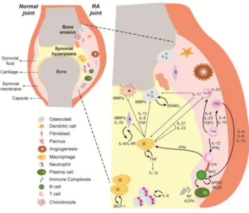
Figure I - Representative scheme illustrating immune cells and cytokine networks in rheumatoid joints ... 3 Figure 1.1 - Evaluation of ethanol as a vehicle for the oral administration of celastrol in the AIA rat model ... 13 Figure 2.1 - In vivo evaluation of celastrol’s anti-inflammatory properties in the AIA rat model ... 15 Figure 3.1 - Histopathological evaluation of paw joints after celastrol treatment ... 17 Figure 4.1 - Immunohistochemical evaluation of paw joint sections after treatment with celastrol .... 19 Figure 5.1 - Quantification of serum levels of bone resorption and bone turnover markers in AIA rats after celastrol treatment ... 22 Figure 6.1 – Evaluation of celastrol’s toxicity: in vivo body weight measurement and blood
VIII TABLES INDEX
IX ABBREVIATIONS
ACR – american college of rheumatology
ACR50 – american college of rheumatology 50% of response rate ACPA – anti-citrullinated protein antibody
AIA – adjuvant-induced arthritis APRIL – proliferation-inducing ligand BLYS – B-lymphocyte stimulator CCP – cyclic citrullinated peptide CIA – collagen-induced arthritis CK – creatine kinase
CRP – c-reactive protein
CTLA4 – cytotoxic T-lymphocyte-associated protein 4 CTX-I – C-terminal telopeptide of collagen type I CXCR4 – C-X-C chemokine receptor type 4 DMARD – disease-modifying anti-rheumatic drug DMSO - dimethylsulphoxide
ECM – extracellular matrix
ESR – erythrocyte sedimentation rate
EULAR – european league against rheumatism FLS – fibroblast-like synoviocytes
HLA – human leukocyte antigen IC – immune complex
IFNγ – Interferon gamma IL – interleukin
IL-6R – IL-6 receptor,
iNOS – inducible nitric oxide synthase Mφ – macrophage
M-CSF – macrophage colony-stimulating factor MCP-1 – Monocyte chemotactic protein-1 MHC – major histocompatibility complex MMP – metalloproteinase
MTX – methotrexate
NFATc1 – nuclear factor of activated T cells-1 NF-κB – nuclear factor κB
NSAID – non-steroidal anti-inflammatory drugs OPG – osteoprotegerin
X PTPN22 – protein tyrosine phosphatase non-receptor type 22
pSTAT3 – phosphorylated signal transducer and activator of transcription 3 RA – rheumatoid arthritis
RANK – receptor activator of nuclear factor NF-kB RANKL – RANK ligand
RF – rheumatoid factor
ROS – reactive oxygen species SF – synovial fluid
SM – synovial membrane TCR – T cell receptor
TE – thunder god vine extracts
TGFβ – Transforming growth factor beta TGV – thunder god vine
TH1 – T helper 1 cell
TH17 – T helper 17 cell
TNF – tumor necrosis factor
TRACP – tartrate resistant acid phosphatase
TRAF1/C5 – tumor necrosis factor receptor-associated factor 1 Treg – regulatory T-cell
1 INTRODUCTION
Rheumatoid arthritis Definition
Rheumatoid arthritis (RA) is a chronic and progressive auto-immune disease which is mainly characterized by polyarticular inflammation with consequent bone and cartilage destruction. It is associated with systemic inflammation which often results in disorders of multiple organ systems. The prevalence of RA varies between 0.3 and 1.1% [1].
Despite being a heterogeneous disease, common symptoms include tender, warm and swollen joints, morning stiffness, edema and weight loss. Smaller joints tend to be affected in the early phase and, as the disease progresses, symptoms often spread to the wrists, knees, ankles, elbows, hips and shoulders [2]. Over time, RA can cause the deformation of joints, secondary osteoporosis and other associated comorbidities which decrease life expectancy [3].
Although it is a complex and yet uncompletely understood disease, the last years’ advances have allowed clinicians and researchers to establish new targets in order to develop more effective therapies. The current treatment methods, which reflect this progress, require an aggressive therapy to start soon after diagnosis and need to be tailored according to the disease activity of each patient, in pursuit of clinical remission [4]. The classification criteria set, which is used to define RA, is based on 2010 American College of Rheumatology/European League Against Rheumatism (2010 ACR/EULAR) and takes into consideration the number of swelled joints, presence of Rheumatoid Factor (RF) and anti-citrullinated protein antibodies (ACPA), levels of c-reactive protein (CRP) and erythrocyte sedimentation rate (ESR), and the duration of symptoms [5].
Etiology
RA results from a complex set of key events that drive a severe autoimmune attack against endogenous tissues. Substantial evidences have been proving that the disease initiates with a minor event in a genetically susceptible individual, such as a specific environmental exposure or a stochastic episode, triggering a positive-feedback cycle that eventually leads to persistent immunopathology [6, 7]. The reactivity against a self-antigen is variable between RA patients however, 70-80% of the patients are seropositive for ACPAs, autoantibodies that recognize peptides containing citrulline (aminoacid generated by a post-translational modification) or for RF, autoantibodies that bind to Fc domain of immunoglobulins from IgG class [8]. Citrullination is a normal intracellular physiologic process [9], however, inflammatory conditions may cause the release of citrullinated peptides to the microenvironment enabling their contact with the immune system and eliciting the production of autoantibodies [10]. The main targets of ACPA in the joint are citrullinated forms of fibrinogen, α-enolase, vimentin and fibronectin [11-14]. Hence, the autoantibodies present in the serum potentiate the formation of immune complexes (ICs) that opsonized with complement system proteins recruit innate and adaptive immune cells [15]. In these cases the autoantibodies can be detected in patients’ serum up to 10 years before the onset of the disease [16] and are related with a more aggressive disease course and reduced rates of remission [17].
The presence of autoantibodies is strongly dependent on the genetic background of the patient [18, 19]. Some polymorphisms in human leukocyte antigen (HLA)-DRB1 gene namely in alleles HLA-DRB1*04, *01 and *10 imply a functional alteration in major histocompatibility complex (MHC) class II, making the molecule capable of accommodate citrulline residues and present them to T cells, which in turn incite B cells to produce more autoantibodies [20, 21].
2 Certain polymorphisms in alleles related with nuclear factor κB (NF-κB) dependent signaling, such as tumor necrosis factor receptor-associated factor 1 (TRAF1/C5) [22], or with T cell stimulation, activation and functional differentiation, such as protein tyrosine phosphatase non-receptor type 22 (PTPN22) [23] and cytotoxic T-lymphocyte-associated protein 4 (CTLA4) [24], can also be associated to a high risk of developing RA.
From all the environmental risks for RA, smoking is the strongest factor [6]. Its attributable risk is 20% for all RA and 35% for ACPA+ RA [25]. Other factors also associated with increased risk for RA include periodontitis [26], air pollution [27] and silica [28].
Pathophysiology
A joint is a structure where two or more bones connect with each other. Cartilage covers the end of each bone and is responsible for lubrication, articulation, loading and resistance of the bone [29-31]. Its properties are consequence of the macromolecular extracellular matrix (ECM) that are synthetized and maintained by cartilage cells, the chondrocytes [32]. Surrounding the cartilage is the synovia constituted by synovial membrane (SM) which lines the cavities of joints, and synovial fluid (SF), that lubricates the articulating surfaces [33]. A healthy SM is a relatively acellular structure consisting of a distinct intimal lining layer and a sublining layer. The intimal lining layer is composed by synoviocytes identified as being from macrophage or fibroblast lineage. The sublining layer contains blood vessels, adipocytes and fibroblasts, with few lymphocytes and macrophages [34]. In individuals with RA the synovia is invaded with leukocytes, proinflammatory cytokines, chemokines, metalloproteases (MMPs) and prostaglandins that drive inflammation and bone and cartilage resorption [35-38].
The migration of leukocytes from peripheral blood to the joints in RA is ensured by the massive amount of chemokines and adhesion molecules produced by vascular endothelium and synovial lining layer cells as a consequence of disease [39]. In fact, there are evidences that the cytokine-rich environment that occurs in RA induces leukocytes to acquire novel chemokine responsiveness prompting their migration to the joints [40]. For instance, blood neutrophils from patients with early RA express de novo CCR2, a receptor expressed in monocytes and lymphocytes, not in neutrophils in the physiologic state. This alteration is important for the further migration of these cells to the inflamed tissue [41]. Moreover, angiogenesis is also an early and critical event in RA and results in an expanded endothelial surface, which may promote more intense inflammatory cell ingress into the synovia [42].
Neutrophils are the most abundant and the first leukocytes to migrate to the SF [43]. Indeed, interleukin (IL)-8, a potent chemotactic agent for neutrophils is significantly increased RA patients’ SF in comparison with peripheral blood [44]. The inflamed synovia induces an alteration of neutrophil’s activation, recruitment and apoptosis [45-47] promoting the production of inflammatory cytokines [48] and the release of high concentrations of reactive oxygen species (ROS) as well as cytotoxic agents that contribute to the cartilage degradation [49].
The inflammatory character of RA is also sustained by macrophages (Mφ). Besides the capacity of presenting autoantigens to T cells, Mφ have the ability to differentiate into osteoclasts (bone resorbing cells), which further contribute to bone damage [50]. Moreover, this type of cells overexpresses a number of cytokines that drive and intensify inflammation. Among them are TNF, IL-1β and IL-6 whose levels are concomitantly increased in RA patients’ serum [51-53] and in SF [54]. TNF and IL-1 potentiate monocyte activation, adhesion molecules expression by endothelial cells [55] and stimulate chondrocyte and fibroblast-like synoviocytes (FLS) to degrade cartilage [55, 56]. TNF also inhibits functions of regulatory T cells (Treg) [57], induces effector T cells resistance to Treg-mediated suppression [58] and induce pain and fever [59]. IL-6 has been shown to increase to the production of autoantibodies by acting on plasmablasts [60]. All these cytokines are also major players dysregulating bone imbalance, enhancing bone resorption [61], as discussed below. In addition, Mφ also secrete
IL-3 23 and IL-12 which are responsible for T helper 17 cells (TH17) and TH1 polarization, respectively [62,
63].
Autoantigen recognition by T cells ensures the self-perpetuation of the disease as the mutual stimulation between T and B cells triggers autoantibodies production [64]. In fact, CD4+ T cell depletion using specific antibodies suppresses autoantibody production and reduces disease severity in collagen- or antigen-induced arthritis models in rodents [65, 66]. TH17, the most abundant subpopulation of T
cells present in the synovia [67], secrete a huge amount of IL-17 which is responsible for FLS, monocytes and neutrophils activation [68] as well as osteoclastic bone resorption [67, 69]. Synovial B cells are mainly localized in T and B aggregates, and are supported by the expression of factors that include B-lymphocyte stimulator (BlyS) and proliferation-inducing ligand (APRIL) which are highly expressed in the synovia [70].
As RA becomes chronic, increased synovial cell proliferation leads to pannus formation. This hypertrophic and hyperplastic synovial tissue is highly invasive and is responsible for bone and cartilage erosions [71].
Figure I - Representative scheme illustrating immune cells and cytokine networks in rheumatoid
joints. In RA, the inflammatory process leads to a cellular infiltration of the synovial membrane and synovial fluid, with consequent pannus formation, cartilage and bone destruction. Immune cells are responsible for the secretion of high amounts of several cytokines that amplify inflammation by the recruitment and activation of immune cells. ACPA – Anti-citrullinated protein antibodies, APRIL – A proliferation-inducing ligand, BLyS – B-lymphocyte stimulator, IFNγ – Interferon gamma, IL – Interleukin, IL-6R – IL-6 receptor, MCP-1 – Monocyte chemotactic protein-1, MHC – Major histocompatibility complex, MMP – Metalloproteinase, RANKL – Receptor activator of nuclear factor ligand, RF – Rheumatoid factor, TCR – T cell receptor, TGFβ – Transforming growth factor beta, Th –
4 Bone remodeling
Bone is a rigid but dynamic tissue composed by an organic matrix, mostly formed by fibers of collagen type I [72] and by a mineral phase of inorganic components, mainly hydroxyapatite [73]. The composition and organization of the bone matrix gives this tissue special mechanical properties such as stiffness, ductility, rigidity and tensile strength. In order to maintain its structural integrity, bone is continuously shaped and repaired in a process called bone remodeling [74, 75].
Physiological bone remodeling is orchestrated by two main cell types with opposing functions: osteoblasts, which form new bone, and osteoclasts, which resorb damaged or old bone. Osteoblasts are cells of mesenchymal origin that secrete bone-matrix proteins such as osteocalcin, a protein that is responsible for the calcium binding in bone matrix, therefore being considered as a specific marker of osteoblastic function [76]. They can transform into osteocytes or in bone lining cells. Osteocytes are osteoblasts trapped in the bone matrix. These type of cells produce signals to regulate osteoclasts and osteoblasts as a consequence of bone loading [77]. Bone lining cells are flat cells that cover available bone surfaces and act as a barrier that separates the bone from interstitial fluids and controls the mineral homeostasis of the bone [78]. On the other hand, osteoclasts are cells from monocyte/macrophage lineage responsible for the acidic decalcification and proteolytic degradation of the bone matrix [79].
The remodeling cycle begins with an initiation phase that includes the recruitment of Mφ to the resorption site. This is followed by their fusion and attachment of the subsequent multinucleated cell to the bone surface. The mature and multinucleated osteoclast is activated by signals which lead to initiation of bone resorption [80].
Osteoclasts differentiation, survival and activity is mediated by macrophage colony-stimulating factor (M-CSF) and receptor activator of nuclear factor NF-kB ligand (RANKL) [81]. Both mediators induce the transcription factor nuclear factor of activated T cells-1 (NFATc1), responsible for the up-regulation of various genes related with osteoclast adhesion, migration, acidification, degradation of inorganic and organic bone matrix [82, 83]. After RANKL-stimulation osteoclasts express tartrate-resistant acid phosphatase 5b (TRAcP 5b), an enzyme released into the serum that is considered a marker of osteoclasts number and, upon further stimulation, cathepsin K, an enzyme responsible for collagen type I cleavage, considered as a marker of osteoclastic resorption [84]. RANKL is tightly regulated by its decoy receptor, osteoprotegerin (OPG), both produced by osteoblasts. OPG prevents the interaction of RANKL with RANK, receptor on the surface of osteoclasts. Thus, the RANKL/OPG system constitutes an important indicator predicting either excessive bone resorption or bone formation [85].
The huge amount of proinflammatory cytokines present in the joint dysregulate osteoclastogenic balance [86-91]. Special attention should be paid to IL-1 that mediates the osteoclastogenic effect of TNF-α and stimulates the differentiation of osteoclasts precursors [86] and to TNF that promotes RANKL production [92], synergizes with RANKL to amplify osteoclastogenesis [93, 94] to intensify osteoclastic resorption [95].
There are biomarkers that reliably detect ongoing bone turnover and are useful to monitoring not only disease progress but also the efficacy of a treatment. Some of them are used to identify severity and risk of progression of joint damage in RA and other diseases such as osteoporosis [96-99]. They are also used in several animal models of arthritis [100, 101]. N-terminal propeptide of type I collagen (P1NP) is a peptide derived from the N-terminal of type I procollagen that is enzymatically cleaved at the moment of bone formation and liberated into the circulation, acting as a marker of bone formation. C-terminal telopeptide of collagen type I (CTX-I) is a portion of the C-terminal part of collagen type I that is cleaved by cathepsin K and is released into the circulation at the time of bone resorption, acting as a marker of bone resorption [102].
5 Treatment strategies
Since RA is still an incurable disease, current treatment strategies are focused on halt the inflammation, prevent joint damage and maintain the patient’s quality of life and ability to function in pursuit of clinical remission [4].
There are three major classes of drugs commonly used in the treatment of RA: non-steroidal anti-inflammatory drugs (NSAIDs), glucocorticoids and disease modifying anti-rheumatic drugs (DMARDs).
NSAIDs are effective agents decreasing pain and suppressing minor inflammation. However, NSAIDs do not modify the disease course nor reduce acute-phase reactants being only used as first-line agents for the symptomatic relief [103].
Glucocorticoids are useful to control symptoms, to preserve function until DMARDs start to exert their effects and also to treat flares of disease while the patient is receiving other treatments [104]. Nonetheless, significant side effects of glucocorticoids require the use of the lowest possible dose for the shortest period of time [105].
DMARDs are used with the aim of promoting a benefic long-term effect since they can modify the disease course improving inflammation and joint damage [106, 107]. This class of drugs can be divided in two groups: the conventional and the biologic DMARDs. Among conventional DMARDs, methotrexate (MTX) is the most frequently used due to its favorable efficacy/toxicity ratio [108, 109]. In addition, MTX can be used as co-treatment in patients treated with other conventional or biologic DMARD.
Besides the good outcomes of conventional DMARDs, biologic agents target strategic molecules in RA network. Biologic DMARDs are often reserved for patients who have not completely responded or cannot tolerate conventional DMARDs in doses large enough to control inflammation [110, 111]. Several biologic DMARDs are being commercialized and others are being tested in clinical trials. Currently, there are five available anti-TNF agents such as infliximab, etanercept, adalimumab, certolizumab pegol and golimumab [112]. Also included in the group of biologic DMARDs are T cell co-stimulation inhibitor abatacept, the anti-B cell agent rituximab, the IL6R-blocking monoclonal antibody tocilizumab and the IL-1 inhibitor anakinra [113].
Therapy with DMARDs should be started as early as possible after the diagnosis of RA [114]. It is recommended the use of MTX as the first-line treatment in patients with active RA, unless contraindicated, and it may be accompanied with low-dose of glucocorticoids [115, 116]. Combination therapies with two or more DMARDs are more effective than monotherapy however, adverse effects may also be greater [117]. If RA is not well controlled with a conventional DMARD, a biologic DMARD should be initiated simultaneously. Several data suggest that biologic therapy combination are not recommended due to a higher rate of adverse events and/or lack of additive efficacy [118, 119].
Despite the clinical efficacy of these therapies, around 30% of patients discontinue treatment due to a weak clinical response or to adverse effects [120-122]. Furthermore, the limited availability of biologics in developing countries and its relatively high cost impair many RA patients to access these therapies [123]. As an attempt to find a more reliable therapy, increasing number of agents derived from natural sources have drawn increasing attention in the last years. These type of compounds are generally less toxic and better tolerated than many of the aforementioned conventional drugs [124].
Celastrol
Celastrol, a pentacyclic-triterpene, is a bioactive component of the medicinal plant Trypterigium
wilfordii Hook F., also known as Thunder God Vine (TGV). It has been widely used in Chinese medicine
anti-6 oxidant, anti-inflammatory and anti-cancer properties, celastrol has been considered a promising candidate for drug development. In fact, its effects have been demonstrated in animal models of Alzheimer disease [126], asthma [127], systemic lupus erythematosus [128] and RA [129-131]. Those experiments and others have disclosed some of the key molecular targets of celastrol. It has been shown that the compound has the ability to modulate the expression of several pro-inflammatory cytokines [132], inhibit inducible nitric oxide synthase (iNOS) [133], adhesion molecules in endothelial cells [134], proteasome activity [135], topoisomerase II [136], potassium channels [137] and heat shock response [138]. Celastrol is also known to inhibit the proliferation, angiogenesis and metastasis of a variety of tumor cells [135, 139-141].
Interestingly, celastrol has been known for its remarkable action in the context of RA. Yu Y. et
al [142] treated purified neutrophils from healthy donors and RA patients with celastrol and discovered
that the compound inhibited neutrophils’ oxidative burst and extracellular traps formation. Zengtao Xu and colleagues [143] revealed that celastrol is capable of counteract FLS hyperplasia inducing its DNA damage, cell cycle arrest as well as apoptosis and Li GC. et al [144] showed that the compound suppressed hypoxia-induced human FLS migration and invasion by inhibiting C-X-C chemokine receptor type 4 (CXCR4) pathway. Furthermore, other groups have reported that celastrol significantly decreased the levels of the pro-inflammatory cytokines IL-17, IL-6, TNF, IL-1β and IL-18 in vitro [145] and reduces the expression of phosphorylated signal transducer and activator of transcription 3 (pSTAT3), an important transcription factor in IL-17 production [129].
An in vitro drug screening performed by our group to select compounds that simultaneously inhibit IL-1β and TNF secretion, two major players in RA physiophatology, identified celastrol as a potent inhibitor of those cytokines and also of NF-κB and caspase-1 secretion [131]. We further tested the anti-inflammatory effects of celastrol using an adjuvant-induced rat model of arthritis (AIA) and reported that this compound was able to significantly suppress inflammatory symptoms including cell infiltration and proliferation in arthritic joints in an early and late stage of disease progression as well as reduce CD68+ synovial macrophages, cells that are reduced in adequated therapeutic responses for RA [146]. Supporting our own results, Venkatesha et al [147], using the same rat model, documented a significant decrease in several chemokines responsible for cell recruitment to the inflammation site in celastrol-treated rats in comparison with arthritic-uncelastrol-treated rats. They also showed that celastrol significantly reduced the levels of anti-cyclic citrullinated peptide (CCP) antibodies, had an inhibitory effect on metalloproteinase (MMP)-9 and decreased Th17/Treg cell ratio in the joints [148].
There are some randomised controlled trials to evaluate the pharmacological effects and clinical toxicity of TGV. A study have reported that TGV extracts (TE) significantly improved the overall assessments of adult RA patients when compared with patients under placebo treatment [149]. Moreover, there are some heterogeneities in literature regarding the comparison between TE and DMARDs, however a recent study with 207 patients with active RA showed that the combination therapy of TGV and MTX led to a 1.6 fold increase in american college of rheumatology 50% of response rate (ACR50) than MTX monotherapy, further suggesting the beneficial effect of TGV in the treatment of RA [150]. From the studies made so far it was reported some adverse effects that should not be overlooked, including mild to moderate gastrointestinal events and amenorrhea in humans [149, 151] and hepato- and nephrotoxicity in mice [152]. However, these toxic events were described as a consequence of the administration of TE, a heterogeneous product that exhibit a high degree of variability in its composition. Thus, the use of a pure compound, as celastrol, is more appealing since it is easier to monitor its quality and concentration. Up to now there are few reports regarding adverse effects of pure celastrol. Jakob Hansen et al [153] have stated that celastrol could reduce lymphoblastoid viability in a dose-dependent way and Sifeng Wang et al [154] have showed that the compound affected the normal development of zebrafish embryo. However, our previous studies conducted in AIA rats, a model of arthritis capable of predict toxicity in humans [155], have proved that celastrol has not
short-7 term toxicity when intraperitoneally administered at 1µg/g/day [146]. In addition, more complete studies regarding long-term toxicity induced by celastrol are being peformed by our group.
8 AIMS
RA is a chronic auto-immune inflammatory disease with irreversible damage in cartilage and bone. Early diagnosis and advances in molecular biology have tailored the current therapy to a complete and long-lasting remission but only partial remission is achieved and frequent relapses as well as safety issues are common. Thus, the development of a therapy based on a small molecule, allowing oral administration, with low production costs, effective and safe remains an unmet medical need.
Previous data from our group have shown in the AIA rat model that celastrol, a bioactive compound from TGV, when administrated intraperitoneally, possesses significant anti-inflammatory and anti-proliferative properties in the early and established phase of arthritis, without adverse effects [146]. However, it still remains to prove its efficacy when orally administered. Moreover, the lack of significant toxicity reports led us to evaluate the toxicological profile of celastrol in this route of administration. Hence, the main aim of this project was to evaluate the anti-inflammatory and bone protective properties of celastrol when orally administered as well as analyze its safety profile in the same administration route. This study intent to provide a rationale for setting dose levels for further preclinical studies.
Specific aims:
Aim 1 – To evaluate ethanol as a possible vehicle for the oral administration of celastrol in the AIA rat model;
Aim 2 – To assess the anti-inflammatory properties of celastrol when orally administered in the AIA rat model in vivo and in joint articular tissues;
Aim 3 - To investigate the effect of celastrol when orally administered in bone turnover markers as well as in a bone resorption marker;
Aim 4 - To analyse toxic effects caused by oral administration of celastrol.
9 MATERIALS AND METHODS
Animal model and experimental design
Eight-week-old Wistar adjuvant-induced arthritis (AIA) female rats were obtained from Charles River Laboratories International (Massachusetts, USA) weighing 230-250 g. Animals were housed under specific pathogen free (SPF) conditions at 22°C with 12-hour light/12-hour dark cycles and food and water were provided ad libitum. Before the initiation of the studies, animals were allowed to acclimatize in the animal facility for 5 days. The animals were euthanized when presenting deformities and functional impairment in more than 2 paws or more than 20% of body weight loss. All experiments were approved by the Animal User and Ethical Committees at the Instituto de Medicina Molecular, according to the Portuguese law and the European recommendations.
The induction of the AIA rat model was performed by Charles River Laboratories and is detailed reported elsewhere [156]. Briefly, Wistar rats were inoculated with an intradermal injection at the right hind paw of heat-killed Mycobacterium tuberculosis suspended in paraffin oil. The preclinical phase of the model extends from the day of arthritogen administration until the day of clinically visible arthritis onset, nearly the 9th day. The next ten days are characterized by the acute clinical phase of the disease
when the body weight loss, progressive inflammation and skeletal erosions are more intense. This phase is are followed by a period of remission that implies the end of the study [157]. The similarity between the AIA rat model and human RA as well as greater disease severity and progression in comparison with other arthritic models makes this model suitable for preclinical studies and extensively used by the pharmaceutical industry [155].
Celastrol (Sigma, Missouri, USA) (purity ≥ 98 %) was dissolved in ethanol 100% at a concentration of 10mg/mL. Subsequently the stock solution was diluted using polyethylenglycol 400 (PEG400) (Sigma, Missouri, USA) for intragastric administration in animals.
To study the anti-arthritic properties and possible side effects of celastrol when orally administered, rats were randomly assigned to four experimental groups: two groups of AIA rats were treated with celastrol, daily by gavage (intragastric delivery), at 5 or 7.5 µg/g body weight (N=6 each group) and a group of AIA untreated rats (N=15) as well as a group of healthy non-arthritic of female age-matched Wistar rats (N=8) received vehicle (ethanol 17% in PEG 400) or water by gavage, respectively. Treatment with celastrol was initiated 8 days after disease induction, following a therapeutic model [158], and was maintained until day 22, when animals were euthanized by CO2
narcosis. The need for daily administrations is supported by Zhang J. et al who showed that the half-life of pure celastrol is approximately 10 hours [159]. In order to evaluate ethanol as a vehicle, a group of AIA rats was submitted to oral administration of ethanol 17% (Arthritic EtOH+) (N=15) and compared with a group of arthtiric animals who did not receive ethanol (Arthritic EtOH-) (N=5). The percentage of ethanol used in this assessment corresponded to the volume of ethanol administered in the highest dose of celastrol (7.5 µg/g/day) preparation.
Ankle swelling, body weight and inflammatory score were measured throughout the experiment. Inflammatory signs were evaluated by counting the score of each joint in a scale of 0–3 (0 — absence of inflammatory signs; 1 — erythema; 2 — erythema and swelling; 3 — deformities and functional impairment). The total inflammatory score of each animal was defined as the sum of the partial scores of each affected joint [160]. At the time of sacrifice the blood was collected from each animal. Serum and plasma were obtained and stored at -80ºC. Left hind paws as well as internal organs such as the liver, spleen, lung, kidney, heart and thymus were also collected and fixed in 10% formalin solution for further analysis.
10 Histopathological and immunohistochemical evaluation of the left hind paws
Paw samples, after being fixed with 10% neutral buffered formalin solution and dehydrated with increasing ethanol concentrations (70%, 96% and 100%), were also decalcified in 10% formic acid. After embedded in paraffin, the samples were serially sectioned at a thickness of 5 μm and mounted on microscope slides. An hematoxylin and eosin staining was applied for morphological examination. Data regarding the synovial lining layer number was scored from 0-3 (0- fewer than 3 layers; 1- three to four layers; 2- five to six layers; 3- more than six layers) [161]. The sublining layer cell infiltration was scored from 0-4 (0- none; 1-lymphoid cell infiltration; 2- lymphoid aggregates; 3- lymphoid follicles; 4- lymphoid follicles with germinal center formation) [161]. Bone erosion was scored from 0-4 (0- no erosions; 1- minimal; 2- mild; 3- moderate; 4- severe) [160]. Global severity of the disease was scored from 0-3 (0- no sign of disease/inflammation; 1- mild; 2- moderate; 3- severe). Paw sections were also used for immunohistochemical staining with CD68, Ki67, osteocalcin (markers of macrophages and fibroblasts, proliferation and bone formation, respectively) (Abcam, Cambridge, UK) and cathepsin K (marker of bone resorption) (Biorbyt, Cambridge, UK) antibodies. Tissue sections were incubated with the primary antibody and with EnVision+ (Dako, Glostrup, Denmark) and color was developed in solution containing diaminobenzadine-tetrahydrochloride (Sigma, Missouri, USA). Slides were counterstained with hematoxylin and mounted. Immunohistochemical evaluation of rat joints was performed using a semi-quantitative score of 0–4 (0- no staining; 1– 0–25% staining; 2– 25–50% staining; 3– 50–75% staining; 4- more than 75% staining) [131]. All images were acquired using a Leica DM2500 (Leica microsystems, Wetzlar, Germany) microscope equipped with a CCD color camera. Quantification of the systemic markers of bone turnover and resorption
Bone turnover markers CTX-I and P1NP (Immunodiagnostic System, Boldon, UK) as well as bone resorption marker TRAcP5b (Immunodiagnostic System, Boldon, UK) were quantified in serum samples using specific rat enzyme linked immunosorbent assay (ELISA) kits according to the provider's recommendations. Standard curves for each marker were generated by using reference concentrations supplied by the manufacturer. Samples were analyzed using a plate reader Infinite M200 (Tecan, Mannedorf, Switzerland).
Toxicological evaluation
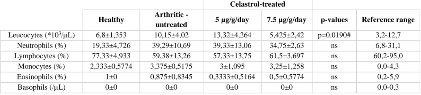
Organs samples were fixed immediately in 10% neutral buffered formalin solution and then dehydrated with increasing ethanol concentrations (70%, 96% and 100%). Samples were next embedded in paraffin, sectioned and stained with hematoxylin and eosin. Tissue histopathological changes were examined by a pathologist blinded to the experimental groups (data not shown). All images were acquired using a Leica DM 2500 microscope (Leica microsystems, Wetzlar, Germany). Moreover, blood toxicological parameters, such as creatine kinase, urea, alanine transaminase, lactate dehydrogenase (BioAssay Systems, California, USA), measured in serum, and cardiac toxicological mediator pro-ANP (Biomedica, Wien, Austria), measured in plasma, were performed by ELISA technique according to the manufacturer’s instructions. Samples were analyzed using a plate reader Infinite M200 (Tecan, Mannedorf, Switzerland). Blood samples were used to obtain white blood cell count (VetinLab, Lisbon, Portugal).
Statistical analysis
Data was analysed using GraphPad Prism 5 Software (GraphPad, California, USA). The normality distribution was assessed by D’Agostino and Pearson omnibus normality test. For populations that did not follow a Gaussian distribution it was performed nonparametric Mann-Whitney tests for
11 comparisons between 2 independent groups. Data are represented as median with interquartile range. Differences were considered statistically significant for p<0.05.
12 RESULTS
1. Evaluation of ethanol as a vehicle for the oral administration of celastrol in the AIA rat model
Celastrol’s poor water solubility implies the selection of an organic solvent capable of solubilize the compound. Dimethylsulfoxide (DMSO), one of the recommended solvents for celastrol, presents intragastric toxicity when orally administered [162]. Hence, our group has considered the use of another recommended solvent for the compound, ethanol. To examine the effect of ethanol as a suitable vehicle in this set-up, a group of AIA rats received ethanol 17% in PEG400 by gavage starting 8 days after disease induction until day 22 (arthritic EtOH+). A group of AIA rats that did not receive ethanol (arthritic EtOH-) and healthy rats were used as controls for comparison. There were no significant differences in inflammatory manifestations of the disease, evaluated during the treatment period (appendix 1), between the two arthritic groups. Twenty-two days after disease induction, both arthritic groups showed a significant increase in the inflammatory score (p=0.0001 and p=0.0019 in arthritic EtOH+ and arthritic EtOH- groups vs healthy controls, respectively, shown in Fig 1.1A) and ankle swelling (p=0.0004 and p=0.0032 in arthritic EtOH+ and arthritic EtOH- groups vs healthy controls, respectively, shown in Fig 1.1B) comparing with the healthy controls. Moreover, we have not found significant differences between the arthritic groups revealing that ethanol does not have an impact on disease activity.
Histopathological examination of left hind paw sections also validated the above mentioned data (illustrative images can be observed in Fig 1.1C). We have also assessed global disease severity score as an indicator of the impact of arthritis on joint articular tissues. The results, depicted in Fig 1.1D, showed that both arthritic groups have a significant increase in global disease severity in comparison with healthy controls (p=0.0001 and p=0.0019 in arthritic EtOH+ and arthritic EtOH- groups vs healthy controls, respectively), with no significant differences between them.
In order to evaluate potential side effects of intragastric administration of ethanol in AIA rats, body weight was measured during the experiment (Apprendix 2). Arthritic animals weighted less on day 22 than healthy rats (p=0.0016 and p=0.0043 in arthritic EtOH+ and arthritic EtOH- groups vs healthy controls, respectively, as shown in Fig 1.1E) as consequence of arthritis. Importantly, comparing the two arthritic groups there was no significant difference.
The similarity in arthritis condition and the absence of differences in body weight, an indicator of toxicity, between the two arthritic groups, validate the further use of ethanol as a vehicle for oral administration of celastrol in AIA rats.
13
Figure 1.1 - Evaluation of ethanol as a vehicle for the oral administration of celastrol in the AIA rat model. At the end of the experiment arthritic EtOH+ and arthritic EtOH- groups did not show differences in inflammatory score (A) and in ankle swelling (B). The similarity between the two arthritic groups in disease condition was validated by the histological representation of the joints (C) and global severity score (D). Ethanol did not provoke a reduction of body weight (E). Erosions are identified with “E” and synovial membrane with “S”. Magnifications of 50× and 100×. Bars: 200 μm. Differences were considered statistically significant for p-values<0.05 (*). Data are expressed as Median with interquartile range. Healthy N=8, arthritic EtOH+ N=15 and arthritic EtOH- N=5.
A
B
C
Healthy EtOH+
EtOH-0 5 10 15 * * Arthritic In fl a m m a to ry s c o re d a y 2 2
D
E
Healthy EtOH+
EtOH-0 100 200 300 * * Arthritic B o d y we ig h t (g ) d a y 2 2
Healthy EtOH+
EtOH-0 1 2 3 4 * * Arthritic Gl o b a l s e v e ri ty s c o re
Healthy EtOH+
EtOH-0 1 2 3 4 * * Arthritic A n k le p e ri m e te r (c m ) d a y 2 2
Healthy Arthritic EtOH+ Arthritic EtOH-
S S S S S S E E E E
14 2. In vivo assessment of the anti-inflammatory properties of celastrol in the AIA rat model
Once validated the use of ethanol as a suitable solvent for celastrol for this set-up, our group focused on evaluating orally administered celastrol, in two different doses, using ethanol 17% in PEG400 as a vehicle for comparison.
AIA rats were treated with 5 or 7.5 µg/g/day of celastrol, starting 8 days after arthritis induction (therapeutic model of administration), until day 22, when the model starts remission. A group of healthy and AIA control rats (arthritic-untreated) received water and vehicle by gavage, respectively. Figure 2.1A illustrates the variation in inflammatory score between all the experimental groups throughout the disease course.
By the fourth day of disease induction, all AIA animals already presented signs of arthritis. In arthritic-untreated animals, the inflammatory manifestations sharply increased from day 10th onwards.
Since the first day of treatment, signs of inflammation in both celastrol-treated groups were attenuated. At day 15, inflammatory signs in celastrol at 5 μg/g/day treated group were mantained and the group treated with the highest dose of the compound (7.5 µg/g/day) even showed a tendency to decrease their inflammatory score. Importantly, the inflammatory signs of arthritis in both celastrol-treated groups were already significantly reduced in comparison with the arthritic-untreated group since day 11 (p=0.0449 in 7.5μg/g/day celastrol-treated group vs arthritic-untreated group) or since day 14 (p=0.0096 in 5μg/g/day celastrol-treated group vs arthritic-untreated group).
By the end of the study, both doses of celastrol were able to significantly decrease the inflammatory score (p=0.0039 and p=0.0032 in 5 µg/g/day and 7.5 µg/g/day celastrol-treated groups vs arthritic-untreated group, respectively, shown in Fig 2.1B) and ankle swelling (p=0.0032 and p=0.0073 in 5 µg/g/day and 7.5 µg/g/day celastrol-treated groups vs arthritic-untreated group, respectively, shown in Fig 2.1C).
15
Figure 2.1 - In vivo evaluation of celastrol’s anti-inflammatory properties in the AIA rat model. (A) Celastrol is able to suppress inflammation throughout time. Celastrol in both doses was able to halt the inflammatory score whereas the arthritic-untreated animals kept increasing the inflammatory score. The arrow indicates the beginning of the treatment, 8 days after disease induction. (B) The inflammatory score and (C) ankle swelling of celastrol-treated animals was maintained significantly decreased at the end of the experiment in comparison with arthritic-untreated animals. Data are expressed as Median with interquartile range. Differences were considered statistically significant for p-values<0.05 (*). Healthy N=8, arthritic-untreated N=15, celastrol at 5 μg/g/day treated group N=6, celastrol at 7.5 μg/g/day treated group N=6.
A
B
C
Healthy Arthritic 5g/g 7,5g/g 0 2 4 6 8 10 * * * * * Celastrol In fl a m m a to ry s c o re d a y 2 2 Healthy Arthritic 5g/g 7,5g/g 0 1 2 3 4 * * * Celastrol A n k le p e ri m e te r (c m ) d a y 2 216 3. Histopathologic evaluation of the left hind paw after treatment with celastrol
The histopathologic evaluation of rats left hind paw sections was performed using a semi-quantitative score as described in materials and methods section (illustrative images can be observed in Fig 3.1A)
Synovial cell infiltration was significantly increased in arthritic-untreated animals when compared with healthy controls (p=0.0002, shown in Fig 3.1B). Celastrol was able to significantly decrease the infiltration into affected joints when compared with arthritic-untreated animals (p=0.0012 and p=0.0008 in 5 µg/g/day and 7.5 µg/g/day celastrol-treated groups vs arthritic-untreated group, respectively). Synovial cell proliferation was assessed by counting the number of the lining layers in SM. This parameter was also significantly increased in arthritic-untreated animals when compared with healthy controls (p=0.0005 in arthritic-untreated rats vs healthy controls, shown in Fig 3.1C). In contrast, celastrol-treated groups have significantly decreased cell proliferation comparing with the arthritic-untreated animals (p=0.0084 and p=0.0070 in 5 µg/g/day and 7.5 µg/g/day celastrol-treated groups vs arthritic-untreated group, respectively). In celastrol 7.5 µg/g/day group cell proliferation in the SM was even similar to healthy rats. Moreover, celastrol-treated groups have significantly reduced bone erosions (p=0.0059 and p=0.0031 in 5 µg/g/day and 7.5 µg/g/day celastrol-treated groups vs arthritic- untreated group, respectively, shown in Fig 3.1D), parameter that were significantly higher in arthritic-untreated animals in comparison with healthy animals (p=0.0001 vs healthy rats). The group treated with the highest dose (7.5 µg/g/day ) has preserved bone structure similar to healthy animals. As an indicator of the impact of arthritis on joint articular tissues, we scored the global severity of the disease. Reflecting the previous observations, untreated rats showed a higher disease severity (p=0.0001 arthritic-untreated group vs healthy controls, shown in Fig 3.1E) but celastrol in both doses have halted that phenotype (p=0.0108 and p=0.0038 in 5 µg/g/day and 7.5 µg/g/day celastrol-treated groups vs arthritic-untreated group, respectively).
17
Figure 3.1 - Histopathological evaluation of paw joints after celastrol treatment. (A) Histological representation of paw joints. Celastrol has inhibited cellular infiltration (B), cellular proliferation (C) and prevented bone erosion occurrence (D), allowing an improvement in disease severity (E). Synovial membrane is identified with “S”. Magnifications of 50× and 100×. Bars: 200 μm. Differences were considered statistically significant for p-values<0.05 (*). Data are expressed as Median with interquartile range. Healthy N=8, arthritic-untreated N=15, celastrol at 5 μg/g/day treated group N=6, celastrol at 7.5 μg/g/day treated group N=6.
S S Healthy Arthritic 5g/g 7,5g/g 0 1 2 3 4 * * * Celastrol Ce ll i n fi lt r a ti o n s c o r e Healthy Arthritic 5g/g 7,5g/g 0 1 2 3 4 * * * Celastrol Ce ll p r o li fe r a ti o n s c o r e Healthy Arthritic 5g/g 7,5g/g 0 1 2 3 4 * * * Celastrol E ro s io n s s c o re Healthy Arthritic 5g/g 7,5g/g 0 1 2 3 4 * * * * Celastrol Gl o b a l s e v e r it y s c o r e H e a lt h y A rt h rit ic -u n tr e a te d C e las tr o l 5 µ g /g C e las tr o l 7 .5 µ g /g
A
B
C
D
E
S S S S S S18 4. Immunohistochemical evaluation of paw articular joints after treatment with celastrol
To further validate the histopathological results, we stained by immunohistochemistry the left hind paw sections for several markers and analyzed the results with a semi-quantitative score (illustrative images can be observed in Fig 4.1A)
Ki67 is a protein expressed in proliferation cells which was used to detect proliferative cells in the synovia. Arthritic-untreated animals have showed a significantly higher score in this marker than healthy rats (p=0.0002, shown in Fig 4.1B). Celastrol significiantly reduced cellular proliferation (p=0.0048 and p=0.0015 in celastrol 5 µg/g/day and 7.5 µg/g/day treated groups vs arthritic untreated group, respectively).
We also used CD68, a biomarker of clinical response (Fig 4.1C). Decreased numbers of macrophages CD68+ in the synovia predict an adequate drug response in RA. Arthritic-untreated rats have increased numbers of synovial CD68+ cells rats in comparison with healthy controls (p=0.0005). Notably, celastrol in both doses was able to significiantly reduce the number of CD68+ macrophages in the synovia (p=0.0211 and p=0.0127 in 5 µg/g/day and 7.5 µg/g/day celastrol-treated groups vs arthritic-untreated group, respectively).
Furthermore, to locally address the effects of celastrol in bone formation and resorption cells, paw sections were stained for osteocalcin and cathepsin k, respectively. Osteocalcin, a marker of osteoblasts, was significantly increased in arthritic-untreated animals (p=0.0002, shown in Fig 4.1D) and completely restored to healthy levels in both celastrol-treated groups (p=0.0015 and p=0.0001 in 5 µg/g/day and 7.5 µg/g/day celastrol-treated groups vs arthritic-untreated group, respectively). The same result was observed in cathepsin k, a marker of osteoclasts, (p=0.0051 in healthy controls vs arthritic-untreated group; p=0.0243 and p=0.0448 in 5 µg/g/day and 7.5 µg/g/day celastrol-treated groups vs arthritic-untreated group, respectively, shown in Fig 4.1E).
19
Ki67 osteocalcin cathepsin K CD68
H e a lt h y A rt h rit ic -u n tr e a te d C e las tr o l 5 µ g /g C e las tr o l 7 .5 µ g /g Healthy Arthritic 5g/g 7,5g/g 0 1 2 3 4 * * * * Celastrol Ki 6 7 Healthy Arthritic 5g/g 7,5g/g 0 1 2 3 4 * * * Celastrol Os te o c a lc in Healthy Arthritic 5g/g 7,5g/g 0 1 2 3 4 * * * Celastrol Ca th e p s in K Healthy Arthritic 5g/g 7,5g/g 0 1 2 3 4 * * * * * Celastrol C D 6 8
A
B
C
D
E
20
Figure 4.1 - Immunohistochemical evaluation of paw joint sections after treatment with celastrol. (A) Representative immunohistochemical sections of rat joints for each biomarker. Arthritic-untreated animals have significantly increased levels of positive cells for all the markers. Celastrol treatment, in both treatment groups, decreased Ki67 positive cells in the synovial membrane (B) and CD68 positive macrophages (C). Both celastrol-treated groups decreased the number of osteoblasts (D) and osteoclasts (E) to healthy levels. Magnifications of 100× and 200×. Bars: 200 μm. Differences were considered statistically significant for p-values<0.05 (*). Data are expressed as Median with interquartile range. Healthy N=8, arthritic-untreated N=15, celastrol at 5 μg/g/day treated group N=6, celastrol at 7.5 μg/g/day treated group N=6.
21 5. Serum bone resorption and bone turnover markers measurement after treatment with
celastrol
Bone turnover markers, such as P1NP and CTX-I as well as bone resorption marker TRAcP 5b, were measured in animals’ serum by ELISA (Fig 5.1).
High levels of the P1NP protein, a reliable marker to predict bone formation, were detected in the serum of arthritic-untreated rats when compared with healthy controls (p=0.0055, as shown in Fig 5.1A). These levels were decreased by celastrol in both doses as proved by the significant difference between the arthritic-untreated and both celastrol-treated animals (p=0.0201 and p=0.0475 in 5 µg/g/day and 7.5 µg/g/day celastrol-treated groups vs arthritic-untreated group, respectively). Of note, celastrol in both doses was able to decrease P1NP to similar levels to healthy rats.
Accordingly, arthritic-untreated animals have significantly higher levels of CTX-I, a marker of bone resorption, than healthy rats (p=0.0186, shown in Fig 5.1B). Both treatment groups showed a tendency towards a decrease in CTX-I, however it did not reached statistical significance.
Moreover, both doses significantly decreased TRAcP 5b serum levels, a bone resorption enzyme produced by osteoclasts (p=0.0022 and p=0.0090 in 5 µg/g/day and 7.5/µg/g/day celastrol-treated groups vs arthritic-untreated group, respectively, shown in Fig 5.1C).
22
Figure 5.1 - Quantification of serum levels of bone resorption and bone turnover markers in AIA rats after celastrol treatment. Celastrol was able to significantly decrease bone formation (A), showed a tendency to decrease bone resorption (B) and significantly reduced the number of osteoclasts (C). Differences were considered statistically significant for p-values<0.05 (*). Data are expressed as Median with interquartile range. Healthy N=8, arthritic-untreated N=15, celastrol at 5 μg/g/day treated group N=6, celastrol at 7.5 μg/g/day treated group N=6.
Healthy Arthritic 5g/g 7,5g/g 0 2 4 6 8 10 * * Celastrol T R A c P 5 b ( U /L ) Healthy Arthritic 5g/g 7,5g/g 0 20 40 60 80 * Celastrol CT X -I ( n g /m L ) Healthy Arthritic 5g/g 7,5g/g 0 20 40 60 80 * * * Celastrol P 1 N P (n g /m L )