The Tocotrienol-Rich Fraction Is Superior to
Tocopherol in Promoting Myogenic
Differentiation in the Prevention of
Replicative Senescence of Myoblasts
Shy Cian Khor, Azraul Mumtazah Razak, Wan Zurinah Wan Ngah, Yasmin Anum Mohd Yusof, Norwahidah Abdul Karim, Suzana Makpol*
Department of Biochemistry, Faculty of Medicine, Level 17, Preclinical Building, Universiti Kebangsaan Malaysia Medical Centre, Jalan Yaacob Latif, Bandar Tun Razak, 56000 Cheras, Kuala Lumpur, Malaysia
Abstract
Aging results in a loss of muscle mass and strength. Myoblasts play an important role in maintaining muscle mass through regenerative processes, which are impaired during aging. Vitamin E potentially ameliorates age-related phenotypes. Hence, this study aimed to determine the effects of the tocotrienol-rich fraction (TRF) andα-tocopherol (ATF) in pro-tecting myoblasts from replicative senescence and promoting myogenic differentiation. Pri-mary human myoblasts were cultured into young and senescent stages and were then treated with TRF or ATF for 24 h, followed by an analysis of cell proliferation, senescence biomarkers, cellular morphology and differentiation. Our data showed that replicative senes-cence impaired the normal regenerative processes of myoblasts, resulting in changes in cellular morphology, cell proliferation, senescence-associatedβ-galactosidase (SA-β-gal) expression, myogenic differentiation and myogenic regulatory factors (MRFs) expression. Treatment with both TRF and ATF was beneficial to senescent myoblasts in reclaiming the morphology of young cells, improved cell viability and decreased SA-β-gal expression. However, only TRF treatment increased BrdU incorporation in senescent myoblasts, as well as promoted myogenic differentiation through the modulation of MRFs at the mRNA and protein levels.MYOD1andMYOGgene expression and myogenin protein expression were modulated in the early phases of myogenic differentiation. In conclusion, the tocotrie-nol-rich fraction is superior toα-tocopherol in ameliorating replicative senescence-related aberration and promoting differentiation via modulation of MRFs expression, indicating vita-min E potential in modulating replicative senescence of myoblasts.
Introduction
Sarcopenia is a geriatric syndrome that is characterized by a dramatic loss of skeletal muscle mass and strength in advancing age. Although the underlying mechanism of these alterations OPEN ACCESS
Citation:Khor SC, Razak AM, Wan Ngah WZ, Mohd
Yusof YA, Abdul Karim N, Makpol S (2016) The Tocotrienol-Rich Fraction Is Superior to Tocopherol in Promoting Myogenic Differentiation in the Prevention of Replicative Senescence of Myoblasts. PLoS ONE 11(2): e0149265. doi:10.1371/journal.pone.0149265
Editor:Michael Kyba, University of Minnesota, UNITED STATES
Received:July 9, 2015
Accepted:January 30, 2016
Published:February 17, 2016
Copyright:© 2016 Khor et al. This is an open access article distributed under the terms of the Creative Commons Attribution License, which permits unrestricted use, distribution, and reproduction in any medium, provided the original author and source are credited.
Data Availability Statement:All relevant data are within the paper.
Funding:This study was funded by the Universiti Kebangsaan Malaysia grants AP-2012-012 and UKM-FF-2013-259. The funders had no role in study design, data collection and analysis, decision to publish, or preparation of the manuscript.
Competing Interests:The authors have declared
is not clear, several risk factors have been considered, such as immobilization, chronic diseases,
hormone and pro-inflammatory cytokine shift and malnutrition in the elderly [1]. Loss of
mus-cle regenerative capacity has been suggested as one of the possible contributory factors of this
age-related muscle deterioration [2].
Skeletal muscle has an established regeneration competency in restoring and maintaining
muscle mass when muscle cells undergo injury [3]. Muscle regeneration essentially involves
four sequential and overlapping phases: degeneration, inflammation, regeneration and remod-eling. Satellite cells are the key regenerative phase and will be activated, proliferate and
differen-tiate in response to stimuli. Proliferating satellite cells are known as myoblasts [4]. In addition
to producing functional progeny via differentiation, satellite cells can replicate to maintain the
satellite cell pool; thus, they are also categorized as muscle stem cells [5]. The heterogeneity of
satellite cells has provoked the rationale of targeting these cells for therapeutic purposes in
ameliorating age-related sarcopenia or pathological dystrophic muscle [6].
In aging, satellite cells malfunction and fail to sustain their normal quiescent state,
irrevoca-bly influencing their regenerative and self-renewal capacities [7]. A decreased number of
satel-lite cells in old age were also observed [6,8]. However, this decrease may not be the sole reason
for the gradual loss of muscle rejuvenation capacity in old age. In fact, a permissive atmosphere is imperative rather than the number of satellite cells, whereby satellite cells from old muscle
can be engaged for myogenic activity when exposed to a young systemic environment [9–12].
Myogenic differentiation is regulated by a family of myogenic regulatory factors (MRFs) that
includes MyoD, Myf5, Myogenin and MRF4. MRFs are transcription factors with a basic helix–
loop–helix (bHLH) central domain that assist protein interactions and DNA binding to activate
muscle-specific genes [4]. The deregulation of Myf5, MyoD and myogenin at an early stage of
dif-ferentiation is interrelated with the difdif-ferentiation capability of senescent myoblasts, resulting in
the formation of smaller myotubes that resemble the condition in sarcopenia [13,14]. Thus,
ongo-ing research in findongo-ing ways to restore the regenerative capacity in old myoblasts will presumably provide precious insight for combating muscle atrophy in aging or degenerative diseases.
Because muscle atrophy or aging itself is closely related to oxidative stress, the re-establish-ment of redox balance should be potentially advantageous in the amelioration of age-related
muscle wasting [15,16]. Vitamin E is a lipid-soluble vitamin that is able to scavenge free
radi-cals, boosts cellular antioxidant competency and prevents oxidative damage. There are two
subgroups of vitamin E: tocopherols and tocotrienols [17]. Howard et al. reported thatα
-tocopherol (ATF) was able to repair the laser-induced disrupted membrane of myoblasts,
which supports a therapeutic effect exerted by vitamin E [18]. A significant correlation between
the ATF level and sarcopenia indicators among the elderly has been reported [19]. Vitamin E
deficiency will not only affect muscle performance but also accelerate the progression of aging
[20]. Therefore, it is rational to introduce antioxidants, such as vitamin E, to prevent
sarcope-nia, even though further studies are still required [16].
Human myoblasts can be isolated and culturedin vitrowith a limited proliferation capacity,
whereby at a certain stage, they will undergo growth arrest, termed replicative senescence [21].
The present study was designed to elucidate the effects of the tocotrienol-rich fraction (TRF)
andα-tocopherol (ATF) in ameliorating senescent myoblasts and promoting myogenic
differ-entiation during replicative senescence.
Methods
Cell Culture and Replicative Senescence Model
Primary human myoblasts (Human Skeletal Muscle Myoblasts; HSMM) at passage 2 from two donors, a 17-year-old Caucasian female and a 16-year-old Caucasian male were purchased
from Lonza (Walkersville, MD, USA). Briefly, myoblasts were cultured in Skeletal Muscle Basal Medium (SkBM) that was supplemented with human epidermal growth factor, fetal bovine serum, dexamethasone, L-glutamine, and gentamicin sulfate/amphotericin B (Lonza, Walkersville, MD USA). Cells were cultivated at 37°C in a humid atmosphere containing 5%
CO2. The myoblasts then underwent serial passaging to reach senescence. For each passage,
the population doublings (PD) of cells was calculated as: In (N/n)/In2, whereNis the number
of cells at harvest stage, andnis the number of cells at seeding stage [14]. The starting PD in
this study was 8. The cells achieved replicative senescence when they were unable to proliferate within 10 days in culture, even with consecutive replenishment.
Analysis of Cell Morphology and Myogenic Purity
Myogenic purity and myoblasts morphology were observed by the immunocytochemistry method using a mouse monoclonal anti-Desmin antibody (D33; Dako, Produktionsvej,
Den-mark). Myoblasts were plated inμ-Slide 8 well (ibidi, Martinsried, Germany) at a density of
1×10⁴cells per well. The cells were fixed in cold ethanol. Then, anti-Desmin antibody (1:50)
and Alexa Fluor 488 goat anti-mouse (Life Technologies, Carlsbad, CA, USA) were used to incubate the myoblasts in sequence. Nuclei were visualized using Hoechst 33342 (Life Technol-ogies, Carlsbad, USA). The slides were then viewed under a Confocal Laser Scanning Micro-scope Leica TCS SP5 II, and data were acquired using LAS AF version Lite 2.6 software (Leica Microsystems, Wetzlar, Germany). To determine the percentage of desmin-positive cells, a minimum of 50 cells were counted in three independent cultures. In addition, the morphologi-cal changes of myoblasts were observed, while the width and length of myoblasts were visual-ized and measured using LAS AF version Lite 2.6 software (Leica Microsystems, Wetzlar, Germany). For each group of cells, at least 30 cells were analyzed.
Determination of DNA Synthesis in Proliferating Cells
The amount of 5-bromo-2’-deoxyuridine (BrdU) incorporation indicates the total proliferating
cells. Thus, the cell proliferation ELISA, BrdU (colorimetric) kit (Roche, Penzberg, Germany) was used to determine the effects of replicative senescence, TRF and ATF on cell proliferation.
This immunoassay was performed according to the manufacturer’s instructions. The cells were
labeled with BrdU, a pyrimidine analog that will incorporate into the DNA and was detected by a microtiter plate reader (VersaMax Molecular Devices, USA) at 450 nm with reference to 690 nm.
Determination of Senescence Biomarkers
The expression of SA-β-gal was determined as described by Dimri et al. [22] in order to
con-firm the presence of senescent myoblasts. This process was carried out using a Senescent Cell Histochemical Staining Kit (Sigma-Aldrich, St. Louis, Missouri, USA) according to the
manu-facturer’s instructions. Cells were incubated in staining solution for 8 hours at 37°C in the
absence of CO2before analysis. At least 100 cells were observed, and the percentage of blue
stained cells was calculated. In addition, the morphological changes of myoblasts were also observed.
Preparation of Vitamin E Treatments
TRF Gold Tri E 70 (Sime Darby Sdn. Bhd., Selangor, Malaysia) and ATF (Malaysian Palm Oil Board, Selangor, Malaysia) were used as treatments in this study. Briefly, stock solutions of
month. A similar process was applied for ATF preparation. TRF and ATF were then incubated overnight with fetal bovine serum at 37°C before use. The cell viability was assessed with a
Cell-Titer 961Aqueous Non-Radioactive Cell Proliferation Assay (MTS; Promega, Madison, WI
USA) according to the manufacturer’s instruction. Various concentrations of TRF or ATF
were used to treat the cells for 24 hours. Then, MTS was added and further incubated for 2 hours. The absorbance of MTS formazan was measured at 490 nm with a microtiter plate reader (VersaMax Molecular Devices, USA). The optimum dose of treatments was used for subsequent experiments.
Induction of Myogenic Differentiation
To induce muscle cell differentiation, the proliferating medium SkBM was replaced with DMEM:F12 (Lonza, Walkersville, MD USA) that was supplemented with 2% horse serum (ATCC, Baltimore, USA). The differentiation medium was changed every 2 days until the desired day of differentiation for parameter measurement.
Analysis of Myogenic Differentiation
To evaluate the efficiency of differentiation, a micro-insert 4 well,μ-Dish was used (ibidi,
Mar-tinsried, Germany) to culture the cells to determine myotubes formation. After 9 days of differ-entiation, myotubes were stained using an anti-Desmin antibody. The fusion index and the size of myotubes were calculated, indicating myotube formation. The formula below was used to calculate the fusion index, and a minimum of 50 nuclei were counted in 3 different randomly chosen optical fields.
Fusion Index¼ The number of nuclei in myotubesð> 2nucleiÞ
The total number of nuclei in desmin positive cells100%
To determine the size of the myotubes, the number of nuclei per myotube was counted in a
minimum of 11 multinucleated cells in 3 different randomly chosen opticalfields.
Determination of MRFs at an Early Phase of Myogenic Differentiation
At days 0, 1 and 2 of differentiation, total RNA was extracted using the TRI reagent (Molecular Research Center Inc., Ohio, USA). For gene expression determination, quantitative real-time
RT-PCR (qRT-PCR) was used. The expression ofMYF5,MYOD1andMYOGmRNA was
quan-titatively analyzed using a one-step qRT-PCR technique. qRT-PCR was performed with 100 ng of total RNA, 400 nM each primer and KAPA SYBR FAST One-Step qPCR kit (Kapa
Biosys-tems, Boston, Massachusetts, USA) according to the manufacturer’s instructions. The primer
sequences areGAPDHforward5’-TCCCTGAGCTGAACGGGAAG-3’,GAPDHreverse5’
-GGAGGAGTGGGTGTCGCTGT-3’,MYF5forward5’-TCACCTCCTCAGAGCAACCT-3’,
MYF5reverse5’-ATTAGGCCCTCCTGGAAGAA-3’,MYOD1forward5’-CGCCAGGATATG
GAGCTACT-3’,MYOD1reverse5’-GAGTGCTCTTCGGGTTTCAG-3’,MYOGforward5’
-CAGTGCCATCCAGTACATCG-3’andMYOGreverse5’-AGGTTGTGGGCATCTGTAGG-3’.
The master mix was prepared, and PCR reactions were carried out in a Bio-Rad iQ5 Cycler (Her-cules, CA, USA) with the following programmed reaction profile: cDNA synthesis for 5 min at 42°C; pre-denaturation for 4 min at 95°C; and PCR amplification for 40 cycles of 3 sec at 95°C and 20 sec at 60°C. These reactions were followed by a melt curve analysis to determine the reac-tion specificity and the expression of each targeted gene. The expression level of each targeted gene was normalized to that of glyceraldehyde 3-phosphate dehydrogenase (GAPDH). The
rela-tive expression value (REV) was calculated using the 2-ΔΔCtmethod of relative quantification and
the following equation: REV = 2Ct value of GAPDH -Ct value of the gene of interest. Then, the fold change of expression was determined.
Determination of Myogenin Expression
At day 3 of differentiation, the number of cells expressing myogenin was estimated using a mouse monoclonal anti-myogenin antibody (F5D, Dako, Produktionsvej, Denmark) at a 1:20 dilution overnight at 4°C. Alexa Fluor 488 was used as the secondary antibody. Nuclei were visualized using Hoechst 33342. The cells were observed under an EVOS FL Digital Inverted Fluorescence Microscope (Life Technologies, Carlsbad, USA).
Assessment of Intracellular Free Radical Generation
In order to measure free radicals generation by myoblasts, we used two types of dyes, i.e.
dihy-droethidium (DHE) and 5-(and-6)-carboxy-20,70-dichlorodihydrofluorescein diacetate
(car-boxy-H2DCFDA) (Molecular Probes, Eugene, OR, USA). The DHE-stained cells indicated
oxidation by superoxide anion, while carboxy-H2DCFDA is oxidized by hydrogen peroxide
(H2O2), peroxynitrite or hydroxyl radical. Superoxide anions may contribute to
carboxy-H2DCFDA oxidation albeit at a lesser degree. Briefly, myoblasts were incubated in 20μM of
DHE and 40μM of carboxy-H2DCFDA for 45 min. After that, the cells were washed with PBS
and recovered in medium for 30 minutes. Then, we measured the intensity by using microplate
reader (Infinite1
200, Tecan, USA) at excitation/emission wavelength (Ex/Em) 518/600 nm and 488/521 nm respectively.
Statistical Analysis
Statistical analyses were performed using SPSS 17.0 software (IBM, NY, USA). All of the data are reported as the means ± standard deviation (SD) from at least three replicates. For all of the
tests, p<0.05 was considered statistically significant. To determine significance between two
treatment groups, comparisons were made using an independent T-test, while ANOVA was
used to analyze multiple groups, followed by apost-hocTukey HSD or LSD (if equal variance
was assumed) and Dunnett T3 (if equal variance was not assumed) tests.
Results
Replicative Senescence Model of Myoblasts: Characteristics and
Proliferation
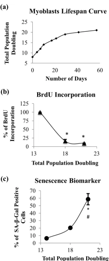
To elucidate the effects of aging on myoblasts, we expanded the cells until replicative senes-cence. The lifespan curve that was plotted based on their cumulative PD showed that myoblasts
have a limited proliferative capacity, and cell growth was halted at 21 divisions in culture (Fig
1a). Based on BrdU incorporation, the proliferation of myoblasts decreased with increasing
total PD, whereby the percentage of BrdU incorporation at PD18 and PD21 was significantly
different compared to the percentage of BrdU incorporation at PD14 (p<0.05) (Fig 1b). The
percentage of SA-β-gal-stained cells increased with the serial passaging of myoblasts, which
was significantly higher at PD21 compared to both PD14 and PD18 (p<0.05) (Fig 1c).
There-fore, myoblasts are considered young at PD<15 and senescent at PD>20. No loss of
myogeni-city was observed during the replicative senescence of myoblasts, as indicated by the presence
of desmin in 96% of the cell population (Table 1), allowing a reliable statistical comparison of
Fig 1. Effects of serial passaging on population doubling, cell proliferation and expression of SA-β -gal in myoblasts.During extensive expansion, myoblasts significantly lost their proliferation capacity as represented by a hyperbolic proliferative lifespan curve (a) and decreasing percentage of BrdU incorporation (b), while the percentage of senescent cells increased, as represented by positive SA-β-gal staining (c). For (b) and (c), the data are presented as the means±SD, n = 3.*p<0.05 compared to myoblasts at PD14 (young),#p<0.05 compared to myoblasts at PD18 (pre-senescent) with apost-hocDunnett T3. doi:10.1371/journal.pone.0149265.g001
Promotion of Cell Viability and Proliferation
Incubation with various concentrations of TRF or ATF for 24 h significantly increased the
via-bility of young and senescent myoblasts (Fig 2a and 2b). Cells that were treated with TRF and
ATF at concentrations of 50μg/ml, exhibited the greatest percentage of viability in both
myo-blasts. Therefore, in the subsequent experiments, 50μg/ml TRF and 50μg/ml ATF were used
for treatment in young and senescent myoblasts. Prolonged treatment (48 hours) using the
optimal dose (50μg/ml) improved cell viability in young cells, but not in senescent myoblasts
(Fig 2c). Subsequently, the optimal dose was applied on senescent myoblasts from the other donor (a 16-year-old Caucasian male). However, there was no significant different observed on the cell viability as compared to the viability of myoblast from the first donor (a 17-year-old
Caucasian female) (Fig 2d). Therefore, for the following experiment, myoblasts from the first
donor was used.
A significantly decreased percentage of BrdU incorporation was observed in senescent
myo-blasts compared to young cells (p<0.05). Treatment with TRF increased the percentage of
BrdU incorporation in senescent myoblasts (p<0.05) (Fig 3), while no significant difference
was observed in young myoblasts that were treated with TRF or ATF.
Improvement in Myoblasts Cellular Morphology with TRF and ATF
Treatment
Myoblasts were spindle shaped when young but transformed into large and flat cells with a
prominent intermediate filament network at the senescent stage (Fig 4a and 4d). Senescent
cells exhibited a significantly higher ratio of cytoplasm to nucleus content than did young cells,
as manifested by increased width during replicative senescence (p<0.05) (Fig 4g). However,
both TRF- and ATF-treated senescent myoblasts retrieved the young-like morphology with the
presence of more spindle-shaped cells (Fig 4e and 4f). Moreover, the average width of senescent
myoblasts that were treated with both TRF and ATF significantly decreased compared to that
of the untreated control (p<0.05) (Fig 4g). The spindle-shaped cells can be maintained in
cul-ture for two days after withdrawal of treatments and being observed in the following passage (Fig 4h).
Reversal of Replicative Senescence by TRF and ATF
Senescent myoblasts were stained positive for SA-β-gal (Fig 5d–5f). The percentage of
SA-β-gal-positive cells is shown inFig 5g. The SA-β-gal-positive cells were markedly increased in
senescent myoblasts (58.67% ± 7.5) compared to young cells (6.33% ± 1.2) (p<0.05). The
per-centage of SA-β-gal-positive cells significantly decreased to 29.67% ± 3.8 and 32.67% ± 4.0
(p<0.05) with TRF and ATF treatment compared to the untreated groups.
Superior Effects of TRF in Promoting Cell Differentiation in Senescent
Myoblasts
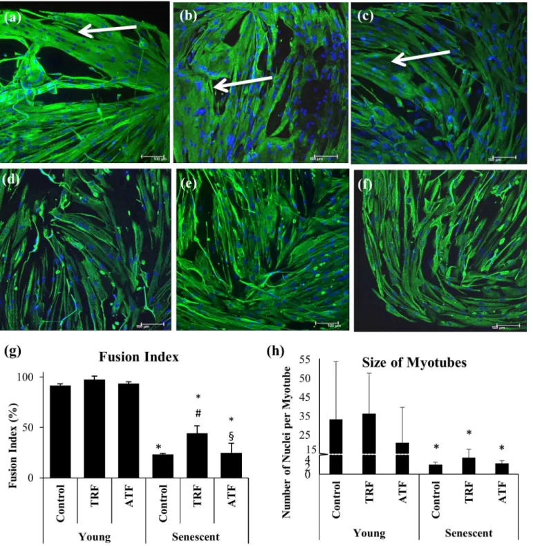
Young and senescent myoblasts were allowed to differentiate for 9 days to form myotubes.
Young myoblasts fused together, forming large and branched multinucleated myotubes (Fig
6a). Senescent cells, however, formed smaller myotubes with fewer branches compared to
Table 1. Percentage of desmin-positive cells in different cell stages.
Myoblasts Young(PD14) Presenescent(PD18) Senescent(PD21)
Desmin +ve 96.67±2.31(n = 3) 98.67±1.15(n = 3) 96.00±2.00(n = 3)
Fig 2. Effects of the TRF and ATF treatments on cell viability and proliferation.Dose-response curve of TRF (a) and ATF (b) treatments (24 h) in young and senescent myoblasts (n = 9). The prolonged treatments of TRF and ATF (48 h) at optimal dose were unlikely to further improve cell viability in senescent myoblasts (c). Therefore, 24 h treatment was used in subsequent experiment. Comparison of cell viability between myoblasts from donor 1, a 17 year-old, female Caucasian and donor 2, a 16 year-old male Caucasian (d). There were no significant different observed between the two cell lines in response to both TRF and ATF treatments based on the viability assessment.Post-hocDunnett T3 test for (a) and (b).*p<0.05 significantly different compared to untreated young myoblasts,#p<0.05 significantly different compared to untreated
senescent myoblasts,§p<0.05 significantly different compared to TRF-treated senescent myoblasts. The
data are presented as the means±SD.
doi:10.1371/journal.pone.0149265.g002
young cells (Fig 6d), indicating an inefficient differentiation process during the replicative senescence of myoblasts, which is similar to sarcopenic muscle.
A significant decrease in the fusion index and size of myotubes was observed in senescent
myoblasts (Fig 6g and 6h). Approximately 91.66% of young myoblasts turned into myotubes,
but only 23.17% of senescent myoblasts were able to fuse and form myotubes (Fig 6g). Large,
branched multinucleated myotubes were formed from young myoblasts with approximately 25 nuclei per myotube, while much smaller myotubes were formed during replicative senescence
with the presence of 2.5 nuclei per myotube (Fig 6h).
The multinucleated myotubes that formed from senescent myoblasts, however, were
improved with TRF and ATF treatment (Fig 6e and 6f), even though they were still smaller in
size and with fewer branches compared to young myoblasts. An analysis of the fusion index demonstrated that TRF treatment significantly promoted cell differentiation during cellular senescence, as indicated by a significantly increased fusion index in TRF-treated myoblasts
(p<0.05) (Fig 6g). ATF, however, did not produce similar effects.
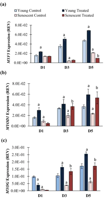
Modulation of MRFs at the Early Phase of Myogenic Differentiation
The modulation of MRFs expression by TRF and ATF during the replicative senescence of
myoblasts was also investigated by determining the expression ofMYF5,MYOD1andMYOG
mRNA at the early phase of differentiation in young and senescent myoblasts (Fig 7a–7c).
MYOGmRNA expression was upregulated at day 2 of differentiation induction in both
untreated young and untreated senescent myoblasts (p<0.05), whileMYOD1expression was
upregulated at day 1 of differentiation in untreated young myoblasts only (Fig 7b and 7c). The
expression ofMYOD1andMYOGmRNA, however, was lower in untreated senescent
myo-blasts compared to untreated young cells (p<0.05).
Treatment with TRF down regulatedMYF5,MYOD1andMYOGin young and senescent
myoblasts at day 0 of differentiation induction compared to the untreated young control
(p<0.05). At day 1 and day 2 of differentiation induction, the expression ofMYF5,MYOD1
andMYOGin both young and senescent myoblasts significantly increased (p<0.05) (Fig
7a–7c).
Fig 3. Effects of TRF and ATF on BrdU incorporation.Cells were treated with optimal dose of TRF and ATF followed by cell proliferation determination based on the percentage of BrdU incorporation (n = 3). Only TRF-treated senescent myoblasts showed increased BrdU incorporation indicating promotion of cell proliferation and DNA synthesis with TRF treatment.*p<0.05 significantly different compared to untreated young myoblasts, #p<0.05 significantly different compared to untreated senescent myoblasts, §p<0.05 significantly different compared to TRF-treated senescent myoblasts, withpost-hocLSD test. The data are presented as the means±SD.
Fig 4. Effects of replicative senescence and vitamin E treatment on myoblasts phenotype.The photomicrographs of myoblasts were taken from a young control (a), TRF-treated young (b), ATF-treated young (c), senescent control (d), TRF-treated senescent (e) and ATF-treated senescent (f) cells (magnification: 400×). Myoblasts were stained for desmin (green) and Hoechst (blue). Both TRF- and ATF-treated senescent myoblasts resembled the morphology of young cells. The width and length of cells were measured (g). The width of senescent myoblasts significantly increased in the untreated control and decreased with TRF and ATF treatment. The spindle-shaped cells can be maintained in culture for two days after withdrawal of treatments and retained in the following passage (h).*p<0.05 significantly different compared to untreated young myoblasts,#p<0.05 significantly different compared to
untreated senescent myoblasts, withpost-hocDunnett T3. The data are presented as the means±SD, n = 30.
doi:10.1371/journal.pone.0149265.g004
No significant changes were observed in the expression ofMYF5,MYOD1andMYOGat day 0 of the differentiation induction of young myoblasts with ATF treatment compared to untreated control. After differentiation induction, ATF treatment caused upregulation to
MYOD1andMYOGmRNA in young myoblasts. OnlyMYOD1was upregulated in senescent
myoblasts at day 1 of differentiation induction (p<0.05) (Fig 7a–7c).
Myogenin protein expression significantly decreased in untreated senescent myoblasts
com-pared to untreated young cells (p<0.05) (Fig 7d and 7e). TRF treatment significantly increased
the expression of the myogenin protein in senescent myoblasts (p<0.05), while no significant
changes were observed in the expression of myogenin with ATF treatment (Fig 7d and 7e).
Modulation of MRFs during Myogenic Differentiation Phase
To further validate the effect of TRF on MRFs during differentiation, expression of MRFs were determined in an extended time course and enhanced treatment protocol, in which myoblasts Fig 5. Effects of replicative senescence and vitamin E treatment on senescence biomarker.The photomicrographs of myoblasts were taken from young control (a), TRF-treated young (b), ATF-treated young (c), senescent control (d), TRF-treated senescent (e) and ATF-treated senescent (f) cells (magnification: 40×). Most of the senescent control myoblasts were stained positive for SA-β-gal (blue stained), as indicated by the arrow. The percentage of blue-stained cells was determined (g). TRF and ATF significantly reduced the number of blue-stained cells of senescent myoblasts.*p<0.05 significantly different compared to untreated young myoblasts,#p<0.05 significantly different compared to untreated senescent
myoblasts, withpost-hocTukey HSD. The data are presented as the means±SD, n = 3.
Fig 6. Effects of replicative senescence and vitamin E treatment on the differentiation capacity of myoblasts.The photomicrographs of myotubes were taken from young control (a), TRF-treated young (b), ATF-treated young (c), senescent control (d), TRF-treated senescent (e) and ATF-treated senescent (f) cells (magnification: 200×). Desmin was stained green, and the nuclei were stained blue (Hoechst). The myotubes that formed from young myoblasts were significantly bigger than the myotubes from senescent myoblasts. The fusion index (g) and the size of myotubes (h) were determined to evaluate the efficiency of muscle differentiation. TRF significantly increased the fusion index (n = 3), which was not shown with ATF treatment. No changes was observed in the size of the myotubes that formed (n = 12) with the TRF and ATF treatments.*p<0.05 significantly different compared to young control,
#p<0.05 significantly different compared to senescent control,§p<0.05 significantly different compared to TRF-treated senescent myoblasts, withpost-hoc
Tukey HSD. The data are presented as the means±SD.
doi:10.1371/journal.pone.0149265.g006
Fig 7. Effects of replicative senescence and vitamin E treatment at the early phase of myogenic differentiation.TheMYF5(a) andMYOD1(b) mRNA expression levels on day 0 and day 1 of differentiation were determined, while theMYOG(c) mRNA expression level was determined on day 0 and day 2 of differentiation. The percentage of nuclei that stained for myogenin (green) on day 3 of differentiation is shown in (d). Photomicrographs were taken from all groups using a fluorescence microscope (magnification: 200×) (e). TRF significantly increased the number of myogenin-labeled nuclei on day 3 of
differentiation, as indicated by arrows.*p<0.05 significantly different compared to young control at corresponding day of differentiation,#p<0.05 significantly
were treated with 50μg/ml of TRF again during differentiation phase. Myoblasts from other
donor (a 16-year-old male Caucasian) was used. The gene expression of MRFs (MYF5, MYOD1andMYOG) at day 1 (D1), day 3 (D3) and day 5 (D5) of differentiation induction
were determined (Fig 8a–8c). The expression ofMYF5mRNA was significantly decreased in
senescent myoblasts at day 3 and day 5 of differentiation induction (p<0.05) (Fig 8a) while
the expression ofMYOD1andMYOGmRNA was significantly lower in senescent myoblasts
at day 1 till day 5 of differentiation induction as compared to untreated young myoblasts
(p<0.05) (Fig 8b and 8c).
The expression of MRFs was modulated with TRF treatment. In TRF-treated senescent
myoblasts, bothMYOD1andMYOGmRNA were significantly upregulated at day 3 and day 5
of differentiation induction as compared to untreated control (p<0.05) (Fig 8b and 8c).
How-ever, the expression ofMYF5mRNA was significantly lower in TRF-treated senescent
myo-blast at day 5 of differentiation as compared to senescent control (p<0.05) (Fig 8a). TRF
treatment also upregulatedMYF5andMYOD1mRNA in young myoblasts at day 1 till day 5
of differentiation induction as compared to untreated control (p<0.05) (Fig 8a and 8b)
whileMYOGmRNA was upregulated at day 3 and day 5 of differentiation induction (p<0.05)
(Fig 8c).
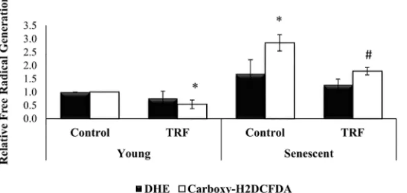
Reduction of Free Radicals Generation by TRF
To further elucidate the antioxidant effect of TRF, generation of free radicals or reactive oxygen species (ROS) was determined, in which the myoblasts were labelled with DHE (in orange) and
carboxy-H2DCFDA (in green). The percentage of cells which were stained positive with
car-boxy-H2DCFDA was gradually increased from young to senescent. Quantitative analysis
showed a significantly increased in carboxy-H2DCFDA-stained senescent myoblasts (2.85
fold) as compared to young myoblasts (p<0.05) (Fig 9). TRF treatment significantly reduced
the amount of intracellular ROS generation in both young and senescent myoblasts, effectively (p<0.05) (Fig 9).
Discussion
The tocotrienol-rich fraction (TRF) has been shown to have not only anti-aging properties in anin vitrostudy [23] but also beneficial effects against aging in animal models [24] and healthy
older adults [25]. Recently, positive effects of TRF were highlighted in stress-induced
prema-ture senescent (SIPS) myoblasts following a 24-hour treatment [26], acquitting its
free-radical-scavenging power. The mechanism by which TRF reduces senescence phenotypes in the muscle may not only involve combating oxidative stress, but is possibly associated with its regenerative capacity. In this study, we revealed novel insight into the potential of vitamin E
for improving myoblasts proliferation and differentiation for the prevention ofin vitro
replica-tive senescence.
A slight modification of skeletal muscle homeostasis may lead to unsuccessful muscle renewal as in aging and pathological dystrophic muscle. In brief, myoblasts undergo the cellu-lar life path starts with an exponential phase that then slows and reaches a finite proliferation
state, indicating the replicative senescence phase [21,27]. However, caution is required when
performing the serial passaging of myoblasts, as fibroblasts may swarm in and affect the
the corresponding day of differentiation,§p<0.05 significantly different compared to TRF-treated senescent myoblasts at corresponding day of differentiation, **p<0.05 significantly different compared to the corresponding treatment at day 0 of differentiation. The data are presented as the means±SD.
doi:10.1371/journal.pone.0149265.g007
Fig 8. Effects of TRF on the MRFs mRNA expression levels during 5 days of differentiation induction.
TheMYF5(a),MYOD1(b) andMYOG(c) mRNA expression levels in senescent control were significantly lower than young control (p<0.05). TRF significantly increased the expression of bothMYOD1andMYOG mRNA at day 3 (D3) and day 5 (D5) of differentiation, resembled the expression in young control, while the expression in senescent myoblasts remained low, even after 5 days of differentiation.ap<0.05 significantly
different compared to young control at corresponding day of differentiation,bp<0.05 significantly different
compared to senescent control at corresponding day of differentiation, The data are presented as the means±SD.
myoblasts culture [28]. Thus, in conducting the experiment, myogenic purity must be main-tained, even when cells reach replicative senescence.
The number of proliferating myoblasts is represented by the total BrdU incorporated into the cells, which gradually decreased with the increased population doubling over time.
Interest-ingly, the amount of these active cells very much depends on the donor’s age [9,21]. Moreover,
old-individual-derived myoblasts had a slower response to proliferative stimuli, which may
contribute to the prolonged proliferation period in senescent cells [9]. In relation to the
prolif-erative capacity, studies have shown that aged myoblasts in culture share the same characteris-tics with old individual-derived myoblasts, thus indicating the perception of regenerative capacity during aging, which can be modulated by intervention. Our data show that TRF not only increases cell viability but also enhances the proliferation capacity of senescent myoblasts. The total number of proliferating cells, however, did not increase with ATF treatment in senes-cent myoblasts, even though the same dosage of ATF retained more viable cells than did untreated control myoblasts. These findings indicate the potential of TRF in improving the proliferation capacity of senescent myoblasts in culture.
To further verify the capability of both vitamin E treatments in rescuing senescent
myo-blasts, the senescence biomarker SA-β-gal was used to identify the presence of senescent cells
[22]. Our results show the association between total SA-β-gal-positive cells and total
popula-tion doubling, indicating the accumulapopula-tion of senescent cells with increased cell expansion as
previous study [29]. Both TRF and ATF dramatically decrease SA-β-gal activity in senescent
cells, signifying aging reversal effects on myoblasts in culture. These findings are supported by previous reports that showed a similar decrease in SA-β-gal expression with TRF treatment in
human diploid fibroblasts [23] and H2O2-induced myoblasts [26].
Apart from a significant reduction in senescence biomarkers, the eradication of typical senescence morphology further demonstrated the anti-aging effects of TRF and ATF. Senes-cent myoblasts normally display an enlarged and flattened morphology with the presence of
more intermediate filament networks [21,29]. Elevated extracellular matrix degradation and
excessive protein loss have been reported to cause failure in ultra-structural preservation,
con-sequently presenting the unique morphology of senescent cells [30]. However, senescent
myo-blasts that were treated with either TRF or ATF showed a cellular morphology that resembled Fig 9. Effects of replicative senescence and TRF treatment on intracellular ROS generation.ROS was normally generated in myoblasts, however the elevated ROS level may disturb proliferation and survival of cells. The amount of intracellular ROS was significantly increased in senescent myoblasts. The fluorescence intensity of positive stained cells in senescent myoblasts was significantly reduced in TRF-treated cells, revealing free radical scavenging properties exerted in TRF.*p<0.05 significantly different compared to young control,#p<0.05 significantly different compared to senescent control, withpost-hocLSD test. Data are presented as mean±SD, n = 3.
doi:10.1371/journal.pone.0149265.g009
that of young cells, whereby more spindle-shaped cells were present, suggesting improvement in cellular physiology by both vitamin E treatments.
There is a connection between the age of the donor and the differentiation process in
myo-blasts [14]. Such imperfection will decelerate muscle regeneration, as observed in aging.
Similar effects were shown in myoblasts that were derived from Duchenne muscular dystrophy
(DMD) patients [31] and patients with myotonic dystrophy type 2 [32]. These reports
sup-ported our findings, which displayed an incomplete differentiation process in senescent
myo-blasts [13,14], and may explain the molecular processes that lead to muscle atrophy in aging
and muscular diseases.
Our data show that the differentiation defect in senescent myoblasts is partially revived by TRF treatment. A similar finding reported that the rejuvenation of aged satellite cells can be
promoted by exposure to youthful niches [10]. In addition, myoblasts that were derived from
old individuals failed to differentiate when exposed to autologous serum but differentiated into myotubes in the presence of young serum, indicating that aged myoblasts were saved by
circu-lating factors that were present in young serum [9]. In our study, senescent myoblasts regained
young features with TRF treatment, which may provide a permissive environment for opti-mum differentiation.
Replicative senescence deregulates the expression of myogenic-differentiation-related key transcription factors, resulting in impaired myogenic differentiation as shown by differentially
expressed MRFs in young and senescent myoblasts [13]. During the early phase of
differentia-tion, the expression of Myf5, MyoD and myogenin is delayed, and their expression levels at peak are still lower than in young myoblasts, signifying the alteration of appropriate signaling
for differentiation in senescent cells [13]. Previous study reported a statistically upregulation of
MYODat day 1 and returned to baseline at day 2, along withMYOGmRNA expression
signifi-cantly elevated from day 2 to day 6 in culture [33]. Thus, in our study, we observed a delay in
MYOD1andMYOGmRNA expression at day 1 and day 2 respectively, as well as decreased myogenin protein expression in senescent myoblasts in the early differentiation state. More-over, we showed that the MRFs mRNA expression remained lower in senescent myoblasts compared to young myoblasts up to 5 days of differentiation induction, which are similar to
findings from a previous study [13]. In another study, a lesser induction of myogenin and
MyoD in old-individual-derived muscle was observed, revealing a setback on MRFs expression
during aging [34].
In our study, surprisingly, the basalMYF5mRNA expression levels in TRF-treated
senes-cent myoblasts were noticeably low. Previously, theMYF5mRNA is found highly expressed in
G0 myoblasts and decreased during the G0/G1 transition and persist during the rest of cell
cycle progression [35]. Therefore, the expression ofMYF5may due to the dynamic of cell
cycle. Besides, satellite cells in skeletal muscle are heterogeneous stem cell pool that consist of
different subpopulations of cells [6] that are regenerated from the asymmetric division of
satel-lite cells [36]. Pax7+satellite cells with Myf5-expression can undergo asymmetric division and
produce both Pax7+/Myf5-and Pax7+/Myf5+satellite cell populations, demonstrating the role
of Pax7+/Myf5-satellite cells in maintaining the muscle stem cell niche [36]. Thus, low level of
MYF5mRNA expression may be explained by this situation, even though further studies may
be required to elucidate the detailed mechanism involved. However, with differentiation
induc-tion, the decreasedMYF5mRNA expression in TRF-treated senescent myoblasts returned to
the normal level as in the young control. This finding agrees with a recent report that found
Myf5 to be expressed in differentiating cells [37].
The results of our study also demonstrate thatMYOD1andMYOGmRNA basal expression
between wild type andMYOD-/-satellite cells revealed that cell cycle progression was sustained
inMYOD-/-satellite cells [38]. On the other hand,MYOGknock-down (MYOGkd) upregulates
genes that are involved in cell proliferation [39]. Thus, the suppression on bothMYOD1and
MYOGmRNA basal expression by TRF may favor myoblasts proliferation in the absence of
differentiation stimuli.
Myogenin expression is chiefly dependent on MyoD [40]. Thus, the modulation of MyoD
expression during replicative senescence as observed in this study would affect myogenin expression. Both MyoD and myogenin have a vital regulatory role in retaining myogenic
differ-entiation [13,38,39]. Thus, a prompt increase inMYOD1andMYOGmRNA expression which
was observed immediately after differentiation induction in TRF-treated senescent cells, may indicate that myoblasts cells were rescued from senescence and more inclined to differentiation in response to stimuli. The increased expression of the myogenin protein that was observed in this study further demonstrates that TRF improves muscle differentiation. In addition, both MYOD1andMYOGmRNA in TRF-treated senescent cells were elevated during the 5 days dif-ferentiation induction strengthening the fact that treatment with TRF during the early stage of differentiation promotes myogenic differentiation.
In this study, we attempted a nutritional approach to ameliorate senescence-associated aber-ration in myoblasts. To date, research findings have shown a link between vitamin E and mus-cle health; for instance, a population-based study and a chronic deprivation rodent model have reported that the adequate daily intake of vitamin E was correlated with muscle performance
[19,20]. Sufficient vitamin E also aided in skeletal muscle survival, even in the presence of
mas-sive ROS. These findings could be attributed to the properties of vitamin E, which acts as a
stabilizer for lipid membrane and scavenges ROS effectively [18]. Thus, vitamin E could be
beneficial for ameliorating muscle degeneration in ageing or muscular diseases. In our study, accumulation of ROS was observed in senescent myoblasts which strengthened the fact that
oxidative stress engendered age-related cell damage [41–43]. Our results demonstrated that
TRF was able to reduce the accumulation of ROS in senescent myoblasts, revealing its potential in protecting myoblasts against oxidative stress during replicative senescence.
Previous studies have reported that tocotrienols have a different structure that makes it pen-etrate the membrane more easily than tocopherols and exerts a more potent antioxidant effect
[44,45]. Compared to tocopherol, tocotrienols can efficiently be recycled and taken up by the
cells [44,46]. These features may contribute to the superior effectiveness of tocotrienols in
some circumstances, as has been shown in a study in which ATF required a higher dosage to
produce similar effects compared toγ-tocotrienols to preserve cell viability from H2O2insults
[47]. In our study, we found that a broad mixture of TRF had better effects than did ATF alone.
This result is comparable to findings from a previous study [24]. Because a discrepancy exists
between isomers, TRF may be more effective than a single isomer of vitamin E.
In summary, our study highlighted the effects of vitamin E in ameliorating senescence-asso-ciated phenotypes and promoting differentiation in myoblasts during replicative senescence. We found that senescent myoblasts exhibited altered morphology and accumulated ROS with impaired proliferation and differentiation capabilities that were distinguishable from young myoblasts. Treatment with vitamin E (both TRF and ATF) was able to retrieve the young-like features in senescent myoblasts. TRF, however, exerted better effects than did ATF in promot-ing myoblasts proliferation, as indicated by BrdU incorporation. TRF possessed higher muscle-differentiation-promoting properties compared to ATF, as shown by the formation of myo-tubes and its modulation of MRFs expression. The antioxidant effect of TRF was also shown in this study. In conclusion, both the TRF and ATF have the potential to protect myoblasts from replicative senescence; however, a superior effect was shown by the TRF. The findings of this study provide the initial benefits of the TRF that may contribute to future clinical translation,
in which TRF can be potentially applied to sarcopenic muscle or dystrophic muscle. Although the TRF improves senescent myoblasts by restoring their regenerative capacity, further studies
are required to determine its effectsin vivo, either in humans or in an animal model.
Acknowledgments
The authors would like to express gratitude to all researchers and staff of the Biochemistry Department, Faculty of Medicine, Universiti Kebangssan Malaysia Medical Centre.
Author Contributions
Conceived and designed the experiments: SM SCK AMR WZWN NAK YAMY. Performed the experiments: SCK AMR. Analyzed the data: SCK AMR SM WZWN. Wrote the paper: SCK AMR SM.
References
1. Fielding RA, Vellas B, Evans WJ, Bhasin S, Morley JE, Newman AB, et al. Sarcopenia: an undiagnosed condition in older adults. Current consensus definition: prevalence, etiology, and consequences. Inter-national working group on sarcopenia. J Am Med Dir Assoc 2011; 12: 249–256. doi:10.1016/j.jamda. 2011.01.003PMID:21527165
2. Carosio S, Berardinelli MG, Aucello M, Musaro A. Impact of ageing on muscle cell regeneration. Ageing Res Rev 2011; 10: 35–42. doi:10.1016/j.arr.2009.08.001PMID:19683075
3. Charge SB, Rudnicki MA. Cellular and molecular regulation of muscle regeneration. Physiol Rev 2004; 84: 209–238. PMID:14715915
4. Yin H, Price F, Rudnicki MA. Satellite cells and the muscle stem cell niche. Physiol Rev 2013; 93: 23– 67. doi:10.1152/physrev.00043.2011PMID:23303905
5. Collins CA, Olsen I, Zammit PS, Heslop L, Petrie A, Partridge TA, et al. Stem cell function, self-renewal, and behavioral heterogeneity of cells from the adult muscle satellite cell niche. Cell 2005; 122: 289– 301. PMID:16051152
6. Collins CA, Zammit PS, Ruiz AP, Morgan JE, Partridge TA. A population of myogenic stem cells that survives skeletal muscle aging. Stem Cells 2007; 25: 885–894. PMID:17218401
7. Sousa-Victor P, Gutarra S, Garcia-Prat L, Rodriguez-Ubreva J, Ortet L, Ruiz-Bonilla V, et al. Geriatric muscle stem cells switch reversible quiescence into senescence. Nature 2014; 506: 316–321. doi:10. 1038/nature13013PMID:24522534
8. Renault V, Thorne LE, Eriksson PO, Butler‐Browne G, Mouly V. Regenerative potential of human skele-tal muscle during aging. Aging Cell 2002; 1: 132–139. PMID:12882343
9. Barberi L, Scicchitano BM, De Rossi M, Bigot A, Duguez S, Wielgosik A, et al. Age-dependent alter-ation in muscle regeneralter-ation: the critical role of tissue niche. Biogerontology 2013: 1–20.
10. Conboy IM, Conboy MJ, Wagers AJ, Girma ER, Weissman IL, Rando TA. Rejuvenation of aged pro-genitor cells by exposure to a young systemic environment. Nature 2005; 433: 760–764. PMID: 15716955
11. Carlson ME, Suetta C, Conboy MJ, Aagaard P, Mackey A, Kjaer M, et al. Molecular aging and rejuvena-tion of human muscle stem cells. EMBO Mol Med 2009; 1: 381–391. doi:10.1002/emmm.200900045 PMID:20049743
12. Bernet JD, Doles JD, Hall JK, Kelly Tanaka K, Carter TA, Olwin BB. p38 MAPK signaling underlies a cell-autonomous loss of stem cell self-renewal in skeletal muscle of aged mice. Nat Med 2014; 20: 265–271. doi:10.1038/nm.3465PMID:24531379
13. Bigot A, Jacquemin V, Debacq-Chainiaux F, Butler-Browne GS, Toussaint O, Furling D, et al. Replica-tive aging down-regulates the myogenic regulatory factors in human myoblasts. Biol Cell 2008; 100: 189–199. PMID:17988214
14. Lorenzon P, Bandi E, de Guarrini F, Pietrangelo T, Schafer R, Zweyer M, et al. Ageing affects the differ-entiation potential of human myoblasts. Exp Gerontol 2004; 39: 1545–1554. PMID:15501025
15. Arthur PG, Grounds MD, Shavlakadze T. Oxidative stress as a therapeutic target during muscle wast-ing: considering the complex interactions. Curr Opin Clin Nutr Metab Care 2008; 1: 408.
17. Sen CK, Khanna S, Roy S. Tocotrienols: Vitamin E beyond tocopherols. Life Sciences 2006; 78: 2088–2098. PMID:16458936
18. Howard AC, McNeil AK, McNeil PL. Promotion of plasma membrane repair by vitamin E. Nat Commun 2011; 2: 597. doi:10.1038/ncomms1594PMID:22186893
19. Cesari M, Pahor M, Bartali B, Cherubini A, Penninx BW, Williams GR, et al. Antioxidants and physical performance in elderly persons: the Invecchiare in Chianti (InCHIANTI) study. Am J Clin Nutr 2004; 79: 289–294. PMID:14749236
20. Rafique R, Schapira AHV, Cooper JM. Mitochondrial Respiratory Chain Dysfunction in Ageing; Influ-ence of Vitamin E Deficiency. Free Radical Res 2004; 38: 157–165.
21. Mouly V, Aamiri A, Bigot A, Cooper R, Di Donna S, Furling D, et al. The mitotic clock in skeletal muscle regeneration, disease and cell mediated gene therapy. Acta Physiol Scand 2005; 184: 3–15. PMID: 15847639
22. Dimri G, Lee X, Basile G, Acosta M, Scott G, Roskelley C, et al. A biomarker that identifies senescent human cells in culture and in aging skin in vivo. Proc Natl Acad Sci U S A 1995; 92: 9363–9367. PMID: 7568133
23. Makpol S, Durani LW, Chua KH, Mohd Yusof YA, Ngah WZ. Tocotrienol-rich fraction prevents cell cycle arrest and elongates telomere length in senescent human diploid fibroblasts. J Biomed Biotechnol 2011; 2011: 506171. doi:10.1155/2011/506171PMID:21541185
24. Lee SP, Mar GY, Ng LT. Effects of tocotrienol-rich fraction on exercise endurance capacity and oxida-tive stress in forced swimming rats. Eur J Appl Physiol 2009; 107: 587–595. doi: 10.1007/s00421-009-1159-6PMID:19705143
25. Chin SF, Hamid NA, Latiff AA, Zakaria Z, Mazlan M, Yusof YA, et al. Reduction of DNA damage in older healthy adults by Tri E Tocotrienol supplementation. Nutrition 2008; 24: 1–10. PMID:17884341
26. Lim JJ, Ngah WZ, Mouly V, Abdul Karim N. Reversal of myoblast aging by tocotrienol rich fraction post-treatment. Oxid Med Cell Longev 2013; 2013: 978101. doi:10.1155/2013/978101PMID:24349615
27. Hayflick L. The limited in vitro lifetime of human diploid cell strains. Exp Cell Res 1965; 37: 614–636. PMID:14315085
28. Alsharidah M, Lazarus NR, George TE, Agley CC, Velloso CP, Harridge SD. Primary human muscle precursor cells obtained from young and old donors produce similar proliferative, differentiation and senescent profiles in culture. Aging cell 2013; 12: 333–344. doi:10.1111/acel.12051PMID:23374245
29. Nehlin JO, Just M, Rustan AC, Gaster M. Human myotubes from myoblast cultures undergoing senes-cence exhibit defects in glucose and lipid metabolism. Biogerontology 2011; 12: 349–365. doi:10. 1007/s10522-011-9336-5PMID:21512720
30. Cho KA, Ryu SJ, Oh YS, Park JH, Lee JW, Kim HP, et al. Morphological adjustment of senescent cells by modulating caveolin-1 status. J Biol Chem 2004; 279: 42270–42278. PMID:15263006
31. Decary S, Ben Hamida C, Mouly V, Barbet J, Hentati F, Butler-Browne G. Shorter telomeres in dystro-phic muscle consistent with extensive regeneration in young children. Neuromuscular Disord 2000; 10: 113–120.
32. Malatesta M, Giagnacovo M, Renna L, Cardani R, Meola G, Pellicciari C. Cultured myoblasts from patients affected by myotonic dystrophy type 2 exhibit senescence-related features: ultrastructural evi-dence. Eur J Histochem 2011; 55.
33. Owens J, Moreira K, Bain G. Characterization of primary human skeletal muscle cells from multiple commercial sources. In Vitro Cell Dev Biol Anim 2013; 49: 695–705. doi:10.1007/s11626-013-9655-8 PMID:23860742
34. Alway SE, Lowe DA, Chen KD. The effects of age and hindlimb supension on the levels of expression of the myogenic regulatory factors MyoD and myogenin in rat fast and slow skeletal muscles. Exp Phy-siol 2001; 86: 509–517. PMID:11445830
35. Kitzmann M, Carnac G, Vandromme M, Primig M, Lamb NJ, Fernandez A. The muscle regulatory fac-tors MyoD and myf-5 undergo distinct cell cycle–specific expression in muscle cells. J Cell Biol 1998; 142: 1447–1459. PMID:9744876
36. Kuang S, Kuroda K, Le Grand F, Rudnicki MA. Asymmetric self-renewal and commitment of satellite stem cells in muscle. Cell 2007; 129: 999–1010. PMID:17540178
37. Londhe P, Davie JK. Sequential association of myogenic regulatory factors and E proteins at muscle-specific genes. Skeletal Muscle 2011; 1: 14. doi:10.1186/2044-5040-1-14PMID:21798092
38. Yablonka-Reuveni Z, Rudnicki MA, Rivera AJ, Primig M, Anderson JE, Natanson P. The transition from proliferation to differentiation is delayed in satellite cells from mice lacking MyoD. Dev Biol 1999; 210: 440–455. PMID:10357902
39. Lee EJ, Malik A, Pokharel S, Ahmad S, Mir BA, Cho KH, et al. Identification of genes differentially expressed in myogenin knock-down bovine muscle satellite cells during differentiation through RNA sequencing analysis. PLoS One 2014; 9: e92447. doi:10.1371/journal.pone.0092447PMID:24647404
40. Cao Y, Yao Z, Sarkar D, Lawrence M, Sanchez GJ, Parker MH, et al. Genome-wide MyoD binding in skeletal muscle cells: a potential for broad cellular reprogramming. Dev Cell 2010; 18: 662–674. doi: 10.1016/j.devcel.2010.02.014PMID:20412780
41. Shigenaga MK, Hagen TM, Ames BN. Oxidative damage and mitochondrial decay in aging. Proc Natl Acad Sci U S A 1994; 91: 10771–10778. PMID:7971961
42. Vasilaki A, McArdle F, Iwanejko L, McArdle A. Adaptive responses of mouse skeletal muscle to contrac-tile activity: the effect of age. Mech Ageing Dev 2006; 127: 830–839. PMID:16996110
43. Fano G, Mecocci P, Vecchiet J, Belia S, Fulle S, Polidori MC, et al. Age and sex influence on oxidative damage and functional status in human skeletal muscle. J Muscle Res Cell Motil 2001; 22: 345–351. PMID:11808774
44. Serbinova E, Kagan V, Han D, Packer L. Free radical recycling and intramembrane mobility in the anti-oxidant properties of alpha-tocopherol and alpha-tocotrienol. Free Radical Biol Med 1991; 10: 263– 275.
45. Suzuki YJ, Tsuchiya M, Wassall SR, Choo YM, Govil G, Kagan VE, et al. Structural and dynamic mem-brane properties of. alpha.-tocopherol and. alpha.-tocotrienol: Implication to the molecular mechanism of their antioxidant potency. Biochemistry 1993; 32: 10692–10699. PMID:8399214
46. Saito Y, Yoshida Y, Nishio K, Hayakawa M, Niki E. Characterization of cellular uptake and distribution of vitamin E. Ann N Y Acad Sci 2004; 1031: 368. PMID:15753172