X-ray structure of full-length
human RuvB-Like 2 – mechanistic
insights into coupling between ATP
binding and mechanical action
Sara T. N. Silva
1,2, José A. Brito
1, Rocío Arranz
3, Carlos Óscar S. Sorzano
3, Christine Ebel
4,
James Doutch
5, Mark D. Tully
6, José-María Carazo
3, José L. Carrascosa
3, Pedro M. Matias
1,2& Tiago M. Bandeiras
2RuvB-Like transcription factors function in cell cycle regulation, development and human disease, such as cancer and heart hyperplasia. The mechanisms that regulate adenosine triphosphate (ATP)-dependent activity, oligomerization and post-translational modifications in this family of enzymes are yet unknown. We present the first crystallographic structure of full-length human RuvBL2 which provides novel insights into its mechanistic action and biology. The ring-shaped hexameric RuvBL2 structure presented here resolves for the first time the mobile domain II of the human protein, which is responsible for protein-protein interactions and ATPase activity regulation. Structural analysis suggests how ATP binding may lead to domain II motion through interactions with conserved N-terminal loop histidine residues. Furthermore, a comparison between hsRuvBL1 and 2 shows differences in surface charge distribution that may account for previously described differences in regulation. Analytical ultracentrifugation and cryo electron microscopy analyses performed on hsRuvBL2 highlight an oligomer plasticity that possibly reflects different physiological conformations of the protein in the cell, as well as that single-stranded DNA (ssDNA) can promote the oligomerization of monomeric hsRuvBL2. Based on these findings, we propose a mechanism for ATP binding and domain II conformational change coupling.
RuvB-Like (RuvBL) proteins are found in the domains Archaea and Eukarya, including humans and fungi, where they exert roles in transcription regulation, DNA damage repair, cell cycle control, stress adaptation and dis-ease1–3. The RuvB-Like family includes two homologous members, RuvBL1 and RuvBL2. RuvBLs assemble as
heteromeric complexes that form an integral component of chromatin remodeling complexes TIP604, INO805
and SWR1/SRCAP6 and of the R2TP chaperone complex7. The RuvBL1/2 complex functions as chaperone itself
during the biogenesis of Telomerase, H/ACA RNPs and other supramolecular complexes2,4,8,9. As constituents
of chromatin remodeling machines, RuvBL1 and RuvBL2 are involved in epigenetic regulation of transcription through the deposition of H2A.Z in nucleosomes flanking nucleosome-depleted regions around promoters (SWR1)6, by remodeling nucleosomes in order to increase DNA accessibility for the transcription and repair
machinery, by enabling progression of stalled replication forks (INO80) and by the acetylation of histones H2A and H4, increasing chromatin accessibility (TIP60)4,10. Conversely, RuvBL1 and RuvBL2 function independently
and even antagonistically in many instances11–13. One example is during hypoxia, in which case methylation of
either RuvBL1 or RuvBL2 by G9a methyltransferase leads, respectively, to the hyperactivation13 or
downregula-tion12 of a different set of hypoxia related genes. In this case, methylation of RuvBL2 during hypoxia leads to its
interaction with HIF-1α, and consequent HDAC1-dependent repression of genes such as pro-apoptotic BNIP3
1Instituto de Tecnologia Química e Biológica António Xavier, Universidade Nova de Lisboa, Av. da República,
2780-157, Oeiras, Portugal. 2iBET, Instituto de Biologia Experimental e Tecnológica, Apartado 12, 2780-901, Oeiras,
Portugal. 3Department of Structure of Macromolecules, Centro Nacional de Biotecnología (CNB-CSIC), Campus
Cantoblanco, 28049, Madrid, Spain. 4Institut de Biologie Structurale (IBS), Univ. Grenoble Alpes, CNRS, CEA, 71
avenue des Martyrs CS 10090, 38044, Grenoble, France. 5ISIS Pulsed Neutron and Muon Source, STFC, Harwell
Science and Innovation Campus, Didcot, OX11 0QX, UK. 6European Synchrotron Radiation Facility (ESRF), Grenoble,
France. Correspondence and requests for materials should be addressed to T.M.B. (email: [email protected]) Received: 22 March 2018
Accepted: 30 August 2018 Published: xx xx xxxx
www.nature.com/scientificreports/
and angiogenesis promoters PGK1 and VEGF112. As another example, RuvBL1 can be methylated by the PRMT5
arginine methyltransferase. This was shown to be critical for the acetyltransferase activity of TIP60, and leads to H4K16 acetylation, which facilitates 53BP1 displacement from DSBs, thus regulating homologous recom-bination14. Besides regulating the cell response to stress conditions, RuvBL1 and RuvBL2 have known
implica-tions in cancer. In cancer cells, expression of the metastasis suppressor KAI1 is regulated by binding of either Tip60:RuvBL1 or β-catenin:RuvBL2SUMO to its promoter. These mutually exclusive processes are directly
corre-lated with RuvBL2 SUMOylation state, and lead respectively to upregulation or repression of KAI1 expression15.
Moreover, both RuvBL1−/− and RuvBL2−/− mice are embryonic lethal16,17, suggesting a pivotal role for these
proteins at the onset of development.
These observations led us to investigate the structural basis for their antagonistic activities. Available atomic resolution structures of RuvB-Like proteins include the human RuvBL1 hexamer18, the human RuvBL2 hexameric
ATPase core19, the RuvBL1:RuvBL2 complex from the thermophilic fungus Chaetomium thermophilum20,21, and
the human RuvBL1ΔDII:RuvBL2ΔDII dodecameric complex22. In the latter, the mobile domain II was removed
for crystallization purposes. The absence of a complete structure of human RuvBL2 poses a serious limitation to the study of its function, since the truncated domain II is fundamental for activity regulation22. Furthermore,
mutations in this domain can lead to serious organ developmental defects, such as heart hyperplasia caused by the lik mutant of RuvBL2. Growing interest on the multiple activities of these proteins has justified the recent inception of a multidisciplinary biennial workshop centered on the study of the multiple cellular roles in which they are involved23,24. In cancer patients, RuvBL2 overexpression is considered a mark of poor prognosis, and it
has been suggested as a potential anti-cancer drug target25,26.
Knowledge on the human RuvBL2 structure and nucleotide-dependent activity is, hence, essential, as a first step towards a targeted approach to drug development. In this work, we set out to analyze the structure and func-tion of human full-length RuvBL2 (hsRuvBL2). We herein present the crystallographic structure of hsRuvBL2 to 2.8 Å resolution, and compare it with the previously described RuvBL2 from Chaetomium thermophilum, and the domain II-truncated human RuvBL2 structure. Analysis of the N-termini and nucleotide binding pockets of the three proteins suggests a mechanism of chemo-mechanical coupling between nucleotide binding and domain II motion. We further tackle the interference of purification tags on the oligomerization state of the protein, which has for long been an open question in human RuvBLs, and shed some light on the mode of ssDNA binding.
Results
Structure of full-length human RuvB-Like 2.
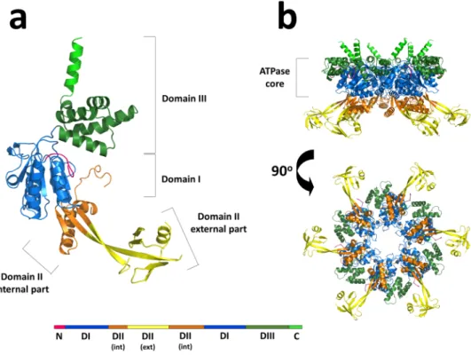
The crystal structure of the full-length human transcrip-tion regulatranscrip-tion factor RuvB-Like 2 (hsRuvBL2) in its apo form was determined to 2.89 Å resolutranscrip-tion (Table 1 and Fig. 1). The structure shows a hexameric arrangement of monomers similar to other structures in the RuvB-Like family18,20–22. The hexameric arrangement of hsRuvBL2 in solution was confirmed by small angle X-ray scattering(SAXS - see below) coupled to size exclusion chromatography, which unequivocally showed the formation of a complex with a mean radius of gyration of ca. 52 Å throughout the elution peak (Supplementary Fig. S1).
In the ring-shaped hsRuvBL2 hexamer, each protomer comprises the typical Rossman-like αβα fold (domain I) and the canonical all-α subdomain (domain III), which form the ATPase core together with the internal region of domain II. Moreover, there is an outward-facing mobile unit composing the external region of domain II (Fig. 1), linked to the ATPase core by two β-sheet linkers. This unit is absent in all previous crystal structures of human RuvBL2 and is observed here for the first time. The interfaces between protomers form a complete nucleotide binding pocket, which includes the canonical nucleotide-binding motifs in domains I and III: Walker A (G77-T84), Walker B (D299-H302), sensor 1 (M326-N329), sensor 2 (T397-A402) and a trans-arginine fin-ger (R353) from the adjacent protomer. Despite the fact that all purification steps were performed in the pres-ence of ADP, no electron density corresponding to this nucleotide (or segments thereof), could be observed in the pocket. Protruding from the C-termini of each protomer in the hexameric-arranged ATPase cores there are six solvent-reaching antennae-like α-helices. Domain II, located between the Walker A and Walker B motifs of domain I, bears no significant sequence similarity to other known domains, except for residues 136–233 which, similarly to RuvBL1 residues 131–227, organise into an oligonucleotide/oligosaccharide-binding (OB)-fold27.
Hence, domain II is unique to RuvB-Like proteins. In addition to the overall mobility of domain II, there are four additional loops that are probably too mobile to produce an electron density: D148 - G159, D185 - V187, S203 - C227 and R253 - Q255. The second and fourth segments are long and located on the external part of the domain II.
The hsRuvBL2 hexamer is 149 Å wide, measured using the Cα of residue Q188 as reference point (versus 147 Å in hsRuvBL1 measured at E212 and 120 Å in the RuvBL1:RuvBL2 dodecamer from Chaetomium thermophilum, which crystallized with a more compact conformation of domain II towards the ATPase core, measured between D343 of RuvBL1 and D338 of RuvBL2). The ATPase core is 51 Å high, similar to 50 Å in RuvBL118 and 51 Å in
the truncated dodecameric complex22. The hsRuvBL2 hexamer has a central channel 23 Å wide (similar to the
CtRuvBL1:RuvBL2 dimensions20,21, and slightly larger than the channel of hsRuvBL1, which is 20 Å wide) on its
narrowest part and although a double-stranded B-DNA molecule could be tightly fitted (not shown), it has been clearly demonstrated biochemically that RuvBL2 can only bind single-stranded DNA28. To the best of our
knowl-edge, similar studies have not been published concerning RuvBL1.
RuvBL1 and RuvBL2 are homologous proteins with 43% sequence identity and 65% sequence similarity that work together as part of chromatin remodeling complexes4,8,29–31. However, they also work antagonistically in
many situations. It has been suggested that the antagonistic activities of RuvBL1 and RuvBL2 may be the conse-quence of interactions with different, more or less specific partners32. Having access to the full-length structure of
hsRuvBL2, and to assess the structural basis for these interaction specificities, we analyzed the structures of both hsRuvBL1 and hsRuvBL2 for differing characteristics at the surface of both complexes. A comparison of the sur-face charge distribution of hsRuvBL1 and hsRuvBL2 hexamers (Fig. 2) shows hsRuvBL2 to have a markedly more
positive inner channel and ATPase core (with small negatively charged patches at the top), and a more negative distribution on the bottom surface of domain II. These charges are mostly inverted in hsRuvBL1, which suggests different mechanisms/affinities for binding DNA or other proteins. Furthermore, hsRuvBL1 and hsRuvBL2 have different oligomerization dynamics, since monomer to hexamer transition occurs at a much higher concentra-tion for hsRuvBL1 than for hsRuvBL2 (ref.22 and this work). The sum of these observations suggests considerable
differences in specificity.
Structural basis for coupling ATP binding to mechanical action in RuvBL2.
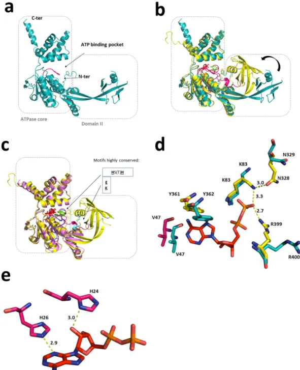
It is a sensible assump-tion that the basis of ATP-dependent activities in RuvB-Like proteins lies upon the mechanical consequences of ATP binding and hydrolysis. In order to elucidate what these consequences might be in the case of RuvBL2, we compared the structure of the full-length human apo RuvBL2 (this work, PDB ID 6H7X) with the ADP-bound variant of RuvBL2 from the fungus C. thermophilum21 (ctRuvBL2, PDB ID 4WW4). Both proteins share 68%identity and 85% sequence similarity, as well as a conserved disposition of the elements that constitute the ATP binding pocket (Fig. 3d). In ctRuvBL2, nucleotide binding leads to a tightening of the binding pocket near the phosphate tail. The β-phosphate of the ADP molecule interacts with neighbouring residues K83, N328 and R399 of ctRuvBL2 (which correspond to K83, N329 and R400 of hsRuvBL2). Concomitantly, nucleotide entrance into the binding pocket causes a displacement of V47 and Y361 (V47 and Y362 in the human protein) due to hydro-phobic interactions with the adenine ring, possibly driving a spatial rearrangement of residues in the vicinity of the nucleotide binding pocket that may be the origin of the downstream rearrangement of the N-terminal loop. We propose that these interactions initiate the coupling between nucleotide binding and mechanical movement of domain II.
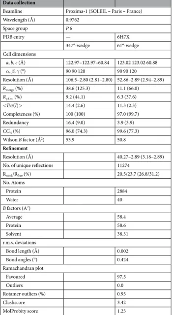
Data collection
Beamline Proxima-1 (SOLEIL – Paris – France)
Wavelength (Å) 0.9762 Space group P 6 PDB entry — 6H7X 347°-wedge 61°-wedge Cell dimensions a, b, c (Å) 122.97–122.97–60.84 123.02 123.02 60.88 α, β, γ (°) 90 90 120 90 90 120 Resolution (Å) 106.5–2.80 (2.81–2.80) 52.86–2.89 (2.94–2.89) Rmerge (%) 38.6 (125.3) 11.1 (66.0) Rp.i.m. (%) 9.2 (44.1) 6.3 (37.6) <I/σ(I)> 14.4 (2.6) 11.3 (2.3) Completeness (%) 100 (100) 97.0 (99.7) Redundancy 16.4 (9.0) 3.9 (3.9) CC½ (%) 96.0 (74.3) 99.6 (77.3) Wilson B factor (Å2) 53.9 50.8 Refinement Resolution (Å) 40.27–2.89 (3.18–2.89)
No. of unique reflections 11274
Rwork/Rfree (%) 20.5/23.7 (26.8/31.2) No. Atoms Protein 2884 Water 40 B factors (A2) Average 58.4 Protein 58.6 Solvent 38.31 r.m.s. deviations Bond length (Å) 0.002 Bond angles (°) 0.424 Ramachandran plot Favoured 97.5 Outliers 0.0 Rotamer outliers (%) 0.95 Clashscore 3.42 MolProbity score 1.23
www.nature.com/scientificreports/
Figure 1. Cartoon representations of the overall structure of hsRuvBL2 monomer and hexamer. (a) The full– length apo hsRuvBL2 monomer. Domains I, II and III are coloured blue, orange/yellow and green, respectively. The visible part of the N-terminal loop and the C-terminal helix are also identified in pink and light green, respectively. A linear schematic representation of the domains of hsRuvBL2, using the same colours, is shown below, highlighting the internal and external portions of domain II. (b) Side and bottom views of the hsRuvBL2 hexamer, highlighting the AAA+ core, using the same colour code.
Figure 2. Surface charge distribution in the hsRuvBL1 and hsRuvBL2 hexamers. Differences in the distribution of amino acid residues at the surface of the rings may underlie their antagonistic activities. Colour by
While the apo hsRuvBL2 structure has no visible electron density up to residue 47 (N-terminus segment shown in dark pink in Fig. 3a), ADP-bound ctRuvBL2 (yellow in Fig. 3b) has electron density for most of the N-terminal loop, supporting a connection between ADP binding and N-terminus mobility, as previously sug-gested20. Furthermore, in ADP-bound ctRuvBL2, domain II is more tightly packed against the ATPase core, a
conformation in which this domain interacts closely with the proximal part of the N-terminus. The apo hsRu-vBL2 structure was further compared with the ATP-bound form in the hsRuvBL1ΔDII:RuhsRu-vBL2ΔDII complex22
(Fig. 3c, light pink; noteworthy, this protein was crystallized with the domain II truncated, PDB ID 2XSZ). This alignment shows that in the absence of an interaction between the N-terminus proximal segment and the exter-nal region of domain II, the N-termiexter-nal loop is stabilized only from the point where it interacts with the bound nucleotide. This nucleotide interaction occurs through the histidine residues of a conserved HSH motif (H25 and H27 in the wild type hsRuvBL2, H40 and H42 in the domain II-truncated hsRuvBL2 and H24 and H26 in ctRuvBL2), while the remaining proximal part remains unstructured. The same is observed in the structure of the human DII-truncated complex associated with the RBD domain of RPAP3 (PDB ID 6FO1)33. These structure
comparisons strongly support a connection between nucleotide binding, N-terminus rearrangement and domain II conformational stabilization in closer proximity to the ATPase core.
These observations support the proposal of the following sequential mechanistic events: in ATP-dependent activities, the nucleotide first binds to the nucleotide-binding pocket, eliciting a rearrangement of the surround-ing residues, which causes the most distal part of the N-terminus to move. Video 1 (Supplementary Information) highlights the predicted movements that would occur in a RuvBL2 monomer, during its transition from the apo to the nucleotide-bound form. Note the movement of the N-terminal loop as it is tethered by interactions with the nucleotide molecule through H24 and H26 in ctRuvBL2. This initial interaction then draws the remaining part of the N-terminus into the close vicinity of the external region of domain II. Here, the conserved N-terminus residues E11, K13 and E14, and domain II residues K183 and I201 (E12, R14, D15 and K184 in hsRuvBL2) seem to be responsible for the electrostatic interactions that sustain the positioning of domain II in close proximity to the ATPase core, as seen in ADP-bound ctRuvBL2 (yellow in Fig. 3). Thus, our apo hsRuvBL2 structure provides a missing link that supports the mechanism proposal whereby the N-terminal loop provides an interface between the external region of domain II and the ATPase core. Key residues involved in the proposed mechanism are not only conserved between the human and fungal forms (Supplementary Fig. S2), but throughout the RuvBL2 family.
On the other hand, nucleotide binding to RuvBL1 does not elicit the same structural changes as for RuvBL2. In fact, despite the initial interaction of the two conserved histidines (H18 and H20 in the human RuvBL1, PDB ID 2C9O) with the nucleotide, both in human and in C. thermophilum RuvBL1 the N-terminal loop is directed towards interactions with domain I of the same protomer: K11 interacts with P95 from hsRuvBL1 and R15 inter-acts with P96 from ctRuvBL1.
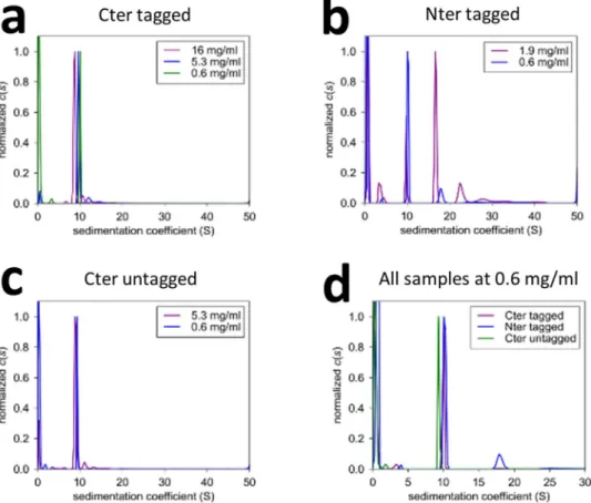
hsRuvBL2 oligomer state and stability can be modulated.
To address whether the location of the affinity tags would affect the oligomeric assembly and stability of human RuvBL2, we expressed recombinant hsRuvBL2 in E. coli, with affinity tags either at the N- or C-terminus. Purifications were performed in pres-ence of ADP, which initial thermal shift assays (TSA) showed to provide the highest degree of thermal stability (as compared to ATP, AMP-PNP and no nucleotide). We analyzed the obtained oligomeric forms by analyti-cal ultracentrifugation (AUC), and analyzed their stability by TSA. Tags placed at the N-terminus destabilized hsRuvBL2 the most, as confirmed by TSA (SM Fig. S3a), both in the native and SeMet forms, and also led to the formation of multiple oligomeric forms (SM Fig. S3b and c). Furthermore, analytical ultracentrifugation showed a concentration-dependent oligomer formation of the N-terminally tagged hsRuvBL2, where higher oligomeric forms and monomers appeared as concentration increased (Fig. 4b). On the other hand, C-terminally tagged hsRuvBL2 consistently formed hexamers at all tested concentrations (Fig. 4a). The effect of the N-terminal tag in dodecamer formation is not clear, but it may be relayed through an influence in domain II, since it has been shown that this domain is important in dodecamer stabilization in the RuvBL1:RuvBL2 complex22,34.To complement the oligomer analysis, a preliminary negative staining EM analysis was performed on the untagged hsRuvBL2 form. Interestingly, about 10% of particles picked from micrographs were shown to be heptamers (Fig. 5), while the remaining were hexameric rings, as expected from observations in solution. This low percentage of heptamers would have gone unnoticed by the other techniques used, since the size difference to hexamers is within their respective error margins. As far as we know, this is the first time heptamers have been observed in the RuvB-Like family. However, this oligomeric form has been observed for some AAA+ ATPases,
such as the NtrC1 of Aquifex aeolicus35.
hsRuvBL2 binding to DNA.
hsRuvBL2 binding to DNA is independent of nucleotide sequence, and restricted to single-stranded DNA22,28. Papin and colleagues have also clearly shown that only monomericRuvBL2 can bind the polynucleotide chain, which suggests a mechanism of action whereby RuvBL2 oligomerizes around a single chain overhang, and only then starts to perform the upstream unwinding of the remaining double helix, in an ATP-dependent process28. In order to assess what structural changes may occur upon DNA binding,
we analyzed this process by negative staining EM. We first observed the dissociation of hsRuvBL2 hexamers into monomers after dilution to a sufficiently low concentration (ca. 100 µg/mL). We subsequently incubated the monomeric hsRuvBL2 with single-stranded DNA, and observed that the presence of ssDNA promoted the re-formation of the previously disassembled rings (Fig. 6a). The low protein concentrations used limits the num-ber of techniques capable of detecting quaternary structure changes taking place upon interaction of hsRuvBL2 with DNA molecules; however, although the circular M13mp18 DNA strand used in these studies could not be observed at the resolution obtained by negative-staining EM, it can be assumed that, if the rings assembled around the DNA chain, the latter would remain associated with the protein complex. Alternatively, DNA binding
www.nature.com/scientificreports/
Figure 3. Comparison between existing structures of RuvBL2 suggests a connection between nucleotide binding and domain II motion. (a) The apo hsRuvBL2 (blue, this work, PDB ID 6H7X) is associated with a stretched, or at least loose conformation of the domain II. (b) The apo RuvBL2 is superimposed with the ADP-bound RuvBL2 from Chaetomium thermophilum (yellow, PDB ID 4WW4). When compared to the apo hsRuvBL2, the ADP-bound ctRuvBL2 displays a positioning of domain II closer to the ATPase core and a more ordered N-terminal loop (pink). ADP is depicted in red sticks. (c) The ATP-bound, domain II-truncated hsRuvBL2 (light pink, PDB ID 2XSZ) is superimposed with the full-length ctRuvBL2 (4WW4). It becomes apparent that the absence of the external part of domain II has an influence in the organization of the N-terminal loop. In both nucleotide-bound forms, the N-terminal loop interacts with the nucleotide through two conserved histidines; however, in the domain II-truncated hsRuvBL2, the N-terminal loop (blue) remains disordered from the first residue up to the place of interaction with the nucleotide. The histidines that interact with the nucleotide are depicted in green and the motif is highlighted; residues involved in electrostatic interactions between the N-terminal loop and domain II are depicted in light blue and highlighted as well. (d) Superimposition of the nucleotide binding pocket of ctRuvBL2 (yellow, 4WW4) with the human RuvBL2 homologous residues (blue, 6H7X – this structure). The ADP molecule (depicted in red) is from the fungal structure. Atoms are coloured as follows: N – dark blue; O – salmon; P – dark yellow. V47 from C. thermophilum is depicted in pink since it is part of the N-terminal loop. (e) Position of conserved histidines 24 and 26 in relation to ADP in the binding pocket of CtRuvBL2. The interatomic distances shown are in Å.
may occur through the external region of domain II, as suggested for RuvBL118, since both proteins display an
OB-fold within this region. However, if binding occurred only through domain II, it would probably have also been observed for the hexameric forms, since this part of the protein is solvent-accessible. However, this binding mode was never observed, neither in this work, nor in previous biochemical studies performed with the human and yeast RuvBL228. Furthermore, helicase activity – for which prior binding to the DNA strand is required - was
shown in complexes where the external region of domain II was deleted22. To further address this question, we
performed electrophoretic mobility shift assays (EMSA) using the same circular M13mp18 DNA (Fig. 6b). We confirmed the lack of binding of hexameric hsRuvBL2 to M13mp18 (lanes 3 and 4), since the observed migra-tion length for ssDNA alone and for ssDNA in presence of hexameric hsRuvBL2 was the same in both lanes. Interestingly, the hexamer was able to indirectly bind DNA, in an interaction apparently mediated by monomeric hsRuvBL1 (lane 2) and ATP-dependent (lanes 5 and 6).
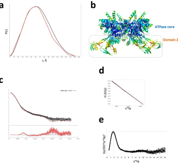
RuvBL2 displays flexibility of domain II.
Molecular dynamics studies previously showed a propensity for the external region of domain II of RuvBL hexamers to acquire a variety of conformations in solution19 whichmay be the reason behind the recalcitrant behaviour towards crystallization. In order to gain further insight into the overall shape and flexibility of RuvBL2 hexamers, we compared the pair distribution function, P(r) profile of hsRuvBL2 in solution (from an experimental SAXS data collection - Fig. 7a, black) with a theoretical plot calculated from the crystallographic coordinates (shown in red). The superimposed plots show that, in solution, hsRuvBL2 dmax is 150 Å, which is similar to that observed in the crystallographic structure of 157 Å. Comparison between the two profiles also shows an overall similar shape with the wider profile for the solution structure in the P(r) suggesting that both are globular with the protruding arms of domain II in different configurations in solution. Since dmax is similar, these results support a change in conformation of the protruding domains, in the
hexamer, as proposed by Petukhov and colleagues19, lending an appearance of homogeneity of structure to the
plot obtained experimentally. This mobility is further supported by the fact that the atomic B-factors are high-est for the proposed β-sheet linker “hinge” residues, which form the bending point between domain II and the ATPase core (Fig. 7b).
Figure 4. Analytical ultracentrifugation analysis of hsRuvBL2 samples. The oligomeric plasticity of hsRuvBL2 varies with the position of the tag: when placed on the C-terminus, hsRuvBL2 forms hexamers in all concentrations tested, even after tag cleavage (a) and (c). However, when expressed with the tag on the N-terminus, hsRuvBL2 oligomeric state varies with concentration (b), forming monomers, hexamers, dodecamers and high molecular weight species.
www.nature.com/scientificreports/
Discussion
We have determined the full-length crystallographic structure of human transcription factor RuvB-Like 2. The collected results suggest a putative mechanism whereby binding of a nucleotide to the binding pocket promotes the stabilization of the external part of domain II against the ATPase core, by eliciting conformational changes of the N-terminal loop. It is in fact possible that the functions of the external region of domain II may be in great part related to its interaction (mediated by the N-terminal loop) with the nucleotide in the binding pocket, with a concomitant motion with as yet undefined consequences. One of these could be the observed disassembly of dodecameric RuvBL1/2 into hexamers upon incubation with ATP29. It is also worth noting that the OB-fold
comprises distinct superfamilies, and while the most common relates to nucleic acid-binding, they can also occur in larger proteins as recognition domains, and have even been found at protein-protein interfaces27. While an
interaction of the OB-fold in domain II with DNA was observed for RuvBL118, it is possible that the OB-fold
in RuvBL2 may also be important for its interaction with other proteins in a supramolecular assembly, such as within the Ino80 complex, where cross-linking/mass spectrometry analysis shows a strong interaction profile between the OB-folds of RuvBLs and Ies25. Interestingly, EM micrographs showed hsRuvBL2 hexamers to be
frequently found in direct lateral contact, forming continuous strings (data not shown), which is also suggestive of a tendency of domain II to act as a mediator in protein-protein interactions.
Recombinant hsRuvBL2 has been used as a tool for in vivo assays, enabling the identification of binding part-ners such as c-myc36. However, affinity tags placed on the N-terminus of RuvBL1 and RuvBL2 were shown to
induce the formation of dodecamers, for example, in the yeast Rvb1:Rvb2 complex37. The influence of tags was
thus suggested to reflect the multiple conformational changes that these proteins can undergo while performing their functions, changes that in vivo are induced by binding partners37. Here we also show that the use of affinity
tags in hsRuvBL2 expression interferes with oligomeric flexibility. Our results also seem to suggest that affinity tags may interfere negatively with hsRuvBL2 activity by decreasing its stability.
An analysis of the surface electrostatic charge distribution in hsRuvBL1 and hsRuvBL2 (Fig. 2) allows an over-all view of distinguishing features between the two homologs that may illustrate the basis for some antagonistic roles. The smaller size of the inner channel of hsRuvBL1 as compared to hsRuvBL2 and the marked differences in the surface electrostatic charge distribution suggest the possibility of a different DNA-binding mechanism between hsRuvBL1 and hsRuvBL2. Since their activities were observed to increase when working together as a heteromeric complex22, it is also possible that their individual characteristics complement each other, with a
consequent increase in DNA processing efficiency. Interestingly, the inner channel of the RuvBL1/2 complex, being composed of alternate positive and negative charges, may also have the function of binding peptide tails; such an interaction occurs between the ankyrin repeat domain of synphilin, which is the domain responsible for its aggregation, and K372 of RuvBL1, which is located on the surface of the ATPase core and in close proximity to the central channel38. The differences observed in charge distribution may also reflect distinguishing motifs that
justify antagonistic activities by different interactions with protein binding partners, even when in the form of a heteromeric complex. Such an example is the preferential binding of RPAP3 C-terminal fragment to the ATPase core of hsRuvBL2 of a RuvBL1/2 complex33.
The biological significance of the hsRuvBL2 heptamers, if any, is still to be determined. However, it is tempting to speculate that the capacity to form heptamers could bring extra flexibility in the association to other proteins, and possibly permit an association with dsDNA, since the central channel seems to be wider.
RuvBLs have been identified as taking part in many supramolecular assemblies, where their ATPase activity is not always required. Such versatility has long posed a mystery, since no specific function can be attributed to these proteins, and yet they appear to be critical for the regulation of an array of complexes, particularly related to the control of gene expression and DNA damage response. It is possible that one of the functions of RuvBLs Figure 5. Negative staining EM analysis shows that hsRuvBL2 is able to form heptamers. 10% of the total of picked particles corresponded to this oligomeric form, highlighted in red.
within larger complexes could be to recognize transient client proteins, allowing the recognition of the needs of the cell and translating them to the rest of the complex, thus regulating its activity. The fact that only heteromeric RuvBL1/2 complexes in the cell have been experimentally demonstrated to date adds another layer of complexity to their roles in transcription regulation.
In cancer patients, RuvBL2 overexpression is considered a marker of poor prognosis25. Previous works have
shown that, in hypoxic conditions, RuvBL2 is methylated at K67 (located in domain I, in blue in Fig. 1a), which leads to downregulation of pro-apoptotic BNIP3 and pro-angiogenic phosphoglycerate kinase 112. Combining
these observations, it is conceivable that, when a tumor reaches hypoxic state, overexpression of RuvBL2, and its consequent methylation en masse, may contribute to a large-scale downregulation of the target genes. The consequence would be the already described increased resistance of hypoxic tumors to chemo and radiotherapy treatments39. This detailed structure of hsRuvBL2 can thus contribute to the development of compounds to either
target the surface of RuvBL2-K67Me or aimed at the disruption of the HIF-1α:RuvBL2-K67Me complex. Further studies on distinct post-translational modifications to surface residues of RuvBLs may provide a framework for the development of compounds that can regulate the specific activities of RuvBL1 and RuvBL2, depending on the tissue, cell stage or stress conditions of the target cells. By targeting specific modifications of RuvBLs that result from particular metabolic cellular states, it might be possible to acquire the degree of specificity desired when aiming for the targeting of a specific cell subset.
Figure 6. hsRuvBL2 interaction with DNA. (a) Interaction of monomeric hsRuvBL2 with ssDNA promotes ring reassembly, as observed by negative-staining EM. The black arrow highlights an hsRuvBL2 ring. An electrophoretic mobility shift assay (b) shows that hexameric hsRuvBL2 did not bind to ssDNA (lane 4), as there is no shift in running distance when comparing to the one observed by DNA alone (lane 3). However, when co-incubated in equimolar amounts with hsRuvBL1 (lane 2), there is a shift that indicates protein binding. Since this shift is equivalent to the one observed when incubating a total molar amount of only hsRuvBL1 (lane 1), we surmise that hsRuvBL2 is also bound to DNA in the former, most likely mediated by hsRuvBL1. Lanes 5 and 6 show no such binding in the presence of AMP-PNP, indicating that this non-hydrolysable nucleotide somehow prevents DNA binding.
www.nature.com/scientificreports/
Materials and Methods
Protein expression and purification.
The codon-optimized sequence of hsRuvBL2 with a C-terminus-His6 tag including a 3C protease cleavage site was obtained from Genscript (USA), as the vector pET49b_ruvbl2_Cter_His, which was transformed into E. coli B834. Gene expression was induced at an OD600 of 0.8 by the
addition of 100 µM IPTG, at 30 °C for 19 h. The cells were collected by centrifugation at 11000 × g for 30 minutes and disrupted in lysis buffer (50 mM phosphate buffer pH 7, 500 mM NaCl, 50 mM imidazole, 100 µM ADP, 1 mM MgCl2) supplemented with EDTA-free protease inhibitor tablet (Roche) and benzonase (Novagen).
hsRu-vBL2 was obtained through several purification steps: (i) a Nickel affinity column (HisTrap HP, GE Healthcare), by elution with a gradient of buffer containing 1 M imidazole; (ii) an anion exchange column (Resource Q, GE Healthcare), by elution with a gradient of buffer with 1 M NaCl; (iii) a size exclusion column (HiLoad 16/600 Superdex 200 pg, GE Healthcare), previously equilibrated with 20 mM Na/K phosphate pH 8, 200 mM NaCl, 3 mM MgCl2 and 500 µM ADP. hsRuvBL2 eluted as a hexamer at a concentration of 10 mg/mL.
Differential scanning fluorimetry (DSF).
The protein melting temperature (Tm) determination wasper-formed by monitoring protein unfolding with the fluoroprobe SYPRO Orange dye, in an iCycle iQ5 Real Time PCR Detection System (Bio-Rad) equipped with a charge-coupled device (CCD) camera and a Cy3 filter with excitation and emission wavelengths of 490 and 575 nm, respectively. The plates were heated from 20 to 90 °C with stepwise increments of 0.5 °C with a 60-second equilibration time, followed by the fluorescence read out. To test the effect of nucleotides, the protein was incubated with nucleotides with a molar excess higher than 10-fold (4 mM of nucleotide to ca. 300 µM hsRuvBL2). All assays were performed with pure protein (N = 3), from the peak corresponding to a hexameric oligomer, collected from the last size exclusion purification step.
Figure 7. Domain flexibility of hsRuvBL2. (a) Pair distribution function, P(r) curve for experimental data (black) giving a dmax 150 Å overlaid with the P(r) calculated from the crystallographic coordinates (red) giving a dmax 157 Å. (b) Side view of the hsRuvBL2 hexamer. Atoms are coloured according to B-factor: blue – lowest; red - highest. (c) Small angle scattering Intensity versus scattering vector plot obtained experimentally (black), superimposed with a theoretical plot calculated from the crystallographic coordinates (red). (d) Guinier fit showing straight line over q-range. (e) A dimensionless Kratky plot showing the protein is globular and well folded.
Analytical ultracentrifugation (AUC).
AUC analysis of sedimentation coefficients of hsRuvBL2 was per-formed to determine differences in the oligomerization state of the different hsRuvBL2 constructs in ADP-containing buffer. Sedimentation velocity (SV) was used to determine the proportion of different oligomeric forms for each sample, as well as their approximate molecular weight, and to assess whether the hsRuvBL2 com-plex undergoes concentration-dependent dissociation. SV experiments were performed at 35000 revs per min and 20 °C, in a XL-I analytical ultracentrifuge using an Anti-50 rotor (Beckman Coulter, USA), with 3 and 12 mm path length Ti double-sector centrepieces equipped with sapphire windows (Nanolytics GmbH, Germany), loaded with 110 and 420 µL, respectively, depending on protein concentration, sample and reference solvent. Acquisitions were made using interference optics. The reference channels were filled with the buffer without ADP. Data analyses were done with the Sedfit software40, v14.1, using the program SEDNTERP (http://sednterp.unh.edu/), considering buffer density, ρ = 1.025 g mL−1 and viscosity, η = 1.06 cP. For homogeneous samples, the
non-interacting species analysis provides independent estimates of sedimentation coefficient s and of the diffu-sion coefficient D, which was used in the Svedberg equation s0/D = M (1 − ρv)/RT, to provide an experimental
value for the molar mass, M.
Small angle X-ray scattering (SAXS).
hsRuvBL2 was measured at beamline B21, Diamond Light Source (Didcot, UK; Experiment number SM13193-1), using both static measurement through the sample changer (Maatel, Grenoble) and through in-line size-exclusion chromatography column (SEC-SAXS). Analysis in static mode was performed using Scatter (Bioisis.net) by merging datasets from serial dilutions of a hsRuvBL2 fraction from the centre of the S200 peak, from 0.6 to 4.5 mg/mL. SEC-SAXS analysis was performed by injection of a fraction at 5.5 mg/mL from the middle of the S200 peak, onto a Shodex 403 kw column (Supplementary Fig. S6). Measurements were taken each second across the peak elution. Frames that were shown to have the same Rg were selected and scale-merged using Scatter. Further analysis used Scatter and FoXs41. All samples were filteredthrough a 1 MDa filter prior to analysis to eliminate larger aggregates.
Electrophoretic mobility shift assays (EMSA).
Electrophoretic mobility shift assays were performed to assess hsRuvBLs ability to bind ssDNA in native conditions. Prior to the agarose gel separation, the protein was incubated with M13mp18 in a reaction mixture with a total volume of 20 µL, of which 0.5 µL (1.12 nM) DNA, var-iable amounts of protein (50 or 25 µM) and completed with reaction buffer (25 mM HEPES/KOH pH 8, 2.5 mM Mg(CH3COO)2, 100 mM KCl, 0.2 mM DTT, 4 mM ATP and 2 mM MgCl2). Negative controls were performedby incubating either only DNA or only protein in the reaction buffer. The reaction occurred for 1 h at 23 °C, after which 1 µL of 50% (V/V) glycerol was added and the reaction loaded in a 0.6% (w/V) agarose gel in TBE 1x and run for 2 h30 m at 80 V. The DNA bands were stained with SYBR Gold DNA stain. Fluorescence was detected with a Fuji TLA-5100, using a 473 nm excitation wavelength.
Analysis of hsRuvBL2 binding to DNA by negative staining EM.
Immediately prior to EM data collection, monomeric hsRuvBL2 was incubated with M13mp18 circular ssDNA. A control incubation was pre-pared without DNA. After 30 minutes of incubation at room temperature (about 20 °C) in buffer with ATP, both samples (with and without DNA) were adhered to Rhodium/Copper EM grids (pre-treated with a carbon coating and rendered hydrophilic through an electrical glow discharge), washed with Tris-based buffer, and stained with uranyl acetate. Micrographs were obtained using a Tecnai F20 or a JEOL JEM-1010 TEM, with exposure time per picture of 0.2–0.9 s and a dose of 20–25 e−/Å2 s, and collected using a CCD camera.Crystallization and data collection.
hsRuvBL2 was dialysed to pH 6 immediately prior to crystalliza-tion. Crystallization drops were set up at 30 °C using the vapour-diffusion sitting-drop method in 24-well Linbro plates, with a proportion of 0.7 µL of protein to 0.3 µL of a reservoir solution composed of 250 mM MgCl2 and 2%PEG 3350. Protein was at 10 mg/mL and the reservoir volume was 500 µL. Crystals appeared within 1 day, grew to a maximum size of about 200 µm × 40 µm (hexagonal needles) and were cryocooled in mother liquor supple-mented with 25% glycerol. Diffraction data from a crystal diffracting to 2.89 Å was collected at Proxima-I beam-line, at the Soleil synchrotron source (St. Aubin, Paris, France), using a CCD detector (ADSC QUANTUM 315r).
Structure solution and refinement.
Data were indexed and integrated with XDS42, and the space groupwas determined with POINTLESS43 and scaled with AIMLESS44, all within the autoPROC data processing
pipe-line. Four datasets were collected from the same crystal, in a total of 347 images 1° wide (347° “wedge”). The four datasets were processed individually and scaled together, yielding a data set with high multiplicity in space group P6. Data collection and processing statistics are listed in Table 1. Matthews coefficient calculations45
indi-cated the presence of one molecule per asymmetric unit. The 3D structure of hsRuvBL2 was solved by molecular replacement using PHASER46, with hsRuvBL1 domains I and III (cropped from the PDB ID 2C9O) as the search
model. The total amount of collected images was used to obtain an initial electron density map, making use of the high multiplicity of data, which helped to highlight electron density fragments not clearly visible upon phasing. Electron density was further improved with with BUSTER-TNT47 making use of the “-L” flag within the program.
Iterative cycles of automated model building with BUCCANEER48,49, within the CCP4 suite of programs50, and
refinement with BUSTER-TNT, were performed until electron density was clearly visible for domain II. Iterative cycles of manual model building and refinement were subsequently performed with COOT51 and BUSTER-TNT.
The program HYDROGENATE (distributed with BUSTER-TNT which uses the program REDUCE52 from the
MolProbity suite), was used to add hydrogen atoms (with zero occupancy), to the crystallographic model during refinement. The final refinement round was performed with phenix.refine within the PHENIX53 suite of programs
www.nature.com/scientificreports/
was performed simultaneously with model building and refinement using MolProbity54 as implemented in
BUSTER-TNT and PHENIX.
Electrostatic surface calculations.
The software CHARMM55 was used to calculate the surface chargesdistribution in hsRuvBL1 and hsRuvBL2. Topological visualization of the electrostatic potential was produced in PyMol56 with APBS57.
Data Availability
Coordinates of the refined PDB and structure factors were deposited in the RCSB Protein Data Bank (www.rcsb.org)58
with the PDB ID entry 6H7X. All other data are available from the corresponding author on reasonable request.
References
1. Jha, S. & Dutta, A. RVB1/RVB2: running rings around molecular biology. Mol Cell 34, 521–533 (2009).
2. Venteicher, A. S., Meng, Z., Mason, P. J., Veenstra, T. D. & Artandi, S. E. Identification of ATPases pontin and reptin as telomerase components essential for holoenzyme assembly. Cell 132, 945–57 (2008).
3. Grigoletto, A., Lestienne, P. & Rosenbaum, J. The multifaceted proteins Reptin and Pontin as major players in cancer. Biochim
Biophys Acta 1815, 147–157 (2011).
4. Jha, S., Gupta, A., Dar, A. & Dutta, A. RVBs are required for assembling a functional TIP60 complex. Mol. Cell. Biol. 33, 1164–74 (2013).
5. Tosi, A. et al. Structure and subunit topology of the INO80 chromatin remodeler and its nucleosome complex. Cell 154, 1207–19 (2013).
6. Nguyen, V. Q. et al. Molecular architecture of the ATP-dependent chromatin-remodeling complex SWR1. Cell 154, 1220–31 (2013). 7. Kakihara, Y. & Houry, W. A. The R2TP complex: discovery and functions. Biochim. Biophys. Acta 1823, 101–7 (2012).
8. Nano, N. & Houry, W. A. Chaperone-like activity of the AAA+ proteins Rvb1 and Rvb2 in the assembly of various complexes. Philos
Trans R Soc L. B Biol Sci 368, 20110399 (2013).
9. Machado-Pinilla, R., Liger, D., Leulliot, N. & Meier, U. T. Mechanism of the AAA+ ATPases pontin and reptin in the biogenesis of H/ACA RNPs. Rna 18, 1833–1845 (2012).
10. Ikura, T. et al. Involvement of the TIP60 histone acetylase complex in DNA repair and apoptosis. Cell 102, 463–473 (2000). 11. Rottbauer, W. et al. Reptin and Pontin Antagonistically Regulate Heart Growth in Zebrafish Embryos. Cell 111, 661–672 (2002). 12. Lee, J. S. et al. Negative regulation of hypoxic responses via induced Reptin methylation. Mol. Cell 39, 71–85 (2010).
13. Lee, J. S. et al. Hypoxia-induced methylation of a pontin chromatin remodeling factor. Proc. Natl. Acad. Sci. USA 108, 13510–13515 (2011).
14. Clarke, T. L. et al. PRMT5-Dependent Methylation of the TIP60 Coactivator RUVBL1 Is a Key Regulator of Homologous Recombination. Mol. Cell 65, 900–916.e7 (2017).
15. Kim, J. H. et al. Roles of sumoylation of a reptin chromatin-remodelling complex in cancer metastasis. Nat. Cell Biol. 8, 631–639 (2006).
16. Arnold, C. N. et al. A forward genetic screen reveals roles for Nfkbid, Zeb1, and Ruvbl2 in humoral immunity. Proc. Natl. Acad. Sci.
USA 109, 12286–12293 (2012).
17. Boo, K. et al. Pontin functions as an essential coactivator for Oct4-dependent lincRNA expression in mouse embryonic stem cells.
Nat. Commun. 6, 6810 (2015).
18. Matias, P. M., Gorynia, S., Donner, P. & Carrondo, M. A. Crystal structure of the human AAA+ protein RuvBL1. J Biol Chem 281, 38918–38929 (2006).
19. Petukhov, M. et al. Large-scale conformational flexibility determines the properties of AAA+ TIP49 ATPases. Structure 20, 1321–1331 (2012).
20. Silva-Martin, N. et al. The Combination of X-Ray Crystallography and Cryo-Electron Microscopy Provides Insight into the Overall Architecture of the Dodecameric Rvb1/Rvb2 Complex. PLoS One 11, e0146457 (2016).
21. Lakomek, K., Stoehr, G., Tosi, A., Schmailzl, M. & Hopfner, K. P. Structural basis for dodecameric assembly states and conformational plasticity of the full-length AAA+ ATPases Rvb1. Rvb2. Structure 23, 483–495 (2015).
22. Gorynia, S. et al. Structural and functional insights into a dodecameric molecular machine - The RuvBL1/RuvBL2 complex. J. Struct.
Biol. 176, 279–291 (2011).
23. Matias, P. M. et al. The AAA+ proteins Pontin and Reptin enter adult age: from understanding their basic biology to the identification of selective inhibitors. Front Mol Biosci 2, 17 (2015).
24. Rosenbaum, J. et al. The Emergence of the Conserved AAA+ ATPases Pontin and Reptin on the Signaling Landscape. Sci. Signal. 6, mr1–mr1 (2013).
25. Rousseau, B. et al. Overexpression and role of the ATPase and putative DNA helicase RuvB-like 2 in human hepatocellular carcinoma. Hepatology 46, 1108–18 (2007).
26. Xie, C., Wang, W., Yang, F., Wu, M. & Mei, Y. RUVBL2 is a novel repressor of ARF transcription. FEBS Lett. 586, 435–41 (2012). 27. Theobald, D. L., Mitton-Fry, R. M. & Wuttke, D. S. Nucleic acid recognition by OB-fold proteins. Annu. Rev. Biophys. Biomol. Struct.
32, 115–133 (2003).
28. Papin, C. et al. 3′- to 5′ DNA unwinding by TIP49b proteins. Febs J 277, 2705–2714 (2010).
29. Zhou, C. Y. et al. Regulation of Rvb1/Rvb2 by a Domain within the INO80 Chromatin Remodeling Complex Implicates the Yeast Rvbs as Protein Assembly Chaperones. Cell Rep. 19, 2033–2044 (2017).
30. Gerhold, C. B., Hauer, M. H. & Gasser, S. M. INO80-C and SWR-C: Guardians of the genome. J. Mol. Biol. 427, 637–651 (2015). 31. Jónsson, Z. O. et al. Rvb1p and Rvb2p are Essential Components of a Chromatin Remodeling Complex that Regulates Transcription
of over 5% of Yeast Genes. J. Biol. Chem. 276, 16279–16288 (2001).
32. Diop, S. B. et al. Reptin and Pontin function antagonistically with PcG and TrxG complexes to mediate Hox gene control. EMBO
Rep. 9, 260–266 (2008).
33. Martino, F. et al. RPAP3 provides a flexible scaffold for coupling HSP90 to the human R2TP co-chaperone complex. Nat. Commun. 9, 1501 (2018).
34. Niewiarowski, A. et al. Oligomeric assembly and interactions within the human RuvB-like RuvBL1 and RuvBL2 complexes.
Biochem. J. 429, 113–125 (2010).
35. Chen, B. et al. Engagement of Arginine Finger to ATP Triggers Large Conformational Changes in NtrC1 AAA+ ATPase For Remodeling Bacterial RNA Polymerase. Structure 18, 1420–1430 (2011).
36. Wood, M. A., McMahon, S. B. & Cole, M. D. An ATPase/Helicase Complex Is an Essential Cofactor for Oncogenic Transformation by c-Myc. Mol. Cell 5, 321–330 (2000).
37. Cheung, K. L. Y., Huen, J., Kakihara, Y., Houry, W. A. & Ortega, J. Alternative oligomeric states of the yeast Rvb1/Rvb2 complex induced by histidine tags. J. Mol. Biol. 404, 478–92 (2010).
38. Zaarur, N. et al. RuvbL1 and RuvbL2 enhance aggresome formation and disaggregate amyloid fibrils. EMBO J. 34, 2363–2382 (2015).
39. Brown, J. M. In Methods in enzymology 435, 297–321 (2007).
40. Brown, P. H. & Schuck, P. A new adaptive grid-size algorithm for the simulation of sedimentation velocity profiles in analytical ultracentrifugation. Comput. Phys. Commun. 178, 105–120 (2008).
41. Schneidman-Duhovny, D., Hammel, M., Tainer, J. A. & Sali, A. Accurate SAXS profile computation and its assessment by contrast variation experiments. Biophys. J. 105, 962–974 (2013).
42. Kabsch, W. X. Acta Crystallogr. Sect. D Biol. Crystallogr. 66, 125–132 (2010).
43. Evans, P. An introduction to data reduction: space-group determination, scaling and intensity statistics. Acta Crystallogr. Sect. D 67, 282–292 (2010).
44. Evans, P. R. & Murshudov, G. N. How good are my data and what is the resolution? Acta Crystallogr. Sect. D Biol. Crystallogr. 69, 1204–1214 (2013).
45. Kantardjieff, K. A. & Rupp, B. Matthews coefficient probabilities: Improved estimates for unit cell contents of proteins, DNA, and protein-nucleic acid complex crystals. Protein Sci 12, 1865–1871 (2003).
46. McCoy, A. J. et al. Phaser crystallographic software. J. Appl. Crystallogr. 40, 658–674 (2007).
47. Blanc, E. et al. Refinement of severely incomplete structures with maximum likelihood in BUSTER-TNT. Acta Crystallogr. Sect. D
Biol. Crystallogr. 60, 2210–2221 (2004).
48. Cowtan, K. Fast Fourier feature recognition. Acta Crystallogr. - Sect. D Biol. Crystallogr. 57, 1435–1444 (2001).
49. Cowtan, K. General quadratic functions in real and reciprocal space and their application to likelihood phasing. Acta Crystallogr.
Sect. D Biol. Crystallogr. 56, 1612–1621 (2000).
50. Winn, M. D. et al. Overview of the CCP4 suite and current developments. Acta Crystallogr. Sect. D Biol. Crystallogr. 67, 235–242 (2011).
51. Emsley, P. & Cowtan, K. Coot: Model-building tools for molecular graphics. Acta Crystallogr. Sect. D Biol. Crystallogr. 60, 2126–2132 (2004).
52. Word, J. M., Lovell, S. C., Richardson, J. S. & Richardson, D. C. Asparagine and glutamine: Using hydrogen atom contacts in the choice of side-chain amide orientation. J. Mol. Biol. 285, 1735–1747 (1999).
53. Adams, P. D. et al. PHENIX: A comprehensive Python-based system for macromolecular structure solution. Acta Crystallogr. Sect.
D Biol. Crystallogr. 66, 213–221 (2010).
54. Chen, V. B. et al. MolProbity: All-atom structure validation for macromolecular crystallography. Acta Crystallogr. Sect. D Biol.
Crystallogr. 66, 12–21 (2010).
55. MacKerell, A. D. et al. All-Atom Empirical Potential for Molecular Modeling and Dynamics Studies of Proteins †. J. Phys. Chem. B
102, 3586–3616 (1998).
56. Schrödinger, LLC. The {PyMOL} Molecular Graphics System, Version~1.8. (2015).
57. Dolinsky, T. J., Nielsen, J. E., McCammon, J. A. & Baker, N. A. PDB2PQR: An automated pipeline for the setup of Poisson-Boltzmann electrostatics calculations. Nucleic Acids Res. 32, 665–667 (2004).
58. Rose, P. W. et al. The RCSB protein data bank: Integrative view of protein, gene and 3D structural information. Nucleic Acids Res. 45, D271–D281 (2017).
Acknowledgements
STNS and JAB are recipients of fellowships SFRH/BD/78706/2011/and SFRH/BPD/79224/2011, respectively, from Fundação para a Ciência e Tecnologia (FCT). This work was financially supported by the FCT project PTDC/BBB-BEP/1724/2012; by project LISBOA-01-0145-FEDER-007660 (Microbiologia Molecular, Estrutural e Celular) funded by FEDER funds through COMPETE2020 - Programa Operacional Competitividade e Internacionalização (POCI) and by national funds through FCT; by iNOVA4Health Research Unit (LISBOA-01-0145-FEDER-007344), which is cofunded by Fundação para a Ciência e Tecnologia/Ministério da Ciência e do Ensino Superior, through national funds, and by FEDER under the PT2020 Partnership Agreement. The authors acknowledge the support of employees and the use of experimental resources of Instruct, a Landmark ESFRI project. We acknowledge the platforms of the Grenoble Instruct center (ISBG; UMS 3518 CNRS-CEA-UGA-EMBL) supported by the French Infrastructure for Integrated Structural Biology Initiative FRISBI (ANR-10-INSB-05-02) and by the Grenoble Alliance for Integrated Structural Cell Biology GRAL (ANR-10-LABX-49-01) within the Grenoble Partnership for Structural Biology (PSB).
Author Contributions
S.T.N.S., T.M.B. and P.M.M. designed the study. S.T.N.S. and J.A.B. wrote the manuscript. T.M.B., P.M.M., J.A.B., R.A., C.O.S.S., C.E., J.D., M.D.T., J.M.C. and J.L.C. contributed to revision of the manuscript. S.T.N.S., J.A.B, R.A., C.E., J.D. and M.D.T. performed the experiments. S.T.N.S., J.A.B., C.O.S.S., C.E., J.D., M.D.T. performed data analysis.
Additional Information
Supplementary information accompanies this paper at https://doi.org/10.1038/s41598-018-31997-z. Competing Interests: The authors declare no competing interests.
Publisher's note: Springer Nature remains neutral with regard to jurisdictional claims in published maps and institutional affiliations.
Open Access This article is licensed under a Creative Commons Attribution 4.0 International License, which permits use, sharing, adaptation, distribution and reproduction in any medium or format, as long as you give appropriate credit to the original author(s) and the source, provide a link to the Cre-ative Commons license, and indicate if changes were made. The images or other third party material in this article are included in the article’s Creative Commons license, unless indicated otherwise in a credit line to the material. If material is not included in the article’s Creative Commons license and your intended use is not per-mitted by statutory regulation or exceeds the perper-mitted use, you will need to obtain permission directly from the copyright holder. To view a copy of this license, visit http://creativecommons.org/licenses/by/4.0/.