Universidade de Trás-os-Montes e Alto Douro
Follicular dynamics during the non-reproductive season
jennies of the Miranda donkey breed.
-
Versão Final
-
Dissertação de Mestrado em
Mestrado Integrado em Medicina Veterinária
Sara Ramalheira Martins
Orientador: Doutor Miguel Nuno Pinheiro Quaresma
i
Universidade de Trás-os-Montes e Alto Douro
Follicular dynamics during the non-reproductive season
jennies of the Miranda donkey breed.
-
Versão Final
–
Dissertação de Mestrado em
Mestrado Integrado em Medicina Veterinária
Sara Ramalheira Martins
Orientador: Doutor Miguel Nuno Pinheiro Quaresma
Composição do Júri:
______________________________________________________ ______________________________________________________ ______________________________________________________
iii Declaração
Nome: Sara Ramalheira Martins
Correio eletrónico: sara.ramalheira@hotmail.com
Designação do Mestrado: Mestrado Integrado em Medicina Veterinária
Título da dissertação de mestrado: Follicular dynamics during the non-reproductive season
in Miranda donkey breed jennies.
Orientador: Doutor Miguel Nuno Pinheiro Quaresma
Ano de conclusão: 2017
Declaro que esta dissertação de mestrado é resultado da minha pesquisa pessoal e da orientação do meu supervisor. O seu conteúdo é original e todas as fontes consultadas estão devidamente mencionadas no texto e na bibliografia final. Declaro ainda que este trabalho não foi apresentado em nenhuma outra instituição para obtenção de qualquer grau académico.
Vila Real, 31 de janeiro de 2017
v
“The man who has no imagination has no wings.” Muhammad Ali
vii Agradecimentos/ Acknowledgments
À Universidade de Trás-os-Montes e Alto Douro, a todos os professores, técnicos, auxiliares e demais funcionários, o meu muito obrigada.
Ao Dr. Miguel, pela orientação neste trabalho, pela confiança depositada em mim, pelo exemplo de profissional e ser humano que ele é e sobretudo pela amizade, muito obrigada. A Vila Real, por me ter proporcionado cinco anos da minha vida que jamais esquecerei. To Dr. Pereira and Maria, from Azores Veterinary Practice, for receiving me the way they did, making me feel at home and part of the family. As I said before, you’ve got a friend in me. To Dr. Chris for all the patience, homework, donuts and pancakes! To Dr. Larry, Wendy, Veronica, Leontina and Pat Sweeney, for all the knowledge shared and the good memories I brought with me – a million times “thank you”!
À Dr.ª Patrícia Medeiros pelos meses de trabalho e amizade. Um especial agradecimento ao Dr. Asthon, Dr. Maarten, Dr.ª Maria, Dr. Pedro, Dr.ª Sílvia e Dr. Vidal pelos ensinamentos e simpatia. Um agradecimento também à AASM e a todos os sócios com quem tive oportunidade de me cruzar por tudo o que aprendi com todos e cada um deles.
Ao Professor Mário Silvestre, um grande obrigada pela disponibilidade e preciosa ajuda no tratamento e análise de dados deste trabalho. À Catarina pela linda ilustração que fez para esta dissertação e pelas horas que lhe dedicou.
A todos os meus amigos: à família Estrelinha e ao Tapas, à secção de Boxe do Sport Clube Beira-Mar, pela vossa amizade, força e apoio nos momentos de desânimo. À Megui. Ao Flores.
À minha família.
A ti Raquel, pela amizade que vivemos e por, ainda que fisicamente longe, a tua memória continuar tão viva. Por me ajudares a ver o que de mais importante tenho na minha vida e a esquecer o acessório. Pela tua força e sorriso infinitos, que estão presentes sempre que fecho os olhos para me relembrar de que não existem provas inultrapassáveis.
viii
Por último, à minha mãe. Se devo o que sou a alguém, é sem dúvida a ela. Pelo exemplo de coragem e perseverança, pelo carinho, pela amizade e todo o amor, pelos valores que me transmitiu, pelos sonhos que me permitiu concretizar, e por todas as razões e mais algumas, o maior obrigada de todos.
ix Abstract
The Miranda donkey breed had its origin in the most northeast of the Portuguese country and it is considered in risk of extinction. Studies on this breed have been done towards its better characterization and protection.
It is common the use of mare studies on jennies. However, the increasing number of donkey studies shows that their reproductive characteristics are distinct. The breeding season is with no doubt the most studied reproductive period in mares and jennies. Less seasonal effects in jennies and the necessity of better breeding results, particularly in endangered breed, leads to the interest in studying the non-breeding season.
The follicular diameters existent in both ovaries of fifteen Miranda jennies were measured from September to April, during two consecutive years and the characterization of the follicular dynamics during this period was done. The less marked seasonality of jennies was confirmed. However, a higher rate of seasonal effects, than what was found in literature, was found among our study population. Results on basic characterization of cycle lengths agree with the available literature. When comparing to the breeding season, longer interovulatory intervals were found, as well as longer estrus lengths and larger ovulatory follicles at the onset of the breeding season and spring transition. Diestrus periods were shorter among the group of Miranda jennies that underwent periods of anestrus. Two of the fifteen jennies consistently presented longer diestrus during both breeding and non-breeding season. Anestrus periods lasted longer than what has been previously published. Three anovulatory follicles were recorded, two during spring transition and one during vernal transition – all in jennies that underwent an anestrus period.
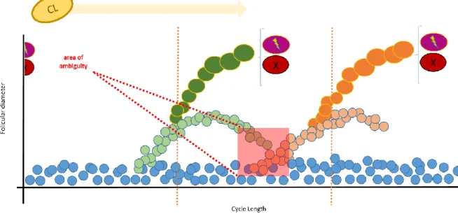
Ovulations from the left ovary were more frequent. More follicles were measured in the right ovary than in the left one during the entire considered period of time. The jenny that presented a persistent CL was the one that presented less follicles per ovary per ultrasound examination. When observing follicular profiles of consecutive waves, an “area of ambiguity” was found that did not allow the distinction between regressing and developing follicles.
x
Indications of lower ovarian activity and smaller follicular diameters during the non-breeding season were corroborated by considerable high rate of follicles under 18 mm in diameter and a higher rate of larger follicles during estrus periods.
Minor waves were significantly more frequent among jennies that underwent an anestrus period. The emergence of consecutive minor waves during anestrus was the most frequent pattern in this group of jennies. Among the remaining jennies, the most common wave pattern was the emergence of only one follicular wave with either one or two ovulatory follicles. The diameter of the largest follicles of minor waves was generally lower than the diameter at divergence of major waves. Also, mean maximum diameter of the largest follicles of major secondary waves was smaller than the maximum diameter of the largest follicles of major primary waves.
It was found the presence of both major and minor secondary waves in all groups of jennies. Jennies that did not present an anestrus period presented, though not significantly, a higher number of follicular waves per cycle. Multiple ovulations with the ovulatory follicles being originated from different follicular waves were also found.
xi Resumo
A raça Asinina de Miranda teve a sua origem na zona do planalto Mirandês, no território mais a nordeste de Portugal. Esta raça encontra-se atualmente na lista de raças autóctones em risco de extinção pelo que estudos têm vindo a ser realizados no sentido da sua melhor caracterização e proteção.
É comum a aplicação de estudos realizados em éguas a burras, no entanto, o gradual aumento de estudo em asininos tem vindo a demonstrar que, nomeadamente a nível reprodutivo, existe um considerável número de diferenças. A época reprodutiva é sem dúvida o principal alvo de estudo da maior parte dos trabalhos publicados. Observações como a sazonalidade menos marcada das burras e a necessidade de otimização dos resultados reprodutivos, particularmente de espécies em risco de extinção, leva ao interesse no estudo da época não reprodutiva.
Este estudo visou a medição dos diâmetros foliculares presentes em ambos os ovários de quinze burras mirandesas durante os meses de setembro a abril durante dois anos consecutivos e a caracterização da dinâmica folicular durante este período. Confirmou-se a sazonalidade menos marcada das burras. No entanto, foi observado um maior número de animais que demonstraram efeitos sazonais que o publicado em estudos anteriores. Os resultados da caracterização dos períodos interovulatórios concordou com a bibliografia disponível. Quando comparados com a época reprodutiva, os ciclos apresentaram-se mais longos, assim como os períodos de estro e os folículos ovulatórios atingiram maiores dimensões no início da época reprodutiva e na época de transição da primavera. As burras que passaram por um período de anestro foram também os animais que registaram diestros mais curtos. Duas das 15 burras a estudo apresentaram diestros sistematicamente mais longos. Os períodos de anestro registados foram mais longos que o publicado em estudos anteriores. Foram registados três folículos anovulatórios, todos em burras que passaram por um período de anestro (dois ocorreram durante a época de transição de primavera e um durante a época de transição de outono.
As ovulações no ovário esquerdo foram mais frequentes. Foi medido um maior número de folículos no ovário direito em todas as burras ao longo de todo o período de estudo em todas
xii
as ecografias realizadas. A burra que apresentou um corpo lúteo persistente foi também a que apresentou um menor número de medições por ecografia.
Ao analisar os perfis foliculares, foi encontrada uma área de ambiguidade que não permitiu a distinção entre folículos em regressão de folículos em crescimento.
Um menor nível de atividade ovárica e menores dimensões foliculares parecem ter estado presentes já que foi encontrada uma grande percentagem de folículos menores que 18 mm e uma grande concentração de folículos de maiores dimensões nos períodos de estro.
Ondas menores foram significativamente mais frequentes durante os períodos de anestro. A emergência de ondas menores consecutivas foi o padrão mais frequente em burras que entraram em anestro. Nas restantes burras a emergência de apenas uma onda com um ou dois folículos ovulatórios foi o padrão mais frequente. O diâmetro máximo do maior folículo de ondas menores foi inferior ao diâmetro à divergência dos maiores folículos das ondas maiores. O diâmetro máximo registado em ondas maiores secundárias foi inferior ao diâmetro máximo registado em ondas maiores primárias.
Foram identificadas ondas primárias e secundárias menores e maiores. Burras que não entraram em anestro apresentaram um maior, mas não significativamente maior, número de ondas foliculares. Verificou-se uma ovulação múltipla em que os folículos derivaram de duas ondas foliculares distintas.
xiii Contents
I. Introduction ... 1
1. The Donkey ... 1
2. The Miranda donkey breed ... 2
3. The estrous cycle of the jenny and the mare as a study model ... 4
3.1 Reproductive activity and cycle length ... 5
3.2 Reproductive behavior... 6
4. Endocrine regulation of the estrous cycle ... 8
4.1 Hypothalamic and pituitary function ... 8
4.2 Follicular development, follicular hormone production and ovulation ... 9
4.2.1 Mares ... 9
4.2.1.1 Follicular recruitment ... 9
4.2.1.2 Ovulation ... 13
4.2.2 Jennies ... 16
4.3 Luteal function and posterior luteolysis ... 19
5. Follicular waves... 22
6. Anovulatory follicles ... 26
7. Seasonality ... 27
7.1 Mares ... 27
7.2 Jennies ... 28
7.3 Endocrine regulation during the anovulatory season ... 29
7.4 Other factors that interfere with seasonality and the estrous cycle ... 32
7.4.1 Photoperiod... 32
7.4.2 Nutrition and body condition... 33
7.4.3 Age and parity ... 34
xiv
8. Ultrasonography and ultrasonographic aspects of ovarian structures ... 36
II. Objectives ... 41
III. Materials and Methods ... 43
1. Animals and management ... 43
2. Assessment of follicular diameters ... 44
3. Statistical analysis ... 45
IV. Results ... 47
1. Ovarian patterns during the non-reproductive season ... 47
2. Follicular profiles of the jennies that underwent anestrus periods ... 52
3. Follicular patterns of the jennies that kept their normal cyclicity ... 54
4. Follicular profiles of the jennies with silent estruses ... 56
5. Follicular profiles of the jenny that presented a persistent CL ... 58
V. Discussion... 59
1. General cycle characterization and seasonal effects ... 59
2. Follicular waves... 65
VI. Conclusions ... 73
xv
Index of figures
Figure 1 Profile illustration of a Miranda donkey jenny in cross-hatching. Black ink pen 0.05 mm on A4 tracing paper. Artist: Catarina Pereira, 2016.
Figure 2 Reproductive exam by ultrasound examination on a Miranda donkey breed jennie (Photo courtesy of Miguel Quaresma)
Figure 3 Miranda breed jenny in estrus and evidencing mouth clapping behavior and with her tail raised (Photo courtesy of Miguel Quaresma)
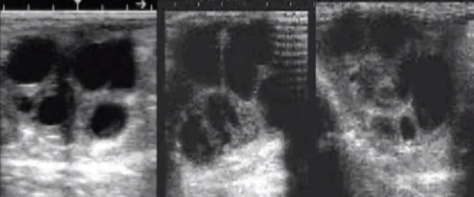
Figure 4 Ovarian ultrasonographic image, showing multiple different sized, anechoic follicles (Photo courtesy of Miguel Quaresma)
Figure 5 Ultrasonographic view of an ovary. The arrows show two corpus luteum (Photo courtesy of Miguel Quaresma)
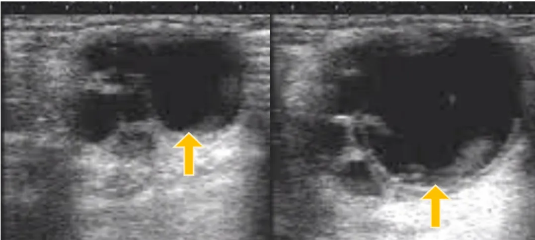
Figure 6 Images from the left ovary of a Miranda jenny at the onset of estrus (left) and close to ovulation (right). The dominant (left) and pre-ovulatory follicle (right) are indicated with a yellow arrow. It is possible to notice the change in size and shape of the dominant follicle until ovulation. It is also observable the presence of subordinate follicles at the left side of the dominant follicle (Photo courtesy of Miguel Quaresma)
Figure 7 Ultrasonographic image of an ovary of a Miranda jenny during the seasonal anestrus (December) with few follicular structures and of small sizes (Photo courtesy of Miguel Quaresma)
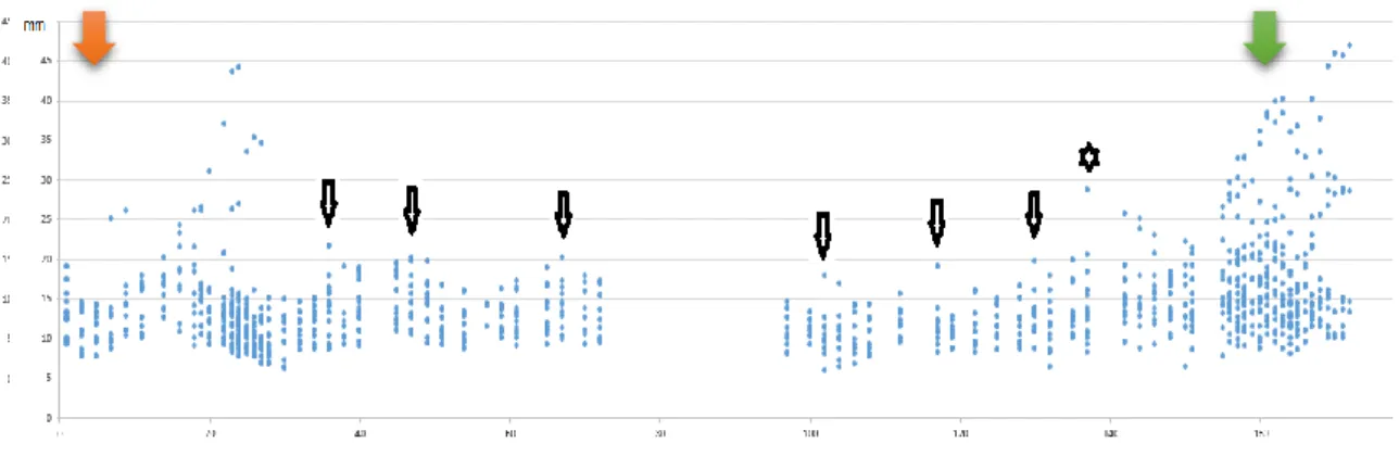
Figure 8 Follicles registered of Jenny 3 (Group A) from 5th October to 25th March.
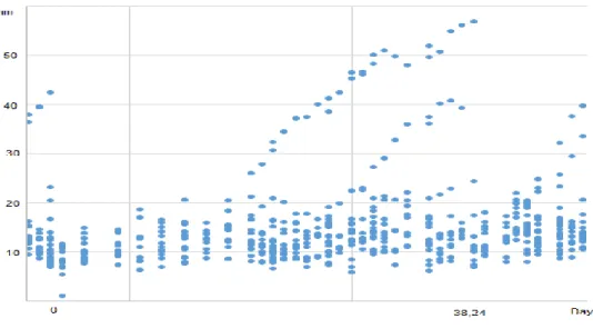
Figure 9 Estrous cycle of jenny 7 (Group E) with two dominant follicles originating from two different major primary follicular waves.
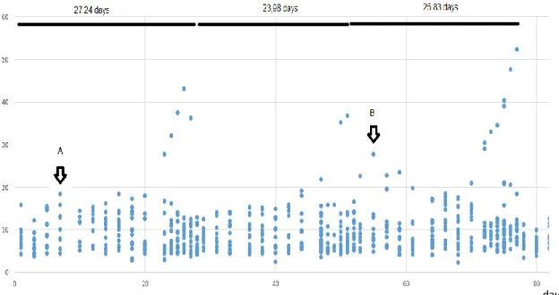
Figure 10 Two consecutive estrous cycles of jenny 8 (Group E), from 3rd October to 22th November.
Figure 11 Three consecutive estrous cycles of jenny 2 (Group SE), from 9th October to 25th December.
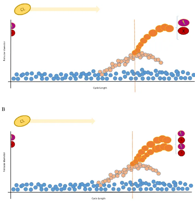
Figure 12 Representation of a cycle with a major secondary (green dots) and primary wave (orange dots), respectively and the resultant area of ambiguity (red square).
xvi Figure 13 Representation of a cycle with minor waves (purple dots) and a primary wave
(orange dots), respectively and the resultant area of ambiguity (red square).
Figure 14 Most frequent one-wave pattern found in mares and jennies. Single ovulation (A) and double ovulation (B).
Figure 15 Representation of the most frequent follicular dynamics pattern within jennies that underwent an anestrus period (Group A).
Figure 16 Representation of a double ovulation observed in jenny 7. The cycle begins with an active CL (yellow dot and arrow) originated from the previous cycle’s ovulation.
xvii
Index of Tables
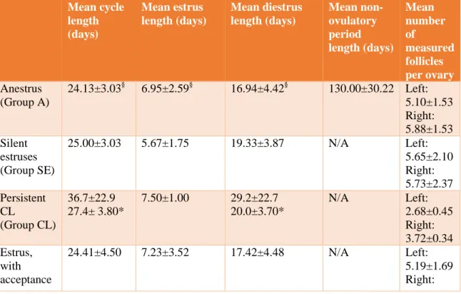
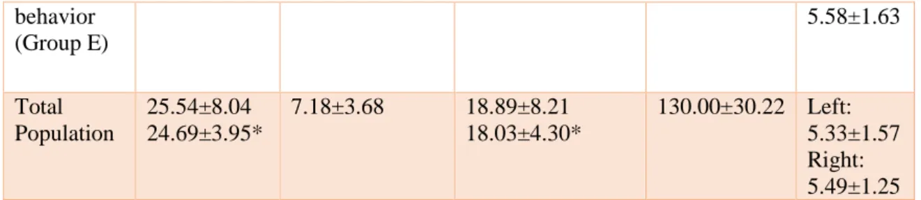
Table 1 Mean cycle, estrus, diestrus and anestrus lengths for all groups and the total
population and number of follicles found per ovary. Mean ± SD.
Table 2 Mean divergence day and follicular diameter, mean interval between divergence
and ovulation, number of follicles found per ovary. Mean ± SD.
Table 3 Left vs right ovary ovulations, single double and triple ovulations; asynchronous
vs synchronous ovulations. Mean ± SD.
Table 4 Number and type of waves present. Interval between waves. Mean ± SD. P -
Primary wave, M – Major secondary wave, m – minor wave.
Table 5 Maximum diameter and respective day in minor waves, mean day of divergence
and largest follicle’s diameter of major secondary waves. Mean ± SD. M1, M2, M3 - successive major secondary waves observed during the same period; m1, m2, m3, m4, m4 m5, m6- successive minor waves observed during the same period.
xix
Abbreviations
AEPGA Associação Para O Estudo E Proteção Do Gado Asinino
AI Artificial Insemination
FAO Food And Agricultural Organization
BCS Body Condition Score
CL Corpus Luteum
COX-2 Cyclooxygenase-2
FSH Follicle Stimulating Hormone
GNRH Gonadotropin-Releasing Hormone
HCG Human Chorionic Gonadotropin
IGF Insulin-Like Growth Factor
IGFPB Insulin-Like Growth Factor Binding Protein
LH Luteinizing Hormone
LHRH Luteinizing Hormone-Releasing Hormone
MRNA Messenger Ribonucleic Acid
P4 Progesterone
N/A Not Applicable
PGF2Α Prostaglandin F2-Alpha
SD Standard Deviation
US Ultrasonography
1
I.
Introduction
1. The Donkey
Domestic donkeys (Equus asinus) and horses (Equus caballus) are the domestic representatives of the Equus genus, known to have some unique reproductive characteristics (Huang et al., 2015; Pugh, 2002). According to recent studies, all the existing equid species are grouped in a single genus – Equus and, under this taxonomy, the domestic donkey (E. africanus asinus) is considered a subspecies of the African wild ass (Rosenbom et al., 2015a). Archaeological findings have led to two different hypotheses about the origin of this species. Some state that the presence of donkey remains from 6000 to 5000 BC in Egypt is an indication of this species being domesticated by the people of Egypt in the Nile Valley; others locate the donkey’s domestication in a northeastern African territory including the Sahara Desert 7000 to 65000 BC (Beja-Pereira et al., 2004; S. Rosenbom et al., 2015b; Scherf et al., 2015).
According to FAO, the donkey world population has increased from around 37 million donkeys in 1961 to around 44 million in 20014, with significant differences between regions; being the semi-arid zones the ones where the donkey concentration is higher. China and Ethiopia are the countries with larger numbers of donkeys (eleven and five million respectively) (Fielding and Starkey, 2004). There has been an increase by 60% in donkey populations in Africa, again with an uneven distribution among the countries in this continent. Latin America also shows an increase in donkey population. Central America has a small and steady growing donkey population. In South America there is a general increasing tendency, with the exception of Argentina and Chile where the populations have been decreasing. At last, in North America, the United States of America have always presented low numbers of donkeys that haven’t increased significantly. Asia in general, Turkey, Iraq, Israel, Jordan and Lebanon have experienced a decrease in donkey populations (with the last three countries suffering little changes in the past decade). On the other hand, Iran, Pakistan and Afghanistan revealed a contrary tendency. There are also countries that maintained their donkey populations relatively stable (Saudi Arabia, Syria, Yemen and Oman). Oceania shows general low numbers of donkeys amongst its countries (Fielding and Starkey, 2004).
2
Pertaining to Europe, there are considerable differences from country to country, with the UK and Germany having small populations of donkeys and the Eastern European countries with a stable population tendency. France and Ireland presented major declines in the past decades, as well as some southern countries like Italy (with a decrease of 96% from 1939 to 1996), Spain and Greece. In Portugal, there has also been a great reduction in the number of donkeys. Between 1999 and 2009 the number of donkeys decreased by 60%, with 30% of all donkeys concentrated in the north of the country (Instituto Nacional Estatística, 2009). This tendency that some countries present in reducing donkeys population and number of breeds endangers World’s domestic animal biodiversity (Aranguren-Méndez et al., 2002, 2001; Collins et al., 2012; Quaresma, 2014; Huang et al., 2015). A remarkable point is that a higher donkey population seems to be related to countries less industrialized, which had, and in some cases still have, agriculture as one of their most important economic activities.
2. The Miranda donkey breed
The Miranda donkey breed is a Portuguese donkey breed (Figure 1) that has its origin in the most northeast of the country, in Planalto Mirandês. As other Iberic breeds, Andalusian, Catalonian, Encartaciones, Mallorquina and Zamorano-Leones, it is facing some serious preservation issues (Aranguren-Méndez et al., 2002, 2001, Quaresma et al., 2014).
The specimens of this Portuguese breed are characterized for having a long brown bay coat, ideally around 130 cm of height and a calm temperament (Figure 2). These animals can be used for agricultural work, leisure, mediated therapy and milk production. This species milk is low fat, low protein and high lactose and can be used for both cosmetic and nutritional purposes (Muehlhoff and FAO, 2013; Quaresma et al., 2014).
3
Figure 1 Profile illustration of a Miranda breed jenny in cross-hatching. Artist: Catarina Pereira, 2016.
Coming from an area with strong agricultural traditions, the Miranda donkey breed was mostly used by farmers to help with their daily workload (Beja-Pereira et al., 2004; Fielding and Starkey, 2004). Resembling many other industrialized countries, with the technological advances, the decreasing popularity of agriculture amongst young people, the emigration and the shifting in many traditions, the donkey has become less and less popular, with the Miranda donkey breed being considered in risk of extinction (AEPGA, 2016).
4
Figure 2 Reproductive exam by ultrasound examination on a Miranda donkey breed jenny (Photo courtesy of Miguel Quaresma)
The Miranda donkey breed studbook comprises 760 animals with a female:male ratio of 9:1. Even though the authors didn’t find many consanguineous animals, the very low number of females and males available for reproduction, the aging of those animals and of their owners, the low breeding rate and the unequal contribution of the herds to the genetic pool are major concerns (Quaresma et al., 2014). Producing scientific knowledge that can help to implement good reproductive strategies is important for breed preservation (Aranguren-Méndez et al., 2002, 2001; Collins et al., 2012). Also, the Miranda donkey breed donkey is a versatile breed that besides agriculture, can still be used as a companion animal, for tourism, asinotherapy and their milk used for cosmetics (Pugh, 2002; Quaresma, 2014).
3. The estrous cycle of the jenny and the mare as a study model
The donkey reproductive cycle is similar in many ways to the horse, but has also some key differences (Huang et al., 2015). Being by far the most studied Equid, the mare is a starting point to obtain more information about the jenny (or jennet) and also other monovulatory species, including humans (Ginther et al., 2005a, 2004b; Mihm and Evans, 2008; Donadeu
5 3.1. Reproductive activity and cycle length
It is known that horses and, to a certain degree also donkeys, are long day breeders. Less domesticated horse breeds and pony mares usually demonstrate reproductive activity from May to October in opposition to more domesticated horses that tend to maintain their reproductive activity even in the winter season (Ginther et al., 1987; Aurich, 2011). On the other side, donkeys tend to show longer breeding seasons and a less marked seasonal anestrus, being most active between the months of March and August, sometimes continuing to ovulate during the rest of the year (Ginther et al., 1987; Pugh, 2002; Quaresma and Payan-Carreira, 2015).
Jennies usually achieve puberty between 1 to 2 years old, being the onset of puberty the result of many related factors such as photoperiod, temperature, individual health status, its BCS and genotype (Fielding, 1988; Pugh, 2002).
The estrous cycle of the donkey is longer than the mares’ (Pugh, 2002). In 1981, a study conducted in Wisconsin with unknown donkey breeds, established a 24.9 days interovulatory interval with a corresponding 6.4 days estrus and a diestrus of 19.3 days (Vandeplassche et al., 1981). Still, only slightly different values were found in more recent studies for distinct donkey breeds and populations’ interovulatory interval length: 24.9 days in the Catalan breed (Taberner et al., 2008) or 23.3 days in Mammoth Asses (Blanchard et al., 1999), 24.25 in Egyptian jennies (Derar and Hussein, 2011), 25.1 in the Tropical jenny (Lemma et al., 2006b) and 23.8 days for Miranda donkey breed jennies (Quaresma and Payan-Carreira, 2015), with indications of the age influencing the length between ovulations. Ovulations occur approximately 15 hours before the end of behavioral estrus, with the persistence of estrous behavior for a variable period of time. (Lemma et al., 2006c; Quaresma and Payan-Carreira, 2015), as for the horse.
During the breeding season, Quaresma and Payan-Carreira (2015) reported for the Miranda donkey breed an estrus length of 6.65 days and a diestrus length of approximately 17.9 days. Similar values were found for Catalonian jennies (5.64 days and 19.83 days for estrus and diestrus, respectively) and for Mammoth Asses jennies (5.9 and 17.4 days for estrus and diestrus length, respectively) (Blanchard et al., 1999; Taberner et al., 2008).
6 3.2. Reproductive behavior
During the period in which the mare is receptive to the stallion, behavioral routines tend to differ from the not receptive period. The time spent eating and resting diminishes and she becomes more active, ending up having some typical estrus behaviors (Clayton et al., 1981). There are three categories in which the reproductive courtship behaviors can be placed: attractivity, proceptivity and receptivity. The first stated category is measured by the behavior of the stallion towards the mare and can be shortly explained as the mare’s value as a sexual stimulus, being involved posturing and olfactory signals, for instance. Proceptivity, in turn, refers to the mare’s response to certain behaviors of the stallion such as vocalizations. At last, receptivity, refers to everything in the mare’s behavior that allows the copulation with the stallion (Beach, 1976).
When more than one jenny is in estrus, they tend to stay together forming a kind of cluster, vocalizing together towards the jack. When the male donkey is teasing one jenny, usually the others stay nearby. McDonnell (1998) compared the sexual behavior of donkeys and ponies and concluded that the jack spends more time teasing the jenny, necessarily taking longer to reach an erection (mounting the jenny multiple times before reaching the erection) when handled than when left to run loose. The same authors and Clayton et al. (1981) also refer that the matting is usually longer in donkeys than in ponies.
Mouth clapping, mouth opening and closing, with the lips relaxed and the head and neck lowered and extended, also known as mouth champing, jawing or yawning, is considered the main behavioral sign of estrus in donkeys (Vandeplassche et al., 1981). Winking and tail raising (Figure 3) were also found to consistently appear among jennies. Mouth clapping is the behavioral sign that appears sooner and it is the last one to disappear. Mouth clapping produces a very characteristic sound audible by humans. Also described in donkeys, are ears back against the neck, hind legs splayed, one foreleg slightly back and the other slightly forward, presentation of the perineum to the jack and urinating small amounts of urine at a time (Vandeplassche et al., 1981; Fielding, 1988; McDonnell, 1998; Pugh, 2002).
7
Figure 3 Miranda breed jenny in estrus and evidencing mouth clapping behavior and with her tail raised (Photo courtesy of Miguel Quaresma)
Other two categories that can be established for reproductive behaviors (and where we can easily fit the behaviors listed above) are the homotypical and heterotypical signs of estrous; the first ones consisting in signs that are distinctive of a certain species, such as demonstration of interest towards the male, posturing, clitoral winking and mouth clapping for the donkey species. As heterotypical signs, there can be found animals sniffing or chasing other females, standing to be mounted or Flehmen responses (McDonnell, 1998). Studies conducted in the Miranda donkey breed (Quaresma and Payan-Carreira, 2015), Catalonian jennies (Taberner et al., 2008) and a group of jennies in Ethiopia (Kebede et al., 2012) registered similar homotypical and heterotypical estrus signs as the ones already described.
Taberner et al. (2008) also characterized the jacks’ behavior towards the females in estrus and agreed with the previously described by Clayton et al. (1981) and McDonnell (1998): the jack vocalized and mounted the mare several times without erection, bit the neck, head and ends of the female donkey, smelt the perineal area and presented the Flehmen response. Also described is the jack covering of the jennies’ excrements with urine (McDonnell, 1998). During diestrus, jennies usually run away from the jack and can even try to bite or kick them to keep them away while keeping their tail closely to the perineum. Curiously, this running
8
behavior has also been described during the estrus phase but ending with the female allowing the jack to mount her (Clayton et al., 1981; McDonnell, 1998).
4. Endocrine Regulation of the estrous cycle
4.1. Hypothalamic and pituitary function
The hypothalamus is fundamental in the control of reproductive function in mares, by secretion of gonadotropin-releasing hormone (GnRH), a decapeptide which regulates the production and secretion of two other hormones: follicle-stimulating hormone (FSH) and luteinizing-hormone (LH). Initially called luteinizing hormone-releasing hormone (LHRH), its name changed when GnRH was found to interfere also with the dynamics of FSH in some species. In the horse, GnRH pulses are followed by LH pulses from the pituitary gland, generally together with a FSH pulse. The frequency of these pulses varies depending on which point in the estrous cycle the mare is. This pulsatile GnRH release is mainly controlled by feedback mechanisms. Structurally similar, LH and FSH, in conjunction, have an important role in fertility of both females and males. Both hormones are produced by a type of cells called gonadotropes from the pituitary gland. Together with thyroid-stimulating hormone (TSH) and equine chorionic gonadotropin (eCG), FSH and LH are part of the same glycoprotein family (Aurich, 2011; McKinnon, 2011).
In the horse, endogenous opioidergic systems are activated by progesterone and estradiol, so that in the luteal phase the hypothalamus is inhibited by these endogenous systems while during the follicular phase they are “inactive”, allowing the pulsatile activity of the hypothalamus (Aurich, 2011; McKinnon, 2011). In the mare, a pronounced differential regulation of LH and FSH secretion is due to the existence of three types of gonadotroph cells in the pars distalis and tuberalis of the pituitary gland: monohormonal gonadotrophs of either FSH or LH and bihormonal gonadotrophs that store both hormones (Aurich, 2011).
9 4.2. Follicular development, follicular hormone production and ovulation
4.2.1. Mares
4.2.2.1 Follicular selection
During the mare’s early fetal life, the primordial germ cells that have migrated, proliferated and become arrested during meiotic division at prophase I as primary oocytes, suffer atresia or develop into primordial follicles. At birth the ovaries contain thousands of these follicles, ready to develop into primary, secondary or antral follicles (Donadeu and Pedersen, 2008). The same phenomenon happens with all farm species (Driancourt, 2001). As in other species, as cattle or even in women, follicles develop in certain patterns as cohorts of follicles or follicular waves (Pierson and Ginther, 1987; Sirois et al., 1989; Bergfelt and Ginther, 1992). Around day 7 of the estrous cycle of mares, being the day of the previous ovulation called day 0, a variable number of small antral follicles, with around 6 mm of diameter start to develop at a similar pace with less than a day of interval between emergence of consecutive follicles (Gastal et al., 2004) in what is called a follicular wave, together with a rise in the peripheral FSH concentrations. The period during which these follicles are recruited can be denominated “recruitment window” and can last 3 days in horses (Driancourt, 2001). In mares, it was reported that a mean of 12 follicles emerge per wave until deviation occurs, though it might vary; after deviation a mean of 4.5 follicles were reported to emerge occasionally until ovulation (Pierson and Ginther, 1987). At this point, several authors use the designation of common growth phase. Only dependent follicles are recruited; gonadotropin-dependency develops when the follicle measures 2 mm in diameter; at this size is when they become. (Driancourt, 2001).
Follicular waves cannot develop without proper gonadotropin stimulation; a surge of circulating FSH precedes each follicular wave (Donadeu and Pedersen, 2008). It peaks about 3 days before the beginning of recruitment or when the largest follicle has around 13 mm of diameter (Irvine et al., 2000; Ginther et al., 2003a). Usually, the future dominant follicle emerges earlier than the remaining follicles, and maintains a 3 mm advantage from the second largest follicle, as the follicles grow at a similar pace at this point of the follicular wave
10
(Gastal et al., 1997, 2004). A similar situation occurs with cattle (Ginther et al., 2003a; Ginther et al., 2001b), but not with women (Baerwald, 2003; Baerwald et al., 2003).
Not only the FSH concentrations influence the developing follicles, but also follicles can influence the FSH concentrations, in this case negatively. It is believed that an increasing concentration of inhibin-A, secreted by granulosa cells and present in the follicular fluid, from mainly the largest follicles, that are around 13 mm at this point, is involved in the decrease of circulating concentrations of FSH (Watson and Al-Zi’abi, 2002a; Ginther et al., 2003b). Not only inhibin, but also androgens and estradiol might contribute to the FSH decline (Evans et al., 1997). Among these follicles, the future dominant follicle or follicles are emerging. Later, around day 13, follicle deviation occurs (Aurich, 2011).
Deviation can be considered a mechanism of monovular species (mares, cattle, women, etc.) to ensure the outcome of a single offspring, increasing this way the chances of survival in these species (Mihm and Evans, 2008). In the mare, deviation occurs when the dominant follicle has reached 21 to 23 mm and consists of a process that will prevent subordinate follicles from growing as much as the dominant (Ginther, 2000; Gastal et al., 2004; Beg and Ginther, 2006). The second largest follicle usually measures around 19 mm at this point of the cycle and might keep its capability to reach dominance for at least 1 day after the beginning of deviation in case the dominant follicle fails (Ginther et al., 2003b; Gastal et al., 2004). In some mares, follicles as small as D5 or D7 (being D1 the largest, D2 the second largest and so on) managed to reach dominance after ablation of the largest follicles, demonstrating that, in some way, the capacity to reach dominance is prevalent among all follicles of a wave (Gastal et al., 2004).
An interesting phenomenon happens at this point of the cycle. While levels of FSH have decreased and are insufficient to allow growth of all follicles of the wave, the reduced concentrations of this hormone are still enough to allow the largest follicle to keep its way into becoming a dominant and later on, an ovulatory follicle. This happens due to a rise in gonadotropin receptors by the dominant follicle. The concentrations of inhibin and estradiol have begun to rise 1 day before the beginning of deviation, around 10 days before ovulation (Ginther et al., 2003b; Ginther et al., 2008a) or at the day of deviation (Ginther et al., 2007e). Also, the dominant follicles’ sensitivity to FSH and LH increases in part due to an increase in
11
its gonadotropin receptors in the theca, simulated by elevated intra follicular levels of estradiol (Donadeu and Pedersen, 2008). Ginther et al. (2003b) have demonstrated the appearance of an ultrasonographic anechoic layer in the future dominant follicle, which likely represents an increase in its vascularity leading to the hypothesis that the largest follicle is, in fact, the main source of the higher levels of estradiol around the time of deviation.
The rise in LH circulating concentration, two days before deviation, and the rise of follicular sensitivity to this and other hormones by the rise of its intra follicular receptors is very important (Ginther et al., 2007e). This, allows the largest follicle of the wave to become dominant and for the development of ovulatory competence, for instance, a full responsiveness to LH surges near ovulation (Goudet et al., 1999; Gastal et al., 2000; Ginther et al., 2003a; Donadeu and Pedersen, 2008). In other words, it can be said that the increase in the dominant follicle’s dependency of on LH is critical for ovulation to occur (Gastal et al., 2000). However, there are still some authors that question whether the LH dependency is a cause or a consequence of follicle selection (Mihm and Evans, 2008).
Circulating estradiol concentrations have been reported to fluctuate depending on the phase of the estrous cycle in which the mare is (Gastal et al., 1999; Irvine et al., 2000). More recent studies achieved different conclusions, indicating that estradiol does not actually play a role in the onset of deviation in mares (Donadeu and Ginther, 2002a; Beg and Ginther, 2006). Additional hormones that behave in a similar way are testosterone and cortisol. Testosterone reaches its highest level the day before ovulation and decreases during the initial luteal phase, while cortisol presents itself in high concentrations during luteal phase and low during follicular phase (Ginther et al., 2007e).
Androgens and progestins are known to be direct and indirect substrates for estradiol production, respectively. Following LH stimulus, theca cells produce androgens that are later aromatized into estrogens in granulosa cells (Beg and Ginther, 2006). Also, androgens can enhance the production of progestins at the granulosa cells. These substances are however, probably not involved in the deviation mechanism due to the absence of a discrepancy in progesterone concentrations before and after deviation and between the largest and second largest follicles after the ablation of the first one (Donadeu and Ginther, 2002a; Ginther et al., 2002, 2007e).
12
Activins, like activin-A and follistatin, are present in the equine and bovine follicular fluid. They are different glycoproteins that bind with each other (Austin et al., 2001; Donadeu and Ginther, 2002a). Knight and Glister (2001) have proposed a list of situations were activin participates: 1) it induces granulosa cell proliferation, 2) increases FSH receptor expression, 3) boosts granulosa cell steroidogenesis, basal and gonadotropin stimulated aromatase activity, estradiol production and also 4) delays luteinization and atresia. Another distinctive action of activin is the block of LH and estradiol androgen secretion from theca cells – this is when follistatin, as an activin binding protein, stops this inhibitory effect (Knight and Glister, 2001; Wrathall and Knight, 1995).
Activin receptors are present in both thecal and granulosa cells (Knight and Glister, 2001). Different results were obtained among studies in cattle (Austin et al., 2001; Beg et al., 2002; Donadeu and Ginther, 2002a) though it is believed that only the largest follicle reaches a certain responsiveness to activin-A which might be involved in the changes of concentrations of estradiol and IGF-1. This glycoprotein was found to increase differentially between the largest follicle and the other follicles of the wave like estradiol, free IGF-1 and inhibin-A leading to the possibility of a role in the increased FSH responsiveness seen in the dominant follicle of the wave (Ginther et al., 2003b; Beg and Ginther, 2006).
The IGF system is involved in growth and differentiation of cells and comprehends IGF-1, IGF-2, IGF receptors, binding proteins (IGFBPs) and IGFBP proteases. A study conducted in mares points out the possibility of the IGF system being crucial for the deviation to occur, even though estradiol, inhibin-A and activin-A concentrations are similarly higher in the dominant follicle than in the remaining subordinates (Beg and Ginther, 2006). Actually, it was reported that after ablation of the largest follicle and before the beginning of deviation, the concentration of free IGF-1 improved at the second largest follicle when compared to the third largest follicle, and that the concentrations of estradiol, inhibin-A and activin-A only increased after deviation began (Ginther et al., 2002). The effects of a change in IGFBPs combined with an increase in IGF produced at the ovarian cells are the following: stimulation of granulosa cell proliferation and steroidogenesis and probably an interference in luteal function (Spicer and Echternkamp, 1995).
13
An IGF-1 treatment of the second largest follicle (F2) resulted in 81% of these follicles deboming dominant and 62% ovulating. Previously, IGF had been reported to influence the concentrations of activin-A, inhibin-A and VEGF during follicle selection in mares together with a reduction in the production of androstenedione (Ginther, 2003b). However, IGF-1 was concluded to be an intrafollicular factor that does not affect systemic concentrations of hormones like FSH, estradiol, inhibin or LH. Follicles of a certain diameter or development stage, after the beginning of deviation, might have capability to reach dominance but can’t produce IGF-1 (Ginther et al., 2008b).
Vascular endothelial growth factor (VEGF) is an angiogenic factor that appears to be augmented in the largest follicle when compared to the second largest follicle of the follicular wave. The largest follicle presents a higher blood flow, allowing a better nutrient, hormone and growth factor supply (Mihm and Evans, 2008). Evidence of an increased follicular vascularization is suggested by the anechoic layer at the ultrasound images (El et al., 1999; Ginther, 2003b) and were confirmed later by Doppler studies in horses (Acosta, 2004a), but not in cattle (Acosta et al., 2005).
Genomic studies have been conducted in cows, though other monovular species, as humans and horses, have not been yet studied. Results indicate the presence of 18 different genes that are expressed differentially among the largest and second largest follicles and that are the differentiation genes and not others, until then, associated with bovine dominant follicle differentiation, estradiol production, anti-oxidant events, LH sensitivity, cell proliferation, anti-apoptotic activity and mRNA splicing genes (Evans, 2004; Mihm and Evans, 2008).
2.4.2.2. Ovulation
In mares, at day 17, the dominant follicle usually measures around 35 mm and continues to grow while the subordinate follicles begin to regress. At the beginning of the preovulatory stage there is a pronounced rise in follicular estradiol that only allows a mild rise in LH concentrations, accompanied by a discrete decrease in FSH that will eventually return to its basal levels. Estradiol peaks two days before ovulation. LH will later exert a negative effect on estradiol and on follicle growth and its concentration continues to raise at a more
14
pronounced rate. LH will be responsible for a decrease in estradiol concentrations, the final maturation of the dominant follicle and ovulation when the follicle reaches around 40mm of diameter. FSH will discreetly rise due to the end of the negative effect estradiol has on this hormone and LH (Ginther et al., 2008a).
Near the day of ovulation (day 21), an increase in LH concentration is noticeable occurring also the raise of progesterone levels. Differently from other species, in the mare, the period of elevated concentrations of LH lasts 1 to 2 days and does not assume the shape of a short and pronounced preovulatory peak but rather a plateau. The vascularization of the ovulatory follicle increases (Aurich, 2011).
As already mentioned, the estradiol concentration peaks 1 to 2 days before ovulation and some observations suggest that if the level of estradiol is not enough for a proper positive feedback, the LH surge would not occur (Irvine et al., 2000). This disagrees with other authors, that defend a negative feedback relationship between these hormones (Ginther et al., 2008a). Irvine et al. (2000) also states that the decrease in estradiol concentration probably occurs due to the luteinization of granulosa cells and to the absorption of estradiol by the follicular fluid and its discharge into the abdomen (Ginther et al., 2008a; Aurich, 2011; McKinnon, 2011). This is accompanied by a decrease in uterine edema, typically seen during estrus. Estradiol promotes physical (cervix relaxation, uterine edema and increase in uterine secretions), endocrine (stimulation of hypophyseal LH release) and behavioral changes in the mare (Aurich, 2011; McKinnon, 2011).
Inhibin is a hormone produced by the granulosa cells dominant follicles that reaches its peak at the day of ovulation and remains in low levels during diestrus (Irvine et al., 2000). Higher levels of this hormone are directly related to estradiol and inversely correlated with FSH concentrations. Actually, one acknowledged function is to diminish FSH secretion by the hypophysis, as previously mentioned (Aurich, 2011; McKinnon, 2011).
In regard to ovulation and the pre ovulatory follicle, the size of this follicle can assume a wide interval, still, it has been shown that there is a considerable repeatability within each animal; studying a sample of estrous cycles of a certain female, the average size of the preovulatory follicle becomes an important information to predict the time of ovulation (Cuervo-Arango
15
and Newcombe, 2008). Usually it varies from 40 to 45 mm in diameter, with a growth rate of approximately 3mm/day (Ginther et al., 2008a).
The preovulatory follicle experiences some morphological modifications that can be perceived by a B Mode or Doppler ultrasound exam (Ginther et al., 2007c). Follicle maturity appears to be characterized by the appearance of a serration of the granulosa; irregularities among this layer of cells are obvious between 12 to 1h before ovulation, probably being the most useful sign to anticipate the moment of ovulation. The presence of apoptotic cells among the granulosa layer is positively correlated with follicles near ovulation (Ginther et al., 2007c). Also, decreased turgidity, follicles’ loss of spherical shape, reduced area at one end (apex) and the antrums’ fluid becoming less anechogenic and appearing to have echoic spots are all signs to have in mind when evaluating a pre ovulatory follicle (Gastal et al., 2006; O.J. Ginther et al., 2007a).
The ovulatory follicle will cease its growth rate and reach a plateau close the diameter with which it will ovulate. It will then rupture at the ovulation fossa (Ginther et al., 2008a). The oocyte and corona radiata then enter the oviduct, while the majority of the follicular fluid end up at the peritoneal cavity where hormones will reach systemic circulation – justifying the inhibin increase at the day of ovulation (Bergfelt et al., 1991).
Multiple ovulations can be defined as more than one ovulation occurring in a single ovulatory period or estrous cycle. Also they can be unilateral if all follicles ovulate from the same ovary, whether the left or the right one, or bilateral if the ovulatory follicles come from both ovaries. This phenomenon can also be classified as synchronous – if all follicles ovulate within an interval not larger than 24h, or asynchronous if ovulation occurs among different days. Transretal real-time ultrasonography has shown that 85.9% were single ovulations and 14.1% were multiple ovulations. Moreover, 14 out of 27 double ovulations came from only one ovary (unilateral ovulations) and the remaining 13 were bilateral ovulations with no relevant differences in the number of synchronous and asynchronous ovulations. Another conclusion reached was that the diameter of the pre-ovulatory follicle was different between single (the largest ones), double unilateral and double bilateral ovulations (the smallest ones) (Ginther and Pierson, 1989). Differently from the percentages presented above it was reported a 43% incidence of multiple ovulations (Ginther et al., 2008c) and, in another study during the
16
same year an incidence of 40% of double ovulations in mares, 2% in ponies and 25% in thoroughbreds (Ginther et al., 2008a).
It is known that the occurrence of multiple ovulations is variable, depending on the breed and even on each animal. Some studies concluded to be a variable grade of repeatability of multiple ovulations within mares influenced by the reproductive status, the age and even on the drugs administered to the animal during the cycle (Ginther et al., 2008a; Aurich, 2011;). Until the size of 30 mm, both single and double ovulating mares present dominant follicles with similar sizes, with the known discrepancies appearing in the 2.5 days after this moment probably due to differences in FSH concentrations consequence of higher estradiol concentrations present in double ovulating mares. Moreover, the cessation or reduction of the size of the ovulatory follicle near ovulation was observed in both dominant follicles in double ovulating mares (Ginther et al., 2008c). Concentration of LH is believed to be similar among single and double ovulating females until ovulation, when a higher concentration of progesterone coming from the presence of two CLs will lead to a lower concentration of LH (Ginther et al., 2008c).
4.2.2. Jennies
The information previously mentioned within this chapter was on the mares’ reproduction, as unfortunately there aren’t enough studies about the donkey species that can provide as much information as needed for a full description on follicular development and hormone fluctuations in equids. However, studies on donkeys’ follicular and gonadotropin levels and dynamics were published and their conclusions will be now presented.
Vandeplassche et al. (1981) found to be a significant day effect for FSH and LH concentrations, for diameter of the largest follicle and the number of small, medium and large follicles. Besides, the first significant increase in diameter of the largest follicle takes place 7 days before ovulation. Similar results where later reported also in Egyptian jennies; with a mean maximum diameter of 36 mm, 1 day prior to ovulation (Derar and Hussein, 2011), smaller than what was later reported for Catalonian jennies (Taberner et al., 2008).
17
The ovulatory follicle of Miranda donkey breed jennies measures around 38.4 mm (Quaresma and Payan-Carreira, 2015), smaller than the mean diameter of 46.3 mm of those in Catalonian jennies (Taberner et al., 2008), 41 mm from a group of jennies studied in Egypt (Derar and Hussein, 2011) and 44 mm (maximum diameter registered) in a group of jennies from Ethiopia (Kebede et al., 2012) but higher than the ovulatory follicle of tropical jennies during the short rainy season (37.8 mm) (Lemma et al., 2006c) or Anatolian jennies (32.25 mm) (Kalender et al., 2012).
Also, the Portuguese breed of donkeys presents a greater growth rate after the onset of estrus (3.18 mm/day) than before the onset of estrus and presents a slowdown in its growth on the day before ovulation (Quaresma and Payan-Carreira, 2015) agreeing with what was published in 1981 by Vandeplassche et al. and with what was published for Catalonian jennies (Taberner et al., 2008). In Egypt a slower growth rate was observed in jennies, about 2.32 mm/day, for the same period (Derar and Hussein, 2011).
A study on tropical jennies in Ethiopia assessed the mean number of follicles per wave and found it to oscillate through seasons between 7.3, 9.6 and 11.3 for the dry, short rainy and long rainy seasons, respectively. A higher frequency of medium and large follicles during the short rainy season was observed (from March to May) (Lemma et al., 2006c). Also reported was a comparative abundance of medium follicles (Kebede et al., 2012); all this data presents itself to be slightly different from the results published for mares, emphasizing the differences these two species have, particularly in regard to seasonality (Pierson and Ginther, 1987; Gastal et al., 2004). Another report from Egyptian jennies refers a lower number of follicles per ovary than the ones quoted above, though it is hard to access if the difference in their results is real or influence of the method used to count the follicles (Abdoon et al., 2014). Medium follicles start to appear at day 7 before ovulation and then their number decline at day 4 before ovulation reaching “0” after ovulation. As for the number of small follicles, it decreases significantly as ovulation day approaches (Vandeplassche et al., 1981).
Available data on the number of follicular waves per cycle reports the existence of 2 to 3 waves per cycle in jennies under controlled management in Ethiopia (Kebede et al., 2012), contrarily to a single wave per cycle reported in jennies from Egypt (Derar and Hussein, 2011).
18
Deviation take place near day 9 before ovulation in Miranda donkey breed jennies, when the largest follicle measures a mean of 19.18 mm (smaller in case of multiple ovulations) (Quaresma and Payan-Carreira, 2015), earlier than what was reported in jennies from Egypt; 5 to 6 days before ovulation the dominant follicle measures approximately 25 mm (Derar and Hussein, 2011).
The occurrence rate of single versus multiple (double or triple) ovulations is a contentious point among the estrous cycle of donkeys with different studies reaching different percentages. The previously quoted study of Vandeplassche et al., (1981), refers that only one double ovulation occurred in 1 out of 7 jennies, among all the estrous cycles that were studied, which reflects a low rate of multiple ovulations. However, this study was published before ultrasonography started to be associated with reproductive exams, which by itself can explain such different results from the more recently published. Also worth mentioning is the tendency that these authors found in multiple ovulations being repeatable within jennies, as previously mentioned for mares (Aurich, 2011) and confirmed in the Miranda donkey breed (Quaresma and Payan-Carreira, 2015) and Catalonian jennies (Taberner et al., 2008). Differently from donkeys, mules did not present multiple ovulations (Volpe et al., 2005). For Miranda donkey breed jennies, Quaresma and Payan-Carreira (2015) reported single ovulations to be more common followed by double and triple ovulations respectively, which agrees with Vandplassche et al. (1981) results. In 33 cycles, 57.58% were single ovulations, 36.36% were double ovulations and the remaining 6.06% correspond to triple ovulations. In Catalonian jennies the numbers are quite similar with 55.66%, 42.45% and 1.89% of single, double and triple ovulations respectively (Taberner et al., 2008). Another study on Spanish donkey breeds, analyzed 258 estrous cycles and of these 49.2% corresponded to multiple ovulations (43.4% of double ovulations and the remaining 5.8% of triple ovulations, with a higher frequency of multiple ovulations at the beginning of the breeding season) (Galisteo and Perez-Marin, 2010). T.L. Blanchard et al. (1999) have reported higher rates than the presented above for Mammoth jennies, with 11 out of 18 jennies presenting multiple ovulations.
Ovulations from the right and left ovary assumed similar number of occurrences in Miranda jennies (Quaresma and Payan-Carreira, 2015) and in Catalonian jennies (Taberner et al., 2008), even though it was observed a slightly bigger number of ovulations from the left
19
ovary, it was not significant. Studies on Tropical jennies also agree with these observations (Lemma et al., 2006b).
FSH concentrations during the jenny estrous cycle were recorded to be significantly high from day 10 to 12 before ovulation (> 11,0 ng/ml) and minimum 3 days before ovulation (7,4 ng/ml), in general agreement with the previously quoted for mares (Vandeplassche et al., 1981; Donadeu and Pedersen, 2008). In a study that used a sample of 14 Miranda donkey breed jennies, serum concentrations of progesterone were measured throughout the estrous cycle. Progesterone remained in very low concentrations at 24h before ovulation and then after ovulations. P4 levels raised and remained high until day 15 after ovulation (luteal phase) to posteriorly start to decrease 2 to 3 days before the onset of estrus. Multiple ovulations were associated with higher concentrations of progesterone (Quaresma and Payan-Carreira, 2015). In relation to E2 levels, these were measured in Anatolian jennies and were at 3.78 ng/ml on the day of ovulation (Kalender et al., 2012).
4.3. Luteal function and posterior luteolysis
The equine corpus luteum (CL) presents itself usually pear-shaped and it is characterized by growing towards the inside of the ovary, contrary to what happens in the majority of other mammals (Kimura et al., 2005). Equine CL is formed by small and large luteal cells and non-luteal cells (fibroblasts, smooth muscle cells, macrophages and endothelial cells) all originated from follicular cells of the already ovulated follicle, as thecal cells are progressively substituted by fibroblasts after ovulation and within only 24h after ovulation they will all be in an advanced stage of degeneration (Aguilar et al., 2006; van Niekerk et al., 1975). Luteal and non-luteal cells will start to develop about 10h after ovulation and will later reach their maximum size (van Niekerk et al., 1975). Also, progesterone producing cells and angiogenic factors (VEGF) present a marked activity (Al-zi’abi et al., 2003; Aguilar et al., 2006).
The main hormones controling of the luteal phase are LH and progesterone, allowing CL function. In equids, progesterone concentrations start to increase right after ovulation, reaching maximum values 8 days after they began to rise and decreasing between 8 until day
20
14, when luteolysis ensues, with the expression of progesterone receptors in large luteal cells (da Costa et al., 2005; Aguilar et al., 2006). Anatolian jennies present a mean concentration of 0.81 ng/ml of at the day of ovulation P4 (Kalender et al., 2012) and Miranda donkey breed jennies present mean serum progesterone levels 24h after ovulation of 0.48 ± 0.14 ng/ml that reach 5.56 ± 0.86 ng/ml 72h after ovulation continuing to increase during mid luteal phase (Quaresma and Payan-Carreira, 2015). Some studies refer that maximum P4 concentrations reach values above 10 ng/ml (Quaresma and Payan-Carreira, 2015). Others, refer higher values as 27.72 ± 14.02 ng/ml at the 14th day after ovulation at the first cycle after parturition (Kalender et al., 2012). Kebede et al. (2012) verified that this hormone starts to drop at day 15 to 16 after ovulation and will be kept at its nadir until next ovulation occurs.
After ovulation, LH presents high concentrations, suggesting an important role in luteal development (Ginther, 1992). The dynamics between LH, progesterone and estradiol were studied in 9 mares during interovulatory period, and similar results were obtained with progesterone reaching its maximum concentration on day 6 after ovulation and decreasing after that interval (Ginther et al., 2006). P4 concentrations of about 2 ng/ml at the beginning and end of the luteal phase were responsible for negative effects on LH concentration, this way, justifying the progressive decrease in LH concentrations after ovulation (Ginther et al., 2008a). Mules present lower maximum concentrations of P4 (5 to 7 ng/ml) when comparing to the mare (Volpe et al., 2005).
It was also observed a decrease in concentrations of these hormones at a similar pace from day 6 until day 14 when luteolysis was expected to commence, consistent with declining positive effect of LH concentrations of progesterone. The area of the CL and plasma progesterone concentrations were compared, showing that they behaved in parallel throughout luteal phase but were asynchronous during the luteolytic period where CL area decreased at a lower rate than progesterone concentration (Ginther et al., 2007d).
Besides LH and Progesterone, IGF system is believed to be responsible as well for the control of the luteal phase, being the increased presence of IGFBP-2, most likely, a cause for luteolysis by inhibition of IGFs and their action on preventing apoptosis and stimulating steroidogenesis (Watson et al., 2005).
21
After day 14 of the cycle, the concentration of progesterone starts to decrease and, later, morphological changes of the CL will occur, with death of luteal cells. A decrease in the expression of P450scc, a steroidogenic enzyme, was found, where it was also studied the
expression of IGFBP-2 mRNA (Watson et al., 2005). VEGF will also decline but it is not believed to contribute in the apoptosis of the CL cells (Aguilar et al., 2006; Ginther et al., 2007d). A study that involved 18 mares, suggested that the developing follicles of a new follicular wave at the ovary might have a role in morphological regression of luteal structures, as they took longer to regress when follicles were ablated before they reached 10 mm in diameter (Ginther, 2005b). When multiple ovulations take place, the progesterone concentration will be higher, most likely because there are two functional CLs instead of only one. This was demonstrated in donkeys, in Portugal, by comparing the areas under the progesterone curves of single and multiple ovulations (Quaresma and Payan-Carreira, 2015). Luteolysis is a very important phenomenon in the estrous cycle of the mare, as it will allow another cycle to begin when a pregnancy does not occur. As in other domestic species, luteolysis in the mare is first signalized by the secretion of PGF2α by the endometrium, which
is believed to be controlled by COX-2 (Boerboom, 2004). Studies in sheep (Charpigny et al., 1997) and cows (Arosh et al., 2002), had already concluded that temporary increases in the expression of COX-2 mRNA happen concurrently with luteolysis.
Oxytocin is a neuropeptide hormone produced, synthetized and kept in the hypothalamus and posterior pituitary together with neurophysin, its “carrier protein”. Within its reported roles are: uterine contractility for drainage of cellular remains and uterine fluids and also contractility during parturition directly and indirectly by stimulation of the release of PGF2α; it
will also interfere in late diestrus with luteolysis (Brownstein et al., 1980; Melrose and Knigge, 1989).
Oxytocin has been identified in several organs and animals, such as human placentas (Chibbar et al., 1993), rat uterus (Larcher et al., 1995), cow (Wathes and Swann, 1982; Fields et al., 1983), goat (Kiehm et al., 1989), sheep (Rodgers et al., 1983), rat (Ho and Lee, 1992), pig (Jarry et al., 1990; Nitray and Sirotkin, 1992) and baboons (Khan-Dawood, 1986). It was identified in human ovaries (Schaeffer et al., 1984; Guillou et al., 1992; Maas et al., 1992;) but not in the mares´ ovaries (Stevenson et al., 1991; Stock et al., 1995). It can be found
22
oxytocin in the mare’s ovaries but not as neurophysin, what indicates that it is probably not secreted there (Watson et al., 1999). However, another study has identified luminal epithelium and superficial endometrial glands as oxytocin secretors in mares (Bae, 2003). The oxytocin secreted by the endometrium and PGF2α will be part of a paracrine-autocrine system that
accelerates luteolysis in mares that are not pregnant (Watson et al., 2005).
After luteolysis, both LH and FSH concentrations increased, probably due to the removal of progesterone’s negative influence; it is also observable an increase in GnRH and gonadotropin pulses (Irvine et al., 2000). Considering FSH concentrations before and at ovulation, they are significantly higher 3 days after ovulation (12 ng/ml) than at the day of ovulation (Vandeplassche et al., 1981).
As for prolactin, there were noted some prolactin pulses after luteolysis followed by an increase in oestrone concentration suggesting that this substance might be involved in follicle maturation (Irvine et al., 2000).
5. Follicular waves
Before the development of the ultrasound technique, studies of follicular dynamics were done using transretal palpation. The ultrasound technique quickly proved itself to be valid and, by far, more accurate (Palmer and Driancourt, 1980; Ginther and Pierson, 1984; Pierson and Ginther, 1987). Other valuable procedures for the study of follicular waves are the distribution of follicles among size categories, study of excised ovaries, ovariectomies and steroid treatments with the consequent emergence of a new wave (Driancourt et al., 1982a, 1982b; Ginther, 1993).
Besides the ultrasonographic day-to-day studies (Bergfelt and Ginther, 1992), other experiments were conducted without maintaining track of each follicle identity, using mathematical methods and reaching similar conclusions (Donadeu and Pedersen, 2008; Ginther and Bergfelt, 1992). Mathematical methods have some advantages – require less skilled operators, data collection and interpretation is easier and quicker, and the emergence of a follicular wave can be detected earlier (Ginther and Bergfelt, 1992). Concrete data demonstrate that by the identity method, divergence was occurring 2 days before its detection,