Elucidating the
catalytic mechanism of
human renin with
hybrid Quantum
Mechanics and
Molecular Mechanics
studies
Ana Rita Almeida Calixto Silva
Mestrado em BioquímicaDepartamento de Química e Bioquímica 2013
Orientadora
Doutora Maria João Ramos, Professora Catedrática, Faculdade de Ciências da Universidade do Porto
Coorientadora
Doutora Natércia Brás, Investigadora
Todas as correções determinadas pelo júri, e só essas, foram efetuadas. O Presidente do Júri,
Oral Communication
Ana Rita Calixto, Natércia Fernandes Brás, Pedro Alexandrino Fernandes and Maria João Ramos. “The catalytic mechanism of human renin protease – a quantum mechanics /molecular mechanics study”, IJUP 2013 – 6º Encontro de Investigação Jovem da Universidade do Porto, 13th-15th February 2013.
Poster presentation
Ana Rita Calixto, Natércia Fernades Brás, Pedro Alexandrino Fernandes and Maria João Ramos, “Elucidating the catalytic mechanism of human renin with a QM/MM study” XXIII Encontro Nacional da SPQ - Desafios em Química, Universidade de Aveiro, 12th-14th June 2013
Ana Rita Calixto, Natércia Fernades Brás, Pedro Alexandrino Fernandes and Maria João Ramos, “A QM/MM investigation of the catalytic mechanism of human renin protease” , XVIII Encontro Luso Galego de Química, Universidade deTrás-os-Montes e Alto Douro, Vila Real, 28th-30th November 2012
Ana Rita Calixto, Natércia Fernades Brás, Pedro Alexandrino Fernandes and Maria João Ramos, “Computational study of the catalytic mechanism of human renin protease”, 5º Encontro de Investigação Jovem da Universidade do Porto, 22nd-24th February 2012.
Acknowledgments
“Appreciation is a wonderful thing: It makes what is excellent in others belong to us as well.”
Voltaire
First of all, I would like to express my sincere gratitude to my supervisor, Professor Maria João Ramos, for giving me the opportunity to work with her, for her excellent guidance, continuous support, patience and constant motivation. Thank you for all the opportunities and suggestions during this work and for sharing your wealth knowledge with me.
I am also heartily thankful to Professor Pedro Alexandrino Fernandes, whose guidance, enthusiasm and encouragement to recover when my steps faltered were essential to finish my dissertation. His suggestions were always an asset to progress of this work. Thank you for have introduced me to the fascinating world of Computational Biochemistry and for the opportunity to work in your group.
I would also like to thank Natércia, my co-supervisor, for guiding me throughout the whole work and for all precious teachings and advices. I am deeply grateful for your support, patient, understanding and generosity. Thank you very much for your availability for everything and for keeping my hopes up when the results did not seems good.
I extend my appreciation to all Theoretical Chemistry and Computational Biochemistry group for providing me an excellent atmosphere to work. I am very thankful for the way I was received in the group. I would like to thank Rui and João for all the fun times we have had during the last year that distracting me from my work, for all the advices and help. I am also thankful to Rui Sousa, Cátia, Rita, Eduardo, Sílvia, Óscar and Zé, not only for all the support in this work, but also for the excellent work environment that you promote and, of course, for all the fun coffee breaks that increase my productivity every day.
I would also like to thank Silvia and Lau for all their friendship, understanding and support during these years.
I owe my deepest gratitude to Damien, who has always stood by me in the best and worst moments. Thank you for your care, patience and support every time I needed.
I am most grateful to my mother and brother for all the sacrifices during the last years and for all the unconditional love and support they have always give me. Without whom this work would not have been possible.
Finally, I would like to thank Foundation of Science and Technology of Portugal (FCT) for the financial support (project PTDC/QUI-QUI/121744/2010).
Abstract
Hypertension, also referred as high blood pressure, is a chronical condition that affects nearly 25% of adults worldwide. The increasing prevalence of this illness has contributed to the pandemic of cardiovascular and renal diseases, which are responsible for a large number of deaths every year [1]. Therefore, it is fundamental to develop new antihypertensive drugs, in order to control and prevent the complications associated with high blood pressure.
The Renin-Angiotensin System (RAS) is the major regulating system of the blood pressure and it consists of a two-step cascade. Firstly, the aspartic protease renin cleaves its only known substrate, angiotensinogen, in a rate limiting step, leading to the formation of the decapeptide angiotensin I (Ang I). Secondly, Ang I is transformed by angiotensin-converting enzyme (ACE) to produce the octapeptide angiotensin II (Ang II), which binds to angiotensin II receptors (AT receptors) and mediates the blood pressure control. Effective antihypertensive drugs involving different RAS targets have already been developed. However, it is believed that the inhibition of renin would offer better results in blood pressure control, due to its specificity for only one substrate and the fewer adverse treatment side effects [2, 3].
Therefore, the main goal of the present work is to describe, with computational methods, the atomistic details of the catalytic mechanism of human renin, to allow future studies on its inhibition. In order to achieve the purpose of this work the initial system (renin + angiotensinogen) was divided in two layers that were studied at different theoretical levels (Density functional theory (DFT) and Molecular Mechanics (MM)). The geometries were optimized with the ONIOM methodology, at the B3LYP/6-31G(d):Amber) level. The energies of the stationary points were recalculated with single-point calculations, using a large basis set (6-311++G(2d,2p)) and a progressive increase in the number of atoms in the DFT layer. Molecular dynamics (MD) simulations were also performed in order to understand the behavior of the system along the time.
Our results suggest that the angiotensinogen hydrolysis by renin occurs by an acid/base mechanism, through three elementary steps. It begins with the formation of a stable gem-diol intermediate, followed by the scissile bond nitrogen protonation and it ends with the
completely cleavage of the peptide bond. The final reaction products are two peptides with carboxylic acid and amine extremities.
We observed that the formation of the gem-diol intermediate is rate limiting, with a barrier near 20 kcal.mol-1. We also conclude that the residues around the active center greatly influence the catalytic mechanism of this enzyme. In addition, we also confirm that human renin catalytic mechanism is very similar to the mouse one [4].
The data obtained in the present work provides important clues about renin catalytic mechanism, which will enable further studies on its inhibition and, consequently, the development of new antihypertensive drugs.
Key words: Hypertension, Renin Angiotensin System, Renin, Angiotensinogen,
Resumo
1
A hipertensão arterial, também conhecida como pressão sanguínea elevada, é uma doença crónica que afeta cerca de 25% da população mundial. A prevalência desta condição patológica tem contribuído para um aumento significativo de doenças cardiovasculares e renais, sendo estas responsáveis por uma grande percentagem de mortes todos os anos [1]. Tendo isto em conta, de forma a controlar esta condição e a prevenir possíveis complicações a ela associadas, torna-se fundamental o desenvolvimento de novos fármacos anti-hipertensivos que apresentem uma melhor resposta, face aos já existentes.
O sistema renina-angiotensina (RAS) é o principal sistema de regulação da pressão sanguínea e consiste numa cascata que pode ser resumida em dois passos. A primeira enzima envolvida nesta cascata é uma protease aspártica, a renina, sendo esta responsável pela clivagem do angiotensinogénio, o seu único substrato conhecido. Este é um passo limitante de toda a cascata e origina a formação do decapeptido Angiotensina I (Ang I). Num segundo passo, a Ang I é clivada pela enzima conversora da angiotensina (ACE) levando à formação do octapeptido angiotensina II (Ang II) que, por sua vez, interage com os recetores de Ang II, mediando, assim o controlo da pressão sanguínea. Existem no mercado vários fármacos que atuam sobre diferentes alvos do RAS, no entanto, acredita-se que o melhor alvo a inibir nesta cascata será a enzima renina, dada a sua especificidade para apenas um substrato e, uma vez que a sua inibição parece estar associada ao aparecimento de menos efeitos adversos [2, 3] .
Tendo isto em conta, o principal objetivo do presente trabalho passa por tentar descrever, através de metodologia computacionais e com detalhe atomístico, o mecanismo catalítico da enzima renina de humano, de forma a permitir e facilitar futuros estudos da sua inibição. Sendo assim, de forma a atingir este objetivo, o sistema em estudo foi divido em duas camadas que, por sua vez, foram estudadas com diferentes níveis teóricos (Teoria do funcional de densidade (DFT) e mecânica molecular (MM). As geometrias foram otimizadas com o método ONIOM com nível teórico B3LYP/6-31G(d):Amber. Nos pontos estacionários,
1
as energias forma recalculadas usando uma base de funções maior (6-311++G(2d,2p)) e incluído, progressivamente, na camada DFT um maior numero de átomos. Foi ainda realizada uma simulação de Dinâmica Molecular, com o intuito de perceber o comportamento do sistema ao longo do tempo.
Os resultados obtidos sugerem que a hidrólise do angiotensinogénio, pela enzima
renina, ocorre através de um mecanismo acido-base, baseado em três passos elementares. Num primeiro passo, ocorre a formação de um intermediário gem-diol, seguida por um segundo passo de protonação do azoto da ligação a quebrar. Num terceiro, e último passo, ocorre a clivagem completa da ligação peptídica, sendo os produtos finais da reação dois péptidos com extremidades ácido carboxílico e amina.
Pode ainda ser verificado que a formação do intermediário gem-diol é limitante da velocidade da reação e que está associada a uma barreira energética de cerca de 20 kcal.mol-1. Conclui-se ainda que os aminoácidos em torno do centro ativo parecem influenciar, de forma significativa, a energia associada a esta reação. Para além disso, verificou-se, ainda, que o mecanismo pelo qual a enzima humana cliva o seu substrato é semelhante ao seguido pela enzima de ratinho [4].
Os resultados obtidos neste trabalho contém informações essenciais sobre o detalhe do mecanismo da renina, que poderão ser úteis em futuros estudos de inibição desta enzima e, consequentemente, no desenvolvimento de novos fármacos anti-hipertensivos.
Palavras-chave: Hipertensão, Sistema Renina Angiotensina, Renina,
Table of contents
Acknowledgments... iv Abstract ... vii Resumo ... ix Abbreviations ... xviii CHAPTER 1 - INTRODUCTION ... 1 1.1 Introductory note ... 1 1.2 Hypertension ... 2 1.3 Renin-Angiotensin System ... 31.3.1 Renin, Angiotensinogen and Angiotensin I ... 3
1.3.2 Angiotensin Converting Enzyme and Angiotensin II ... 5
1.3.3 Angiotensin receptors ... 6
1.4 Anti-hypertensive Agents ... 7
1.4.1 Diuretics ... 7
1.4.2 β-blockers ... 8
1.4.3 Calcium channel blockers ... 8
1.4.4 Vaccination ... 8 1.4.5RAS inhibition ... 8 1.4.5.1Renin inhibitors ... 9 1.4.5.2ACE inhibitors ... 9 1.4.5.2AT1 receptor antagonists ... 9 1.5Renin ...11
1.5.1Relation between structure and function ...11
1.5.1Proposal for renin catalytic mechanism ...13
1.5.3Development of renin inhibitors ...14
1.6Computational enzymatic catalysis and drug design ...15
1.6.1The role of computational chemistry in enzymatic catalysis ...16
1.6.2Rational drug design ...17
CHAPTER 2 - THEORETICAL BACKGROUND ... 19
2.1Introduction ... 19
2.2 Molecular Mechanics ... 21
2.2.1Basic Theory – The force field energy ... 22
2.2.1.1The Stretch bond energy ... 23
2.2.1.2The bending energy for angles ... 24
2.2.1.3The dihedral / torsion energy ... 24
2.2.1.4The van der Waals energy ... 25
2.2.1.3The electrostatic energy ... 26
2.2.2Molecular dynamics ... 27
2.3Quantum Mechanics ... 28
2.3.1The Schrödinger equation ... 29
2.3.2The Hamiltonian ... 30
2.3.3The Born-Oppenheimer Approximation ... 31
2.3.4 Hartree-Fock Theory ... 32
2.3.5Linear combination of atomic orbitals ... 34
2.4Density Functional Theory ... 35
2.4.1The Hohenberg-Kohn Theorem ... 35
2.5.2The Konh-Sham Theorem ... 36
2.5 Basis set ... 38
2.5.1 Slater type orbitals ... 38
2.5.2Gaussian type orbitals ... 38
2.6Hybrid QM/MM methods ... 40
2.6.1The ONIOM method ... 41
2.6.1.1Mechanical embedding scheme ... 43
2.6.1.2Electrostatic embedding scheme ... 43
2.6.1.3QM/MM boundary and link atoms ... 44
CHAPTER 3 - COMPUTATIONAL METHODOLOGY ... 45
3.1Introductory note ... 45
3.2Preparation of the system ... 46
3.2.2 Ren:Angdodecapetide:W1:W2 model ...46
3.2.3 Ren:Angmutated model ...46
3.3Molecular dynamics simulations ...48
3.4QM/MM calculations ...49
3.4.1Determination of the potential energy surface ...49
3.4.2Increase of the QM region ...51
CHAPTER 4 - RESULTS AND DISCUSSION ...53
4.1Introductory note ...53
4.2MD simulation ...53
4.2.1Root Mean Square Deviation ...54
4.2.2 Root Mean Square Fluctuation ...54
4.2.3Active site interactions ...55
4.3The catalytic mechanism of human renin ...58
4.3.1The reactants structure ...58
4.3.2The first reaction step ...59
4.3.3The second reaction step ...61
4.3.3The third reaction step ...62
4.3.4Summary of human renin reaction ...63
4.4 The role of a structural water molecule ...64
4.4.1The reactants structure ...65
4.4.2The first reaction step ...65
4.4.3The second reaction step ...66
4.4.4The third reaction step ...68
4.5Human renin catalytic mechanism with a mutated substrate ...70
4.5.1The reactants structure ...71
4.5.2The first reaction step ...71
4.5.3 The second reaction step ...72
4.5.4The third reaction step ...73
4.6Energies associated with human renin mechanistic pathway ...75
4.6.1Energies along the reaction mechanism ...75
4.6.1.1Ren:Angdodecapetide:W1 model ...75
4.6.1.3Ren:AngMutated model ... 77
4.6.2Single Point energy calculations ... 78
4.6.2.1Single point calculations using electrostatic embedding scheme ... 78
4.6.2.2Single point calculation using large QM regions ... 79
4.6.2.3 Single point calculation using a large basis set... 83
4.7Human Renin vs. Mouse Renin ... 85
CHAPTER 5 - CONCLUSIONS AND FUTURE PERSPECTIVES ... 87
5.1 Conclusions ... 87
5.2 Future Perspectives ... 88
List of Figures
Figure 1. Classical renin-angiotensin system ... 4
Figure 2. Human renin structure complexed with its substrate ... 12
Figure 3. General proposal mechanism of amine bond hydrolysis by aspartic proteases. ... 14
Figure 4. The Lennard-Jones potential as a funtion of interatomic distance. ... 25
Figure 5. Representation of all interactions present in the potential energy expression... 26
Figure 6. Representation of the ONIOM two-layer method ... 42
Figure 7. Representation of the different models used in the present work. ... 47
Figure 8. Representation of the system used in Molecular Dynamics Simulation. ... 48
Figure 9. Representation of the system used in QM/MM calculations ... 50
Figure 10. Representation of the atoms included in the different QM regions. ... 52
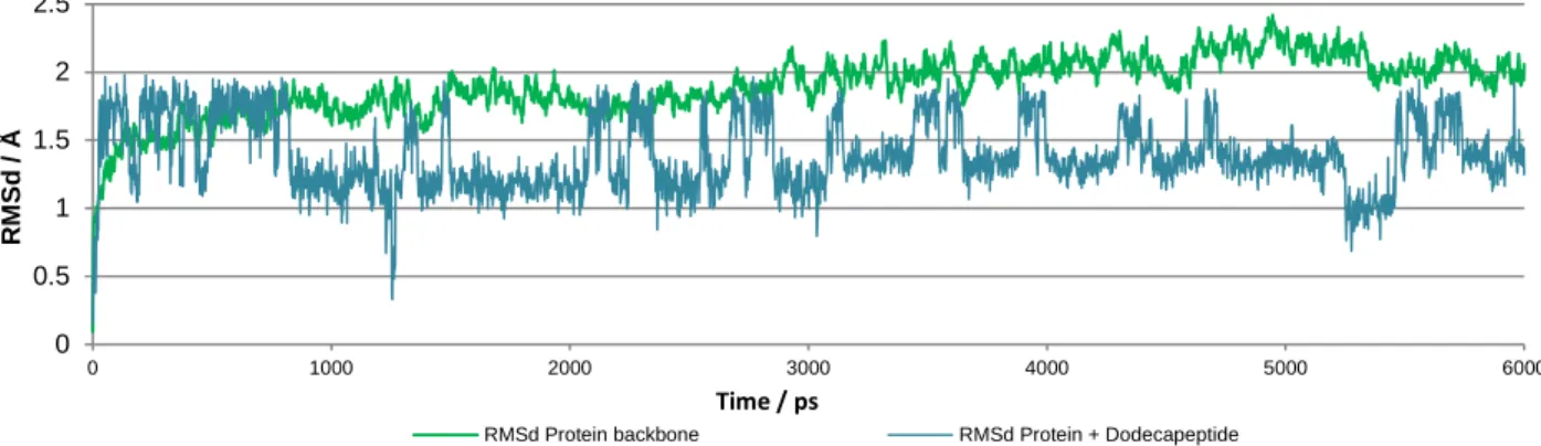
Figure 11. Renin and Renin:dodecapeptide RMSd along the MD simulation ... 54
Figure 12. RMSf of Renin and Angiotensinogen residues along the MD simulation. ... 55
Figure 13. Representation of the main active site interactions throughout MD simulation. ... 56
Figure 14. Representation of the renin:angiotensinogen system before and after the MD simulation. ... 57
Figure 15. Reactants, TS1 and INT1 obtained geometries for Ren:Angdodecapeptide:W1 model. ... 60
Figure 16. TS2 and INT2 geometries obtained for Ren:Angdodecapeptide:W1 model. ... 62
Figure 17. TS3 and Products geometries obtained for Ren:Angdodecapeptide:W1 model. ... 64
Figure 18. Reactants, TS1 and INT1 obtained geometries for Ren:Angdodecapeptide:W1:W2 model ... 67
Figure 19. TS2 and INT2 geometries obtained for Ren:Angdodecapeptide:W1:W2 model. ... 68
Figure 20. TS3 and Products geometries obtained for Ren:Angdodecapeptide:W1:W2 model. ... 69
Figure 21. Reactants, TS1 and INT1 obtained geometries for Ren:AngMutated:model. ... 72
Figure 22. TS2 and INT2 geometries obtained for Ren:AngMutated:W1 model ... 73
Figure 23. TS3 and Products geometries obtained for Ren:AngMutated:W1 model. ... 74
Figure 24. Energetic pathway for angiotensinogen hydrolysis - Ren:Angdodecapeptide:W1 model ... 76
Figure 25. Energetic pathway for angiotensinogen hydrolysis – Ren:Angdodecapeptide:W1:W2 model ... 77
Figure 26. Energetic pathway for angiotensinogen hydrolysis - Ren:AngMutated model ... 78
Figure 27. Single point calculation with B3LYP 6-31G(d) level and electrostatic embedding scheme. ... 79
Figure 29. Single Point QM/MM calculations for a model with 100 atoms in high layer ...80
Figure 30. Single Point QM/MM calculations for a model with 135 atoms in high layer ...81
Figure 31. Single Point QM/MM calculations for a model with 210 atoms in high layer ...81
Figure 32. Single Point QM/MM calculations for a model with 322 atoms in high layer ...82
Figure 33. Single Point QM/MM calculations with a large basis set (6-311++G(2d,2p)) ...84
List of equations
Equation 1. Force field potential energy ...23Equation 2. Bond Stretching potential energy ...24
Equation 3. Bond bending potential energy ...24
Equation 4. Dihedral angles potential energy ...24
Equation 5. van der Waals potential energy ...25
Equation 6. Electrostatic potential energy ...26
Equation 7- Postulate of quantum mechanics...29
Equation 8. Time-dependent Schrödinger equation ...29
Equation 9. Time-independent Schrödinger equation ...30
Equation 10. Hamiltonian operator expression ...31
Equation 11. The Born-Oppenheimer approximation. ...32
Equation 12. Slater determinant ...33
Equation 13. The Hartree-Fock equations...33
Equation 14. Fock operator expression ...34
Equation 15. Energy functional equation (DFT) ...35
Equation 16. Konh and Sham formalism ...36
Equation 17. Exchange-correlation energy ...36
Equation 18. Hybrid QM/MM energy ...41
List of tables
Table 1.Similarities and differences between Renin Inhibitors ACE inhibitors and AT1 receptors
antagonists. ... 10 Table 2. Representation of the main interactions between the active residues and the residues around
the active site during the reaction mechanism for Ren:Angdodecapeptide:W1 model. ... 64
Table 3. Representation of the main interactions between the active residues and the residues around
the active site during the reaction mechanism for Ren:Angdodecapeptide:W1:W2 model. ... 70
Table 4. Representation of the main interactions between the active residues and the residues around
Abbreviations
ACE – Angiotensin Converting Enzyme
AMBER – Assisted Model Building with Energy Refinement
Ang I – Angiotensin I
Ang II – Angiotensin II
ATreceptor – Angiotensin receptor
DFT – Density Functional Theory
GGA – Generalized Gradient Approximation GTO – Gaussian Type Orbital
H-GGA – Hybrid Generalized Gradient Approximation HM-GGA – Hybrid Meta Generalized Gradient Approximation HF – Hartree Fock
INT – Reaction Intermediates Kcat – First order rate constant
KM – Michaelis-Menten constant
LCAO – Linear Combination of Atomic Orbitals
LDA – Local Density Approximation
M-GGA – Meta Generalized Gradient Approximation MD – Molecular Dynamics
MM – Molecular Mechanics
ONIOM – Our own N-layered Integrated molecular Orbital and molecular Mechanics P - Product
PDB – Protein Data Bank PES – Potential Energy Surface
PGTO – Primitive Gaussian Type Orbital QM – Quantum Mechanics
QM/MM – Hybrid Quantum Mechanics and Molecular Mechanics R - Reactants
RAS – Renin Angiotensin System RMSD – Root Mean Square Deviation RMSF – Root Mean Square Fluctuation SCF – Self Consistent Field
SP – Single Point
STO – Slater Type Orbitals TS – Transition State
Amino acid abbreviations
Ala – Alanine Arg – Arginine Asn – Asparagine Asp – Aspartate Cys – Cysteine Gln – Glutamine Glu – Glutamate Gly – Glycine His – Histidine Ile – Isoleucine Leu – Leucine Lys – Lysine Met – Methionine Phe – Phenylalanine Pro – Proline Ser – Serine Thr – Threonine Trp – Tryptophan Tyr – Tyrosine Val – ValineC
HAPTER
1
INTRODUCTION
“Silent killer, public health crisis”
World Health Organization (WHO)
A global brief on hypertension – 2013
1.1 Introductory note
This work is being developed as part of a project with the title "New drugs for hypertension”. One of the initial goals of this project is the study of the enzyme renin as a target for the control of hypertension. As such, this dissertation will describe the catalytic mechanism of this enzyme.
In this introductory chapter a brief review of the literature on some key topics useful for this work, will be made. Multiple subjects will be discussed, since the blood pressure, renin-angiotensin-aldosterone system, to the antihypertensive therapy currently used. A special attention will be given to the crucial biological role of the enzyme renin in blood pressure control, its structural analysis, and its mechanism as an aspartic protease. Renin will be analyzed according to a systemic and biological vision and subsequently by a molecular and atomic point of view. Also in this chapter the enzymatic catalysis and the rational development of new drugs will be addressed, as well as the importance of Theoretical and Computational Chemistry as an essential tool for their study.
1.2 Hypertension
Hypertension, or high blood pressure, is an important worldwide public health challenge because of its high incidence (more than 25% of adults worldwide) and association with risks of cardiovascular and kidney diseases. It has been identified on the ranking of higher risk factor for mortality and a disease that causes disability-adjusted life-years [1]. Hypertension is a chronic medical condition in which blood pressure in the vessels is elevated. The term blood pressure usually refers to the arterial pressure of the systemic circulation and it can be summarized by two measurements, systolic (when muscle’s heart is contracted) and diastolic (when muscle’s heart is relaxed), which corresponds to a maximum and a minimum pressure, respectively. Hypertension is said to be present when the blood pressure is persistently above 140 mmHg for maximum pressure and above 90 mmHg for minimum pressure. The statistic studies predict that in 2025 about 1.5 billion of people will have hypertension, which corresponds to an increase of 60% on the actual number. This increasing prevalence of hypertension worldwide has contributed to the pandemic presence of cardiovascular diseases that are responsible for 30% of all worldwide deaths [5, 6].
Interventions that have proven effective in hypertension include weight loss, reduced intake of dietary sodium, moderate alcohol consumption, modification of eating habits and increased physical activity. However, sometimes these prevention measures are not sufficient, and secondary prevention efforts require a detection, appropriate treatment and correct control of hypertension. Furthermore, in spite of the enormous advances in antihypertensive drug therapies, the number of people with uncontrolled hypertension has continued to rise and, thus there is a huge need to improve these therapies [7, 8].
The increasing number of patients with hypertension is also attributed, in part, to the growing problem associated to risk factors such as obesity and diabetes. Despite its prevalence, only 5% of patients with hypertension have an identifiable cause [9].
Hypertension is a product of dynamic interactions between multiple factors: genetic, physiological, environmental and psychological factors. Autonomic nervous system and kidney are the major drivers of hypertension, and recently some reviews reveal that the immune system play also an essential role in the hypertension pathophysiology [10, 11]. Independent of the organ system involving in blood pressure elevation, it is clear that modifications in vascular function and structure are paramount in a physiological point of view. At the molecular level, abnormal signal transductions are responsible for modifying
cellular function involving different signaling pathways, such as phospholipase C, inositol phosphate, diacylglycerol, mitogen-activated protein kinase, tyrosine kinases /phosphatases, Rho kinases, transcription factors and NAD(P)H oxidase-derived reactive oxygen species. These important signaling pathways are stimulated by pro-hypertensive peptides like angiotensin II (Ang II) through their interaction with membrane associated G protein-coupled receptors [9, 12] . The Ang II molecule is the final product of the renin-angiotensin system (RAS) that is the main regulator system of blood pressure. The next section will discuss this system, its constituents, regulation and importance.
1.3 Renin-Angiotensin System
The RAS is the master regulator of blood pressure and fluid homeostasis. This is an endocrine system and it is activated by various signals including the reduction of blood pressure, decrease in circulating blood volume and plasma-sodium concentration [13].
As representing in Figure 1, in a summary view, this system is a multi-enzymatic cascade that starts with angiotensinogen hydrolysis. This protein is cleaved in two steps, first by renin and then by angiotensin converting enzyme (ACE), resulting in two sequential peptides, the angiotensin I (Ang I) and angiotensin II (Ang II).
The appropriate activation of the RAS is essential for preventing circulatory collapse and maintaining fluid balance. A deregulation or a persistent RAS activation leads to a blood pressure increase.
1.3.1 Renin, Angiotensinogen and Angiotensin I
The first step of RAS
The synthesis and release of renin are the key and early steps of the RAS cascade. In spite of the very early discovery of renin, in 1898, by Tigerstedt and Bergman [14], we only have recently gained a deeper understanding of the mechanisms responsible for its synthesis and release [15]. Human renin is a 340 residues protease and it can be secreted by two cellular pathways: 1) a regulated way with secretion of mature renin or 2) a constitutive way with secretion of pro-renin. Mature renin is secreted into the blood stream only from juxtaglomerular cells of the kidney, in response to different cellular stimuli: a decrease of arterial blood pressure detected by baroreceptors, a decrease in sodium
chloride levels in the nephron’s ultra-filtrate or sympathetic nervous activity, acting through β1 adrenergic receptors. On the other hand, the precursor of renin, pro-renin can be synthetized, not only in juxtaglomerular cells, but also in other tissues like adrenal, collecting duct, eyes, Müller cells, ovary, theca cells, uterus, myometrium, placenta, chronically cells, testis and submandibular gland.
Angiotensinogen, mainly expressed in the liver, is cleaved by renin to release Ang I in plasma. Then, ACE metabolizes Ang I to Ang II, which can also be converted by other non-ACE pathways. This peptide interacts mainly with the AT1 receptors in numerous organs such as kidneys, heart and vessels, and it is responsible for the increase of blood pressure. This system can be inhibited byrenin inhibitors, ACE inhibitors or AT1 receptor blockers (antagonists).
Renin is a member of the well-known family of aspartic proteases and it is responsible for the control of the first and rate-limiting step of RAS. This enzyme cleaves the 10 amino acids from the N-terminus of angiotensinogen, between Leu10 and Val11, to form Ang I.
Angiotensinogen is the only known physiological substrate for renin. In that way, renin has an essential and extremely specific role in RAS, in contrast to the ACE, as we will see later. Heart Vessels Kidney Renin Inhibitors ACE Inhibitors AT1 receptor antagonists Non-ACE pathways
Human angiotensinogen is a 452 residues long peptide mainly produced in the liver. It is also produced by other tissues as brain, immune system and kidney, but in very small quantities. RAS is regulated by a negative feedback and, therefore, plasma angiotensinogen levels are increased by a decrease in the Ang II levels. In addition, plasma corticosteroid, estrogen, and thyroid hormones levels also promote an increase of angiotensinogen levels [16].
The cleavage of angiotensinogen by renin leads to the generation of both Ang I and des(Ang I)-angiotensinogen that corresponds to the remaining residues of angiotensinogen (more than 95%). This portion doesn’t have any associated biological properties to date.
It is well known that renal renin is secreted into plasma to cleave angiotensinogen release from liver and no other function is associated with this enzyme. In addition to renin, other enzymes such as cathepsin D or G, pepsin, tonin, tripsin, have been demonstrated to convert angiotensinogen into Ang I. However, these enzymes are abundant only in specific tissues or in some specific cell types and only have this kind of activity under strict conditions. Therefore, they do not have any significant impact on the systemic production of angiotensinogen peptides [17].
The plasma concentration of angiotensinogen in humans is approximately 1 μM [18], which is close to the Michaelis-Menten constant (KM) of renin (1.25 μM) [19]. This indicates that both angiotensin and renin concentrations are important for the rate of Ang I generation. The Ang I molecule, resulting from the first step of RAS, after the cleavage of angiotensinogen by renin, appears to have no biological activity. It exists only as a precursor to Ang II formation.
1.3.2 Angiotensin Converting Enzyme and Angiotensin II
The second step of RAS
ACE is a zinc dependent carboxyl dipeptidase. It is responsible for the cleavage of 2 residues from the C-terminus of inactive Ang I and it generates the Ang II, in the classical RAS axis (Figure 1).
The Ang II levels are also detectable in the ACE gene knockout mouse, which is indicative that enzymes other than ACE may contribute to Ang II formation. However, these studies also show that Ang II formation predominantly occurs via ACE [20, 21]. Along with Ang II, ACE also cleaves other peptide substrates, including the vasodilator peptide
bradykinin, which is degraded to an inactive peptide [22, 23] . Recently, ACE has also been implicated in the regulation of the immune system [24].
In the non-classical RAS system pathway another bioactive angiotensin, Ang-(1-7), and an ACE homologue, ACE2, add further complexity to the proteolytic cascade. This path is less efficient and it consists in a first hydrolysis of Ang I, by ACE, to form Ang-(1-9), a peptide that is not known to have biological activity. Then, this peptide can be converted to Ang (1-7). ACE2 can also cleave directly Ang II to form Ang-(1-7). This last peptide has important biological activities due to its cardiovascular and baroreflex actions counteract those of Ang II [22, 25, 26].
Although we cannot set aside the existence and importance of these new pathways, they will not be subjects of the present work. Ang II peptide is then the main and higher concentrated product of this second step of RAS. This is an active peptide that binds to the angiotensin receptors to perform their functions, as we will see in the next sub-section.
1.3.3 Angiotensin receptors
The third step of RAS
The actions of Ang II are mediated by specific populations of Ang II receptors. It is known that Ang II interacts at least with two distinct Ang II receptors subtypes, designed by AT1 (Angiotensin receptors type 1) and AT2 (Angiotensin receptors type 2). These subtypes of receptors can be distinguished and characterized according to inhibition by specific selective antagonists. There are also other subtypes of angiotensin receptors (AT3 and AT4) that have already some described functions. However, their activities and behavior are still poorly understood and characterized.
The main known biological actions of Ang II are the regulation of blood pressure, release of aldosterone, electrolyte and water balance, stimulation of sympathetic transmission, cellular growth. All of them are exclusively mediated by its interaction with the AT1 receptor [27-29]. The AT2 receptor, in turn, provides a counter regulatory role to AT1 receptor over activity. Recent evidences suggest also that AT2 receptor exhibits favorable organ protection in the context of the heart, kidney and brain, mediating anti-proliferation, apoptosis and differentiation [22, 29, 30].
When Ang II binds to the AT1 receptor, which belongs to the G protein-coupled receptor superfamily, typically activates phospholipase C (coupled to a Gq protein) and leads to an increase in intracellular inositol 1,4,5-triphosphate and intracellular calcium. These intermediates are responsible for vasoconstriction effects and sodium and water retention. Downstream, protein kinase C, proteins as Ras and RhoA and tyrosine kinase cascades are amplified, affecting members of the mitogen-activated protein kinase family (ERK1/2 and p38) and JAK-STAT pathways leads to vascular and cardiac effects [31].
AT1 receptors are located in the brain, adrenal cells, heart, vasculature and kidney. In blood vessels its activation by Ang II causes vasoconstriction leading to an increase in peripheral vascular tone and systemic blood pressure [29, 31, 32].
Contrarily to the AT1 receptors, the signaling pathway mediated by AT2 receptors leads to inactivation of ERK1/2, opening of potassium channels and inhibition of T-type calcium channel. Although various signaling pathways have been assigned to the AT2 receptor, it still not clear which of these is the most important.
In summary, Ang II is a multifunctional hormone that exerts diverse physiological effects and their actions are mainly mediated by AT1 and AT2 receptors, leads to blood pressure effects.
1.4 Antihypertensive Agents
Drugs that inhibit RAS are gaining increasing popularity as initial medications for the management of hypertensive patients. Other antihypertensive agents are diuretics, beta adrenergic receptors blockers and calcium antagonists. Antihypertensive agents are used as monotherapy only in a small number of patients. The majority of people require two or more agents.
1.4.1 Diuretics
Diuretics, such as thiazides or related drugs, are some of the most frequently employed antihypertensive agents. The mechanism of action of these agents is related to the ability to increase sodium excretion as a result of binding to the thiazide receptor in the kidney. This therapeutic have, however, some side effects associated such as hypoatremia, hypokalemia, impotence and diabetes [33].
1.4.2 β-blockers
Other therapeutic agents are the β-blockers that have been used for more than 30 years. Some of the β-blockers are selective for β1 adrenergic receptors, others are non-selective. Bradycardia, atrioventricular block or arrhythmias are normally associated to β-blockers. They may also adversely affect the lipid profile with increase of triglycerides in serum and are associated with diabetes. The mechanism of action of β-blockers may depend on the reduction in cardiac output [33-35].
1.4.3 Calcium channel blockers
The calcium channel blockers can also be used as antihypertensive agents. They act as peripheral vasodilators as they block the entry of calcium through voltage dependent calcium channels and they include dihydropyridines and non-dihydropyridines. These agents induce oedema, flushing and sometimes headache and palpitations [36] .
1.4.4 Vaccination
An old concept that has now resurrected is the use of vaccination to treat hypertension [37]. Some studies shows that a vaccine that induces antibodies against Ang II has yielded promising results in terms of blood pressure reduction [32]. However, the specificity and controllability of this kind of treatment needs to be evaluated.
1.4.5
RAS inhibition
The drugs that act in RAS system are also used as antihypertensive drugs. The pharmacological inhibition of classical RAS can be achieved through 3 different basic mechanisms, like we can see in Figure 1:
1) Inhibition of Ang I generation from angiotensinogen. This can be achieved by direct inhibition of renin.
2) Inhibition of Ang II generation from Ang I. This can be achieved through inhibition of ACE that, as we saw previously, is responsible for the formation of the octapeptide Ang II.
3) Inhibition of the action of Ang II at the level of its receptors.
1.4.5.1
Renin inhibitors
The most logical point to block pharmacologically the RAS is the rate limiting step catalyzed by renin enzyme. Intervention at this point is more specific than ACE inhibitor or AT1 antagonists, because almost any angiotensin peptide can be generated, and no other peptide system would be affected. The renin inhibitors, due to its advantages, have been studied and developed decades ago, but the first commercialized renin inhibitor, the aliskiren, came onto the market recently [38]. Aliskiren blocks the RAS at the origin of the cascade inhibiting the generation of Ang I, and consequently the generation of Ang II. As the main goal of this work is the study of the catalytic mechanism of renin, to allow future studies on its inhibition, it is essential to understand its role, and therefore more emphasis to this enzyme and its inhibitors will be given later.
1.4.5.2
ACE inhibitors
The first RAS inhibitor was an ACE inhibitor. Its intervention began in 1960’s with the discovery of venom of the snake Bothrops jararaca, which contains a substance that inhibits ACE. By modelling the active site of ACE, several new drugs that potentially bind this site were designed, and the first orally available ACE inhibitor was captopril [39]. Today more ACE inhibitors are available on market. These drugs proved to have a strong beneficial effect on morbidity and mortality in congestive heart failure and renal diseases. As we mentioned before, ACE is also a bradykinin degrading enzyme, therefore its inhibition also increases bradykinin concentration that is a vasodilator peptide. It has also been show, by other studies, that bradykinin potentiation is involved in the cardio and renal protection. However, this peptide may also be the major reason for adverse side effects of these inhibitors, as cough and angioedema [33].
1.4.5.2
AT
1receptor antagonists
A third group of drugs that interfere with RAS are specific antagonists for the AT1 receptor. The first example of this class was losartan, which was followed by several other sartans. These drugs exert a more complete angiotensin blockade because other pathways
of angiotensin generation, such as cathepsins, tonin or chymase, that are not affected by ACE inhibitors, become ineffective by AT1 antagonism. These drugs are also more specific for the RAS, than ACE inhibitors, because other peptide systems are not affected. The compensatory increase in renin concentration after AT1 blockade leads to an accumulation of Ang II, which activates the AT2 receptor. However, it is yet unknown whether this AT2 stimulation often followed by bradkynin generation that is involved in the action of AT1 antagonists [32, 40]. AT1 blockers and ACE inhibitors work by different ways and both have effects on lowering the blood pressure, however, ACE inhibitors are currently best documented then AT1 receptor antagonists. The choice of the best drug depends also on the patient.
The similarities and differences between renin inhibitors, ACE inhibitors and AT1 antagonists are summarized on the Table 1. In this table, it is evident that the renin inhibition is the most rational target to control the RAS system and consequently the blood pressure. It is important to refer that by inhibiting ACE or blocking the AT1 receptors, the negative feedback control, exerted by angiotensin II in the renin concentration, disappear and consequently an elevation on plasma and tissue renin activities are observed. Renin inhibition leads also to an increase on renin secretion and production but, in contrast to others RAS blockers, the newly release active renin is immediately neutralized by inhibitors present in the systemic circulation [41].
Table 1.Similarities and differences between Renin Inhibitors ACE inhibitors and AT1 receptors antagonists.
(+) symbol represents an increase of concentration and (-) symbol corresponds to a decrease of concentration. This table was adapted from [16].
Renin Inhibitors ACE Inhibitors AT1 Antagonists
Plasma Renin Concentration
+
+
+
Plasma Renin Activity
-
+
+
Angiotensin I
-
+
+
Angiotensin II
-
-
+
Bradykinin Unchanged
+
UnchangedAT1 Receptor Not stimulated Not stimulated Blocked
AT2 Receptor Not stimulated Not stimulated Stimulated
Making a point of the situation of what has been described hitherto, the hypertension was initially introduced as a concern disease due to its direct relationship with cardiac and renal diseases. Then, the main regulatory system of blood pressure (RAS) was briefly
presented, with some emphasis to the first step. Finally, the most common antihypertensive therapeutics was summarized.
From now on, the renin enzyme will be the main focus of this work. Its structure, function and inhibition will be discussed, due to this enzyme appears to be the most logical target for the development of new antihypertensive agents.
1.5
Renin
1.5.1
Relation between structure and function
As previously mentioned the renin enzyme cleaves angiotensinogen to release the N-terminal decapeptide Ang I. Efforts into the elucidation of the molecular architecture of renin only succeed when they could be expressed and purified from recombinant sources.
Renin (EC 3.4.23.15) is a mono-specific 340-residues protease polypeptide, with approximately 40 000 KDa that belongs to aspartic protease superfamily [42].
Structurally, renin consists of 2 lobes with different residues sequence but similar structure. These lobes fold mainly β sheet conformation, with a deep cleft between them that is characteristic from the aspartic proteases class. The residues around the cleft correspond to the active site and it accommodates seven residues from its substrate, angiotensinogen (Figure 2C). It has been shown that the minimal substrate sequence that has good affinity and reactivity is an octapeptide constituted by the residues between 6 and 13 of angiotensinogen. The catalytic activity of renin is due to two aspartic acid residues (32 and 215 or 38 and 236 in pepsin and human renin numbering, respectively), each located in a single lobe and held coplanar by a network of hydrogen bonds involving the surrounding main chain and conserved residues side chain groups. Like happens in other aspartic proteases, there are a water molecule in the active center of renin that hydrogen bonding with all four carboxyl oxygen atoms of aspartic catalytic dyad, and it has been implicated in catalysis. It has been suggested that this water molecule is partly displaced near the substrate and it is polarized by one of the catalytic aspartate residues.
The structure of renin has both hydrophobic and hydrophilic residues. The hydrophobic ones are more concentrated inside the protein and form hydrophobic cavities in the active site [41, 43, 44].
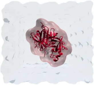
A) Cartoon representation of human renin. B) Cartoon representation of human angiotensinogen interacting with the deep cleft of renin (C). The structure was adapted from 2X0B PDB file.
In the same way, as in the other aspartic proteases, renin possesses a flexible “flap” constituted by residues in a β-hairpin that is close to the active site and allows cleft covering. Recently, the dynamical behavior of this flap was studied by Molecular Dynamics (MD) simulations, and it was verified that it oscillates between open, semi-open and closed conformations could allow the binding of a wide range of inhibitors [45].
The active site of renin was studied by other authors and it is composed of 8 sub-pockets. The characteristics of each pocket was reviewed by Webb [46]. The S1 and S3 pockets forms a continuous and hydrophobic cavity and they contains the S3sp sub-pocket, which is specific to renin and unique among the aspartic proteases and have also hydrophobic and hydrophilic characteristics. The S2 and S4 pockets have also hydrophobic characteristics. The S1’ are predominantly hydrophobic, while S2’ have polar character (hydrophilic). This kind of information has a very important role for the future design of new renin inhibitors.
It is known that renin is very specific for its unique substrate and an important question that arises is that, if the main chains of proteases are conserved, how are the differences in
Figure 2. Human renin structure complexed with its substrate
A
B
specificities achieved? Probably the specificities derive from slight differences in the sizes of residues in the specific pockets (Sn) to complement the corresponding side chain of the substrate. For example some studies have predicted the specificity site S3 to be larger in renin than in other aspartic proteases and that a movement of a helix makes the pocket quite compact and complementary to aromatic rings. Therefore, the position of an element of secondary structure differs between renin and other aspartic proteases with important differences in the pocket specificity. As it happens in S3 pocket, the differing positions of secondary structure elements may also account for the specificities of the other pockets. Previous studies were also suggested that this high specificity is due to the rigidity of its binding pocket. However, the reason of the high renin’s specificity is still unknown [47, 48].
1.5.1
Proposed for Renin catalytic mechanism
The main objective of this work is to clarity with atomistic detail the catalytic mechanism of human renin by a computational approach. The mechanism of action of other aspartic proteases such as HIV-1 protease, presinilin and β-secretase was previously studied with MD simulations and combined Quantum Mechanics and Molecular Mechanics calculations (QM/MM) [49-53]. All of these computational techniques will be discussed in the following chapter.
There are proposals for the catalytic mechanism of renin but, its atomistic details remain unclear. Our group also published a proposal for the catalytic mechanism of mouse renin [4], but for the human enzyme, the mechanism has not been described yet.
The proposal mechanism of action of human renin involves an acid-base mechanism as it was described for other aspartic proteases. One of two catalytic aspartic residues is protonated, whilst the other, with a negative charge, acts as a general base, activating a water molecule tightly bound between both aspartate residues. Then, the water molecule makes a nucleophilic attack on the carbonyl carbon of the scissile bond resulting in the formation of a tetrahedral gem-diol intermediate (Figure 3A-B). The second step involves a protonation of the peptidic nitrogen by one of the catalytic aspartate and a deprotonation of one of the gem-diol hydroxyl group by the other aspartate. Subsequently, this step leads to the cleavage of the peptidic bond (Figure 3C-D).
In the beginning of the reaction the two catalytic aspartate residues have an adjacent position and they have different pKa values, one being protonated and the other ionized at
neutral pH. This result was previously demonstrated by experimental and theoretical studies [54].
A) Representation of the aspartic proteases active site with two Asp residues and a catalytic water molecule. In first step of the proposal catalytic mechanism a water molecule makes a nucleophilic attack on the carbonyl carbon of the scissile bond resulting in the formation of a gem-diol intermediate (B). C) In a second step, the nitrogen of the scissile bond is protonated by the catalytic Asp residue and this step ends with the peptidic bond cleavage (D).
It is also important to say that, in aspartic proteases, the residues around the catalytic center play an essential role in the catalytic mechanism. Furthermore a second conserved water molecule seems to have a structural role due to its participation in an H-bond network (Asp-Ser-W2-Tyr-Trp) that stabilizes the flap conformation during the catalysis [54]. However the role of this water molecule in the catalysis is still unknown.
1.5.3
Development of renin inhibitors
Like we mentioned before, the use of the drugs that inhibit the RAS system is an effective way to intervene in the control of hypertension. Considering the higher specificity of renin, the inhibition of this enzyme has benefits by decreasing its activity without interference with other metabolic pathways [55].
Pepstatin was the first renin inhibitor. It was found to be a competitive inhibitor of most aspartic proteases.
Figure 3. General proposal mechanism of amine bond hydrolysis by aspartic proteases. C
D
B A
Then, considering that it was widely recognized that direct renin inhibition held great therapeutic potential, the design of new molecules that interact with the catalytic center of renin has been studied. First generation of renin inhibitors were peptide analogues (peptidomimetics) of the amino-terminal sequence of angiotensinogen. These analogues contain the renin cleavage site which, with further chemical modifications, led to the development of compounds as CGP29387 that had greater stability and long duration action, but it only have effect in high doses [56]. The second generation of renin inhibitors is characterized by a molecular weight of a tetrapeptide such as enalkiren, CGP38560A, ramikiren and zankiren. When given orally, these compounds had low bioavailability (< 2%), a short half-life and weak blood pressure lowering activity [57]. Later, the aliskiren was discovered as an orally active renin inhibitor by Ciba-Geigy (now Novartis). After successful preclinical and clinical testing it was approved and commercialized [58, 59]. Actually, with the help of crystallography and molecular modeling, the substituted piperidine renin inhibitors have been developed. However, the majority of them are abandoned after preclinical tests and no one was approved until now [55]. The Aliskiren, the unique drug commercialized for renin, interacts with several binding pockets in distinct regions around the active site. In particular it was found to bind the S3sp specific sub-pocket. This inhibitor has been shown, in experimental and clinical studies, to be effective in lowering the blood pressure and in organ protection. The Aliskiren effects are similar to those of other RAS inhibitors and normally it is used in combination with other antiherpentensive drugs.
Given the continuous prevalence of hypertension is still need to develop new drugs for this disease. With the advancement of computational techniques in enzymatic studies, the design of new effective drugs may be easier.
1.6
Computational enzymatic catalysis and
drug design
The aim of this work involves the study of the catalytic mechanism of the renin enzyme. Therefore, a brief approach to enzymatic catalysis is also necessary in this introductory chapter. In fact, living cells and in turn multicellular organisms, depend on chemical transformations for every essential life processes. If these essential biochemistry reactions proceed at non catalyzed rates they are too slow (million years!) to sustain life. It is evident
that a very slow reaction cannot sustain life without some very significant rate enhancement. The enzymes are responsible for this enhancement and, therefore, enzymatic catalysis is essential for all life.
1.6.1
The role of computational chemistry in enzymatic catalysis
The first question to be asked is ‘What is the catalytic mechanism of a reaction?’ Establishing a mechanism of a reaction means identifying which groups in the protein are involved in the reaction, and clarifies their precise roles during catalysis. There are many experimental methodologies such as protein crystallography, NMR, site-directed mutagenesis, kinetics, isotopic labeling and spectroscopic those provide fundamental insights to the study of enzymatic catalysis. Research in the establishment of enzymatic mechanisms has suffered great advanced with new efforts in computational chemistry, which allows the atomistic description of all mechanistic details.
Predicting an enzymatic catalytic mechanism is generally a long and complex job. A complete description involves the identification of reactants (R), intermediates (INT), transition states (TS) and products (P) along the reaction coordinate, as well as the free energies of activation and reaction for each of the reaction steps.
In some cases, experimental methodologies allow the determination of the initial enzyme-substrate complex, the identification of some intermediates and the determination of the free energy of activation of the rate-limiting step. However, the structure of TS and the energies associated to them are impossible to determine by these techniques, because of the reduced lifetime of these species (10-13 seconds that corresponds to the time for a single bond vibration).
On the other hand, computational methodologies offer opportunity to study with atomic detail the enzymatic mechanism, providing atomistic insight in very important processes. All these facts indicate that computational studies have an essential and very important role in mechanistic studies. They are able to correctly identify and describe in a structural and energetic view the Transition states, independently of their lifetime, which complement the information obtained from experimental works [60-62]. In addition, computational methodologies make possible the study of large systems, like enzymes, where the reactive region is only a small part of the system. Thus the remaining structure of the enzyme and its role as a modulator of the active center, either stabilizing or destabilizing along the potential energy surface (PES) is also evaluated.
To study the correct mechanism of an enzymatic reaction, we need to search for the minimum energy pathway in the PES of the study system. The realism and success of the computational data for a mechanistic pathway depends on the system’s Hamiltonian accuracy (the Hamiltonian will be explained latter), the sampling of the reactional space and the sampling of the conformational space [60, 63]. One of the key to elucidating the catalytic mechanism of an enzymatic reaction is the understanding of specific interactions between the substrate and its enzyme environment and their effect on the reaction rate.
1.6.2
Rational drug design
In drug discovery, enzymes are important as targets for new drugs (most part of drugs are enzyme inhibitors). The identification and description of enzymatic catalytic mechanisms add another dimension to structure based ligand design for enzyme targets. Modeling can also identify catalytically important interactions that could be exploiting in drug design.
All chemical enzymatic reactions pass through an unstable structure called the transition state (TS) with is poised between the chemical structures of the substrates and products. Enzymes stabilize the TS of their reactions. By binding the TS more tightly than the substrates or the products, the enzyme is responsible for lower the energy barrier of the reaction. Consequently a TS analog will bind more tightly to the enzyme than either the substrates or the products, preventing them from binding to the enzyme and reacting.
For aspartic proteases, like HIV-1 protease, there are many inhibitors that were obtained by drug design with TS analogs. Nowadays, HIV-1 protease inhibitors constitute around 40% of available drugs against HIV. Nearly all of them contain hydroxyethylene units as TS analog mimic. Thus, the design and success of HIV-1 protease inhibitors represent one of the most remarkable achievements of drug design [64].
Due to rapid technological progress in chemistry, bioinformatics, structural biology and computer technology, studies of computer aided drug design play more and more important role in this respect.
1.7
Purpose of the present work
As mentioned before, RAS represents the most important regulating system of the arterial blood pressure. The renin enzyme, an aspartic protease, has an exclusive activity
and its determinant velocity on the RAS cascade makes it an ideal target for new antihypertensive drugs. Therefore, the main purpose of the present work is describing with atomistic detail, the catalytic mechanism of this human renin by computational methods.
In addition, this study will also assess the role of a structural water molecule in the catalytic mechanism of this enzyme. Moreover, the role of residues around the active center, in the stabilization of the reaction intermediates, will also be evaluated. Finally, the influence of a mutated substrate, in the reaction catalyzed by renin, will also address.
This kind of knowledge has a very important role in future drug design which will enable the control of the “silent killer” and the “public health crisis” that is hypertension.
C
HAPTER 2
THEORETICAL BACKGROUND
“The underlying physical laws necessary for the mathematical theory of a large part of physics and the whole of chemistry are thus completely known, (…) and the difficulty is only that the exact application of these laws leads to equations much too complicated to be soluble.”
Dirac, 1929
2.1
Introduction
Theoretical chemistry aims to develop algorithms, methodologies and theories that explain and reproduce systems at atomic and molecular levels. When they are applied computationally, the methods of theoretical chemistry, seek the clarification of chemistry and biological systems at an atomistic level.
If we wish to study the catalytic mechanism of an enzyme, as is the main purpose of this dissertation, we must use theoretical and computational methods. Computational biochemistry is able to predict results and deductions that are very expensive and time consuming when they are obtained experimentally. These methods are still able to interpret
experimental results and complement them, as well as suggest new interpretations for a better understanding of these results to clarify relevant doubts that may exist. Therefore, experimental techniques and theoretical techniques go hand in hand to try to explain chemical and biological problems at the molecular level.
Since the discovery of the resolution of the Schrödinger equation for the hydrogen atom in 1926 [65], quantum mechanics has been increasingly used in an attempt to interpret and predict the electronic structure and behavior of bio/chemical systems. A great number of efficient computer programs has been developed, which try to study with increasing accuracy large size systems, such as biological enzymatic systems. Due to the fast progress of computing power, the application of these methods to the study of biological systems has been widely used and the results obtained have been more and more precise. Through computational methods applied to Biochemistry it is possible to determine individual properties of biological molecules such as reaction energies, reaction mechanisms, dipole moments, charge distribution, interaction between different molecules among others. Thus, Computational Biochemistry is an extended scientific area that can be combined with any other area of Biochemistry, promoting the development of useful applications in Chemistry, Biology or Medicine. For example, the clarification of enzymatic reaction with computational methods can be extremely useful for the rational design of new molecules/drugs to receptors or enzymes in order to control, or even cure, some diseases.
Nowadays, Theoretical Chemistry methods are increasingly sophisticated and may be applied to more complex systems. Generally theoretical methodologies can be classified into four main categories:
1. Ab initio methods
2. Density Functional Theory (DFT) 3. Semi-empirical methods
4. Molecular Mechanics (MM)
Ab initio methods do not include any empirical parameters in their equations, and they
are only based on the laws of quantum mechanics (QM). They try to solve the equation of Shrödinger as accurately as possible through a strict set of mathematical approaches. Those methods potentially provide very accurate results. However, they require a high computational cost, and therefore, they are only used for small size systems. DFT methods
are also quatum mechanical but they are bases on the total electronic density of the system.
Semi-empirical methods maintain the quantum nature of the calculation, but, as the name
suggests, parameters from experimental data can be introduced in the calculations. These methods are quite computationally inexpensive and rely on a series of approximations. MM methods are very useful when we have very large systems to study, as is the case of enzymes. In MM, the atom is the fundamental particle and classical mechanics laws are used to predict the behavior of the system, contrarily to the methods presented before. In that way, the MM force fields do not allow for an electronic description of the system and the description of bond breaking and forming is not possible. In addition to the methods presented previously, it is also possible to use hybrid methods in the study of large systems like enzymes. These include a combination of QM and MM methods, where the system is divided in two or more fractions and each part is calculated with one distinct method.
In the following sections the computational methods used in this work, as well as their development and application, will be reviewed briefly. The first section of this chapter will deal with the molecular mechanics and then another section will be dedicated to quantum mechanics
2.2 Molecular Mechanics
Molecular mechanics (MM) is a simplified approach to chemical problems. If we want to explore geometries, relative energies of conformers of the same molecule or study the behavior of docked substrates into active sites, MM methods are very effective tools. They are based on classical mechanics and they are very useful to perform calculations on systems containing a significant number of atoms [66-68].
In MM the smallest particle is the atom, which means that neither electrons nor protons are explicit considered and the potential energy depends only on the atom position. Molecules are treated as a set of charged point masses (the atoms) which are coupled together with springs. The total energy of a structure is calculated using an analytical function that sums the individual energy terms. Generally, the function includes bond stretching, bond angle bending, torsion, and non-bond interaction terms.
MM falls apart when one wants some deeper understanding of the system. If we are studying a reaction mechanism profile, breaking or forming bonds, we cannot use the MM