Marta Salgueiro Alves
Antibiotic resistance in Escherichia coli isolates
from different sources
Resistência a antibióticos em isolados de
Escherichia coli de diferentes fontes
Ano 2013
Marta Salgueiro Alves
Antibiotic resistance in Escherichia coli isolates
from different sources
Resistência a antibióticos em isolados de
Escherichia coli de diferentes fontes
Tese apresentada à Universidade de Aveiro para cumprimento dos requisitos necessários à obtenção do grau de Mestre em Biotecnologia Industrial e Ambiental, realizada sob a orientação científica da Doutora Isabel da Silva Henriques, Investigadora Auxiliar do CESAM, Universidade de Aveiro e co-orientação do Professor Doutor António Carlos Matias Correia, Professor Catedrático do Departamento de Biologia, Universidade de Aveiro.
Apoio financeiro do FEDER no âmbito do programa COMPETE e da FCT- Fundação para a ciência e tecnologia através do projeto SEAGULL
(PTDC/AAC-AMB/109155/2008 e FCOMP-01-0124-FEDER-008640)
“Em cada um de nós há um Egipto e um Faraó e um Moisés e uma Liberdade numa Terra Prometida. E, a cada momento no tempo, há uma oportunidade para outro Êxodo. (…) Mas a Liberdade e a Terra Prometida não são elementos estáticos que ficam ali à espera. São conquistas tuas, que podes criar a qualquer momento, em qualquer coisa que fizeres, só por te livrares de quem eras na véspera.”
o júri
presidente Prof. Doutora Sónia Alexandre Leite Velho Mendo
professora auxiliar com agregação do Departamento de Biologia da Universidade de Aveiro
Doutora Célia Maria Manaia Rodrigues
professora auxiliar da Escola Superior de Biotecnologia da Universidade Católica Portuguesa
Doutora Isabel da Silva Henriques
investigadora auxiliar do Departamento de Biologia da Universidade de Aveiro
Prof. Doutor António Carlos Matias Correia
agradecimentos Agradeço ao Prof. Doutor António Correia por me ter recebido no seu laboratório, no início como voluntária e agora, na minha tese de mestrado. Admiro a forma como ensina, a facilidade com que transmite o conhecimento: inspirou-me e despertou em mim a curiosidade em relação à microbiologia. Se for este o objectivo de um professor, no que me toca, foi cumprido.
À Doutora Isabel Henriques agradeço por me ter recebido no seu grupo de trabalho como voluntária inicialmente e agora pela orientação nesta dissertação. Admiro o gosto e a curiosidade que demonstra ter no seu trabalho. Fico-lhe grata por tudo o que me transmitiu.
Agradeço ao Doutor Bruno Castro a disponibilidade e o que me ensinou sobre estatística.
A todos os colegas do Microlab que directa ou indirectamente me ajudaram, ou com boas dicas para o trabalho ou com boa disposição, obrigada. Tenho uma grande estima por todos e a todos desejo o melhor, vocês ganharam uma amiga, nada a fazer, lidem com isso!
Susana agradeço-te a paciência, a disponibilidade, a amizade e o incentivo. Foi um prazer trabalhar contigo, sempre correcta e profissional! Obrigada pelo que me ensinaste. Não esqueço o companheirismo e estou aqui para te retribuir! Desejo-te tudo de bom, mereces o melhor!
Agradeço ao Paulo o amor. Nada me ensinaste sobre bactérias, não te despertam interesse, nem um bocadinho, torces para que os antibióticos ganhem a batalha. Mas gostas de mim e eu de ti e ao longo deste ano as bactérias lidaram bem com isso.
Agradeço aos meus pais, Lina e Quim, meus companheiros, o apoio, o carinho e o amor que me dão todos os dias. Todos. É incondicional, eu sei disso e isso dá-me força e segurança.
palavras-chave Resistência a antibióticos, contaminação fecal, gaivotas, Escherichia coli, Ilhas das Berlengas
resumo A descoberta e produção de antibióticos foi um grande avanço para a medicina na primeira metade do século passado. No entanto, as bactérias adaptaram-se rapidamente desenvolvendo mecanismos de resistência aos antibióticos cuja disseminação é facilitada pela transferência horizontal de genes. Hoje em dia a resistência a antibióticos constitui um problema de saúde pública sendo detectada não só a nível clínico, mas também em ambientes naturais, com particular destaque para os ambientes aquáticos.
As ilhas das Berlengas são uma reserva natural. No entanto tem sido detectada poluição associada a contaminação fecal na água da praia. Estudos anteriores concluíram que a principal origem desta contaminação são as fezes de gaivotas existentes na ilha.
O objectivo principal deste estudo foi analisar o perfil de resistência a antibióticos de isolados de Escherichia coli obtidos da água da praia da ilha das Berlengas, de fezes de gaivotas e do único efluente de águas residuais de origem humana existente na ilha. Com isto, pretende-se avaliar o risco para a saúde pública da contaminação fecal da água e confirmar a origem dessa poluição. Foi também objectivo deste trabalho identificar marcadores associados a resistência a antibióticos que contribuam para descriminar as fontes de poluição fecal.
Neste sentido procedeu-se à classificação dos 414 isolados de E. coli das diferentes fontes de acordo com os principais grupos filogenéticos (A, B1, B2 e D): mais de 70% dos isolados pertenciam aos grupos A e B1 geralmente associados a estirpes comensais. Nas três fontes estudadas existem isolados do grupo A, B1 e D. Verificaram-se isolados do grupo B2 apenas no efluente (10,1%) e em fezes (3,9%).
Foi avaliada a susceptibilidade a antibióticos usando o método de difusão em disco. Globalmente registaram-se para a ilha das Berlengas elevadas percentagens de resistência aos antibióticos testados, com prevalência a nível das penicilinas, aminoglicosídeos e tetraciclinas. A resistência a cefalosporinas de 3ª geração, ao imipenemo e ciprofloxacina foi rara e mais frequente em isolados do efluente. Registou-se uma elevada taxa de isolados multiresistentes (cerca de 30%). Foram pesquisadas as bases genéticas para os fenótipos obtidos, sendo blaTEM, tet(A) e sul2 os genes mais frequentemente detectados. Verificou-se a
ocorrência de blaCTX-M-1 num isolado de água e blaCMY-2 num de fezes. O contexto
genético determinado para estes genes foi idêntico ao previamente descrito para isolados clínicos.
Obtiveram-se diferenças significativas entre o padrão de resistência da água e do efluente. Além disso, alguns dos fenótipos e genótipos detectados em isolados de água ocorreram apenas numa das fontes: fezes (em maior número) ou efluente. Para a utilização destes fenótipos e genótipos como marcadores seriam necessários mais estudos. Este estudo mostra que a poluição fecal associada a fezes de gaivotas, embora geralmente considerada menos grave que a associada a fezes de humanos, pode constituir um risco para a saúde pública.
keywords Antibiotic resistance, fecal pollution, seagull, Escherichia coli, , Ilhas das Berlengas
abstract The discovery and production of antibiotics was a major breakthrough for medicine in the first half of the last century. However, bacteria have adapted quickly through the development of antibiotic resistance mechanisms that spread easily by horizontal gene transfer. The acquisition and dissemination of resistance were promoted by the intensive (mis)use of antibiotics.
Nowadays, antibiotic resistance is a public health problem and is found not only in clinical isolates but also in natural environments with particular emphasis for the aquatic ones.
The Berlengas Islands are a natural reserve. However it was detected fecal contamination in the beach water, for which the main origin was determined to be, in previous studies, the seagull feces.
The aim of this study was to analyze the antibiotic resistance of Escherichia coli isolates from the Berlengas beach water, gull feces and from the only human-derived wastewater effluent in the Island and so to assess the risk to public health of the fecal contamination and confirm its origin. It was also a goal to identify markers based on antibiotic resistance to fecal pollution sources discrimination.
In this sense we proceeded to the classification of the 414 Escherichia coli isolates from different sources in accordance with the main phylogenetic groups (A, B1, B2 and D), and over 70% of the isolates belonged to groups A and B1, usually associated with commensal strains. These two groups, along with group D, were the most frequent in all sources. Group B2 was only present in effluent (10.1%) and in a lower percentage in feces (3.9%).
The assessment of antibiotic susceptibility was performed for all isolates using the disk diffusion method. Overall, high percentages of resistance to the antibiotics tested were detected in the Berlengas island, particularly to penicillins, aminoglycosides and tetracyclines. Resistance to 3rd generation
cephalosporins, to imipenem and ciprofloxacin was rare but more frequent in effluent isolates. It was also observed a global high rate of multiresistant isolates (around 30%). It was investigated the genetic basis for the phenotypes obtained, and blaTEM, tet(A) and sul2 genes were the most frequently detected
genes. blaCTX-M-1 was detected in one water isolate and blaCMY-2 in one feces
isolate. The genetic context determined for these two genes was identical to what has been described for clinical isolates.
There were significant differences between the resistance patterns of water and the effluent. Some phenotypes and genotypes observed in water were only present in one of the other two sources: feces (in major number) and effluent. The potential use of these phenotypes and genotypes as markers of these pollution sources must be further investigated.
This study demonstrates that fecal pollution associated to gull feces, though generally considered less dangerous than human fecal pollution, may also constitute a risk to public health.
I. INTRODUCTION ... 1
1. Antibiotics ... 1
1.1. Classes of antibiotics and their mechanisms of action ... 2
1.1.1. β-lactams ... 3
1.1.2. Other classes of antibiotics... 3
2. Antibiotic resistance ... 6
2.1. Intrinsic and acquired resistance ... 8
2.1.1. Mechanisms of antibiotic resistance transfer ... 8
2.2. Mechanisms of resistance ... 10
2.2.1. β –lactams ... 10
2.2.2. Sulfonamides and Trimethoprim ... 12
2.2.3. Aminoglycoside ... 12 2.2.4. Chloramphenicol ... 12 2.2.5. Tetracyclines ... 12 2.2.6. Glycopeptides ... 13 2.2.7. MLS ... 13 2.2.8. Quinolones ... 14
3. Environmental spread of antibiotic-resistant bacteria ... 15
3.1. The role of HGT in the spread of ARG in the environment ... 16
3.2. Aquatic ecosystems as reservoirs of antibiotic resistance genes ... 17
3.3. Public health concern: ARGs as environmental pollutants ... 20
4. Antibiotic-resistant bacteria in coastal environments ... 21
4.1. Antibiotic resistance genes in coastal waters ... 21
4.2. Seagulls and antibiotic resistance dissemination ... 22
7. Aims of the work ... 28
II. MATERIAL AND METHODS ... 29
1. Culture media ... 29
1.1. Luria-Bertani (LB) medium: ... 29
1.2. Mueller-Hinton (MH) Agar (acc. to CLSI) ... 29
2. General reagents and solutions ... 30
2.1. 1x Tris-Acetato EDTA (TAE) buffer (5 Prime, Deutschland) ... 30
2.2. Tris-EDTA (TE) ... 30
2.3. 6x Loading Dye (MBI Fermentas, Lithuania) ... 30
3. Establishment of an E. coli collection ... 31
4. Antibiotic susceptibility testing ... 32
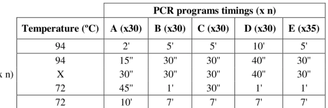
5. General conditions for Polymerase Chain Reaction amplification ... 34
5.1. E. coli phylogenetic group determination ... 34
5.2.1. Detection of blaAmpC-like genes by multiplex PCR ... 38
5.3. Determination of ARGs genomic context ... 39
6. DNA electrophoresis and visualization ... 39
7. PCR products purification and sequencing ... 40
8. Statistical Analysis ... 41
III. RESULTS ... 42
2.1. Antimicrobial resistance vs. phylogenetic group ... 50
2.2. Statistical analysis of the AR phenotypes... 52
3. Antibiotic resistance genes ... 56
3.1. Genomic context of the blaCTX-M-1 and blaCMY-2 genes ... 61
3.2. Statistical analysis for the AR genotypes. ... 62
IV. DISCUSSION ... 67
V. CONCLUSIONS ... 74
VI. REFERENCES ... 75
VII. APPENDICES ... 85
Appendix A - DNA Molecular weight marker ... 85
Appendix B – Sequences annotation ... 86
B.1. blaCTX-M-1... 86
INDEX OF ABREVIATIONS
µL: micro litter
6-APA: 6-amonipenicillanic acid AMC: Amoxicillin/ clavulanic acid AML: Amoxicillin
AMP - Ampicillin
AR: Antibiotic Resistance
ARA: Antibiotic Resistance Analysis ARGs: Antibiotic Resistance Genes
BOX-PCR: BOX elements –
polymerase chain reaction bp: base pare
C: Chloramphenicol CAZ: Ceftazidime CIP: Ciprofloxacin
CLSI: Clinical and Laboratory Standards Institute
CN: Gentamicin CTX: Cefotaxime
d-Ala–d-Ala: d-alanyl–d-alanine
dH2O: distilled water
DHFR: dihydrofolate reductase DHPS: dihydropteroate synthase
DNA: deoxyribonucleic acid
EDTA: Ethylenediaminetetraacetic acid
ESLBs: Extended-Spectrum
β-lactamase(s) g:grams
HGT: Horizontal Gene Transfer IMP: Imipenem
KF: Cefalotin
LA: Luria Bertani Agar LB: Luria Bertani Broth M: Molar
MGEs: Mobile Genetic Elements min: minutes
mL: milliliter
MLS: Macrolides, Lincosamides, Streptogramins
MST: Microbial Source Tracking NA: Nalidixic acid
no: number ºC: degrees Celsius
PBP: Protein Binding Protein
PCR: polymerase chain reaction pmol: picomole
PRL: Piperacillin
RDA: Redundancy Analysis S: Streptomycin
SXT: Sulfonamide+trimethoprim TAE: Tris-Acetato EDTA
TE: Tetracyclin TE: Tris-EDTA
TZP: Piperacillin/ tazobactam
UNESCO: United Nations Educational, Scientific and Cultural Organization UV: Ultraviolet
1
I.
INTRODUCTION
1.
Antibiotics
In 1929 Alexander Fleming discovered that the fungi Penicillium notatum
produced penicillin, a compound with antibacterial properties (Fleming 1929). In 1942
Waksman denominated “antibiotics” all the natural compounds produced by
microorganisms that inhibit microbial growth or have microbiocide effect (Sousa 2005).
Natural antimicrobials are products of the microorganisms’ secondary metabolism
differing from the synthetic ones, or semi-synthetics, like the ones obtained through the core of penicillin 6-amonipenicillanic acid (6-APA), like ampicillin. With the increasing number of synthetic compounds with antibacterial activity, nowadays the term antibiotic denominates all the natural and synthetic antibiotics (Sousa 2005).
Antibiotics are used in human and veterinary medicine to treat and prevent diseases, and are also widely used in agricultural practices. The use of antibiotics to treat infectious diseases had a great impact on human morbidity and mortality rates raising life expectancy (van Hoek, Mevius et al. 2011). Ideally antibiotics should be highly efficient in the elimination of the infectious agent while having low or no toxic effects on eukaryotic cells. So, each group of antibiotics has a target that is specific to prokaryotic cells, thus reducing their negative effects in animals when taken in therapeutic doses (Phillips, Casewell et al. 2004; Sousa 2005; van Hoek, Mevius et al. 2011).
2 1.1. Classes of antibiotics and their mechanisms of action
Nowadays numerous different classes of antimicrobial agents are known and they are classified based on their mechanisms of action and on their spectrum of activity. For example, broad spectrum antibiotics can be effective against positive and Gram-negative bacteria (Neu 1992; van Hoek, Mevius et al. 2011). The different classes of
antibiotics are listed on tables I.1. (β-lactams) and I.2 (other classes); β-lactams are
presented separately due to its relevance and its several sub-classes.
Table I.1 –The class of β-lactam antibiotics and its sub-classes. Between parentheses the year of the discovery of the first antibiotic (Sousa 2005; van Hoek, Mevius et al. 2011).
Classes Antibiotics β-lactams (1929) Penicillins Penicillin G Ampicillin Amoxicillin Ticarcillin Piperacillin Cephalosporins 1º G. Cefaloridin Cefalotin 2º G. Cefaclor Cefamandol 3º G. Ceftazidime Cefotaxime 4º G. Cefepime Monobactams Aztreonam Carbapenems Ertapenem Imipenem β-lactam inhibitors Tazobactam
3 1.1.1. β-lactams
The β-lactams play a major role in therapeutics due to their low toxicity and high
efficiency (van Hoek, Mevius et al. 2011).
These antibiotics inhibit cell wall synthesis, as summarized in table I.3., by binding to the so-called penicillin-binding proteins (PBPs) and by interfering with the structural cross linking of peptidoglycans preventing terminal transpeptidation in the bacterial cell wall. As a consequence the cell wall is weakened and the final result is cytolysis or death due to osmotic pressure (Kotra and Mobashery 1998; van Hoek, Mevius et al. 2011).
The β-lactam family of compounds is characterized by a β-lactam nucleus in the molecular structure and includes the sub- classes listed in Table I.1. A short description of those that were used in the present work is given below:
Penicillins and derivatives: reduced spectrum of activity and susceptible to
hydrolysis by the bacterial enzymes β-lactamases. The development of new molecules
derived from 6-APA made possible to obtain semi-synthetic penicillins, like ampicillin, amoxicillin and piperacillin (Sousa 2005).
Cephalosporins: Semi-synthetic antibiotics grouped in first, second, third, and forth generation cephalosporins according to the timing of introduction in in therapeutics and to their spectrum of activity. In what concerns the spectrum of activity, fourth generation cephalosporins, display a broader spectrum of activity (Sousa 2005).
Carbapenems: are broad spectrum antibiotics, the most active ones belong t the β-lactam family (Sousa 2005).
lactam inhibitors: These compounds are usually combined with other
β-lactams to combat strains that express β-lactamases (van Hoek, Mevius et al. 2011).
1.1.2. Other classes of antibiotics
Besides β-lactams, there are other antibiotic families like sulfonamides (with or without trimethoprim), macrolides, lincosamides, streptogramins, (the MLS group), fluoroquinolones, tetracyclines, aminoglycosides and glycopeptides, listed in table I.2.
4 Table I.2 - Antibiotics and their classes (except β-lactams which are listed in table I.1.). Between parentheses the year of the discovery of the first antibiotic of each class (in bold) (Sousa 2005; van Hoek, Mevius et al. 2011).
Classes Antibiotics Sulfonamides (1932) Sulfadiazine Sulfamethoxazole + Trimethoprim Aminoglycosides (1940) Streptomycin Kanamycin Neomycin Gentamycin Phenicols (1947) Chloramphenicol Tetracyclines (1948) Chlortetracycline Minocycline Tetracycline Glycopeptides (1950) Vancomycin Teicoplanin MLS (1950) Macrolides Erythromycin Clarithromycin Azithromycin Lincosamide Clindamicin Lincomicin Streptogramin Quinolones (1962) Nalidixic Acid Ciprofloxacin
In table I.3. are listed the mechanisms of action of the antibiotics of each of those classes.
The inhibition of protein synthesis is achieved thanks to the affinity of
antibiotics such as aminoglycosides (Vakulenko and Mobashery 2003),
chloramphenicol (Schwarz, Kehrenberg et al. 2004), MLS and tetracycline, for the peptidyltransferase of the 50S ribosomal subunit of 70S ribosomes. Aminoglycosides interfere also with bacterial cell membranes inhibiting the translation of key proteins (Davies and Wright 1997; van Hoek, Mevius et al. 2011).
Quinolones inhibit nucleic acids synthesis by inactivating DNA gyrase and topoisomerase IV (Hooper 2000; van Hoek, Mevius et al. 2011).
5 Bacterial cell wall has a terminus of d-alanyl–d-alanine (d-Ala–d-Ala) to which glycopeptides bound inhibiting the transglycosylation reaction and consequently the cell wall synthesis (Gao 2002; van Hoek, Mevius et al. 2011).
Sulfonamides and trimethoprim inhibit the bacterial growth by modifying the energy metabolism of the microbial cell. These two compounds competitively bind to two enzymes of the folate biosynthetic pathway, dihydropteroate synthase (DHPS) and dihydrofolate reductase (DHFR) respectively, inhibiting the enzymes activity: the products of the enzymes activity, such as thymine for bacterial cell growth and folic acid, aren’t produced. Since 1968, the combination of trimethoprim and sulfamethoxazole has been used extensively because at certain concentrations it results in a synergetic bactericidal effect and reduces selection of antibiotic resistance (AR) consequently reducing associated costs (Roberts 2002; van Hoek, Mevius et al. 2011).
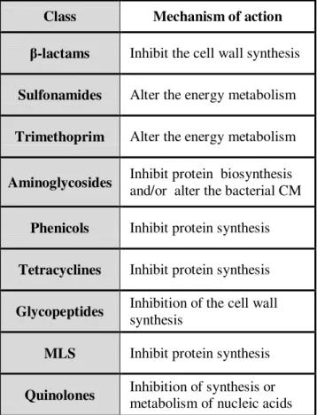
Table I.3. - Mechanisms of action of the most relevant antibiotic classes (Sousa 2005; van Hoek, Mevius et al. 2011).
Class Mechanism of action
β-lactams Inhibit the cell wall synthesis
Sulfonamides Alter the energy metabolism Trimethoprim Alter the energy metabolism
Aminoglycosides Inhibit protein biosynthesis and/or alter the bacterial CM Phenicols Inhibit protein synthesis Tetracyclines Inhibit protein synthesis Glycopeptides Inhibition of the cell wall
synthesis
MLS Inhibit protein synthesis Quinolones Inhibition of synthesis or
6
2. Antibiotic resistance
The introduction of penicillin in the clinics altered the infections therapeutics reducing morbidity and mortality. However a rapid emergence of resistance in
Staphylococcus aureus due to a plasmid-encoded penicillinase was soon observed
(Jevons, Coe et al. 1963). The production of this β-lactamase was then detected in
clinical isolates of several Staphylococcus species (Bradford 2001; van Hoek, Mevius et
al. 2011).
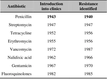
In fact, AR was identified before the release of the first antibiotic for therapeutics:
the first β-lactamase was identified in Escherichia coli prior to the release of penicillin
for use in medical practice (see Table I.4. in bold) (Abraham and Chain 1940).
Tabela I.4. - Antibiotic discovery and resistance development (Products 1999).
Antibiotic Introduction into clinics Resistance identified Penicillin 1943 1940 Streptomycin 1947 1947 Tetracycline 1952 1956 Erythromycin 1955 1956 Vancomycin 1972 1987 Nalidixic acid 1962 1966 Gentamicin 1967 1970 Fluoroquinolones 1982 1985
The aminoglycoside class was, along with the β-lactams, one of the first groups of
antibiotics to which resistance emerged (see table I.4.): initially, the resistance that
emerged in organisms such as Mycobacterium tuberculosis was restricted to
modification of the antibiotic targets, however, enzymatic inactivation of the antibiotic has emerged as an important mechanism in both Gram-positive and Gram-negative bacteria (Davies and Wright 1997; Wright 1999).
Over the years it has been shown by numerous studies that antibiotic use/consumption contributes to the devellopment of AR in bacteria (Goossens, Ferech
7 et al. 2005; Hulscher, Grol et al. 2010; van Hoek, Mevius et al. 2011). Vancomycin-resistant enterococci (VRE), isolated in 1988 constitute an example of the link between antibiotic usage and resistance development: dissemination of the resistance gene to vancomycin in humans happened not just because of the large use of the antibiotics in the clinical practice but also because glycopeptide avoparcin (an analogue of vancomycin) was used in animal food as a growth promoter. In 2006 the use of all antibiotics as growth promoters in animals was forbidden in the European Union (Commission 2008) for the reason that employing antibiotics as growth promoters largely contributes to the emergence of AR (Sousa 2005; van Hoek, Mevius et al. 2011). The presented examples show that microbes reacted to the changed environment (selective pressures imposed by the use of antibiotics) adapting by developing resistance to antibiotics using a variety of mechanisms. Moreover, their ability of interchanging genes by horizontal gene transfer (HGT) has an important role in the evolution and dissemination of resistance as it will be described next.
8 2.1. Intrinsic and acquired resistance
An example of intrinsic resistance is the resistance displayed by antibiotic producing strains that already have a natural resistance to that specific antibiotic being a part of their genetic inheritance. In other microorganisms intrinsic resistance mechanisms encoded in their resistome - the set of antibiotic resistance genes (ARGs) - and expressed at a basal level, confer to them a naturally reduced susceptibility to the drugs (Sousa 2005; van Hoek, Mevius et al. 2011; Lupo, Coyne et al. 2012).
In contrast to intrinsic resistance, acquired AR is not part of the natural characteristics of a species/genus but appears as a result of genetic information transfer induced by selective pressure in some strains of a species/genus. One of the main selective forces has been the human use of antibiotics (Martinez 2009).
HGT events play the major role in the rapid emergence of AR among bacteria being responsible for the transfer of resistance genetic determinants from antibiotic producers or species provided with intrinsic AR mechanisms to commensal and pathogenic bacteria. Besides clinics, other activities where the use of antibiotics is intensive contribute to the emergence and dissemination of AR, namely veterinary, aquaculture, and agriculture (EARS-Net 2011; Lupo, Coyne et al. 2012).
2.1.1. Mechanisms of antibiotic resistance transfer
Antibiotic resistance genes (ARGs) are frequently located in mobile DNA elements. These elements share the ability to move within a genome and/or to other bacterial cells. DNA transfer is usually mediated by mobile genetic elements such as plasmids, transposons and genomic islands or by bacteriophages. However free naked DNA can also be captured and incorporated into the genome in a process designated transformation.
If a resistance gene is on a conjugative (contain all the genetic information required to transfer from one bacterium to another) or mobilizable plasmid (use the conjugation functions of co-resident conjugative elements to transfer to another host) then it has the potential to be transferred to new hosts. Broad host range plasmids can transfer to several phylogenetically distant species and are particularly effective in the dissemination of AR (van Hoek, Mevius et al. 2011; Lupo, Coyne et al. 2012).
9
Integrons are not mobile elements sensus stricto but genetic platforms that are
responsible for integration and rearrangements of resistance determinants called gene cassettes (Koczura, Mokracka et al. 2012). Genes involved in resistance to almost all
antibiotic families have been found in integrons, including β-lactams, aminoglycosides,
trimethoprim, chloramphenicol, fosfomycin, macrolides, lincosamides, rifampicin, and quinolones (Stalder, Barraud et al. 2012). Integrons linked to mobile DNA spread horizontally through bacterial population and so disseminates all the resistance genes contained in their gene cassettes (Stalder, Barraud et al. 2012).
Based on the amino acid sequence of the IntI protein, five classes of AR integrons have been described (Cambray, Guerout et al. 2010). Classes 1, 2, and 3 are the most commonly detected. For instance it has been reported that multidrug resistance among
Enterobacteriaceae is associated with the presence of class 1 integrons (Leverstein-van Hall, Blok et al. 2003). Class 1 integrons have been extensively studied due to their broad distribution among Gram-negative bacteria of clinical interest and are the most reported in bacteria associated to humans or other animals (Stalder, Barraud et al. 2012). It’s easy to conclude that, given all the mentioned mechanisms, bacterial cells have a large potential to adapt by acquiring genes of resistance to antibiotics from other cells or from the environment. This makes the dissemination of resistance to antimicrobial agents an emergent problem with some microorganisms becoming extremely resistant to existing antibiotics. AR and virulence can, in extreme cases, be shared by the same strain raising a public health concern: an example was the strain that
caused a large epidemic outbreak of E. coli O104:H4 in Europe (Brzuszkiewicz,
10 2.2. Mechanisms of resistance
Bacteria have become resistant to antimicrobials through a number of mechanisms (see Table I.5.).
2.2.1. β –lactams
The production of β-lactamases is the most effective and widely disseminated
mechanism of resistance to β-lactams. These enzymes inactivate the antibiotics by
hydrolyzing the β-lactam ring present in their chemical structure. More than 900
different β-lactamases (Jacoby and Bush), encoded by bla resistance genes, have been
identified and they differ in their molecular characteristics and in the number of
different β-lactams that they hydrolyze (spectrum of activity) (van Hoek, Mevius et al.
2011).
Ambler classes (the four classes A, B, C and D) (Ambler 1980) classify
β-lactamases according to their molecular characteristics: β-lactamases from classes A, C
and D have serine at their active site; class B β-lactamases are metallo-enzymes that
require zinc for their catalytic activities (Li, Mehrotra et al. 2007; Bush and Jacoby 2010; van Hoek, Mevius et al. 2011).
β-lactamases have different spectrum of activity: broad spectrum β –lactamases
provide resistance to penicillins and older cephalosporins; extended-spectrum
β-lactamases (ESBLs) confer resistance to penicillins, first-, second-, and third-generation
cephalosporins and monobactams, but not to carbapenems and are inhibited by
β-lactamase inhibitors (van Hoek, Mevius et al. 2011).
Among the class A β-lactamases, the ESBLs represent a public health concern. The main families are TEM, SHV, CTX-M, VEB, and GES enzymes. Among them, the highest number of variants described in the last years corresponds to the CTX-M family (Canton, Gonzalez-Alba et al. 2012).
The mechanisms of lactam resistance include, besides deactivation by β-lactamases, inaccessibility of the antibiotics to their target enzymes, modifications of target enzymes and efflux bombs (Li, Mehrotra et al. 2007; Bonnedahl, Drobni et al. 2009).
11 Table I.5. – Examples of acquired ARG and mechanisms (Sousa 2005; van Hoek, Mevius et al. 2011).
Antibiotics Resistance Genes Resistance mechanism
β-lactams oxa, ges, imp, vim, tem,
shv, ctx-m, Enzymatic hydrolysis by β-lactamases Sulfonamides dfr sul Mutation on
DHFR (drf) and DHPS (sul) genes
Aminoglycosides
aac Enzymatic modification by:
aminoglycoside acetiltransferases,
aad adenylyltransferases,
aph, ant phosphoryltransferases;
amr, rmt, npm methyltransferases
str streptothricin acetyltransferase
sat streptothricin acetyltransferase
npt neomycin phosphotransferase Phenicols cat cml cmr Chloramphenicol acetyltransferase efflux Tetracyclines
tet(A), (B), (C),(D),(E) Efflux proteins
tet(M), (O),(S), (K),(W) Ribosomal protection proteins
otr Oxytetracycline resistance
Glycopeptides vanA, B,C, D,E,G Modified peptidoglycan precursors
MLS
erm
mph, ere, inu, vat mef, msr rRNA methylase Inactivating enzyme Efflux Quinolones qnr aac(6')-Ib-cr qepA Protector proteins Aminoglycoside acetyltransferase Efflux
12 2.2.2. Sulfonamides and Trimethoprim
Resistances to sulfonamides and trimethoprim are encoded by mutations located
on highly conserved areas of DHPS genes (sul) and DHFR genes (dfr) related with the
antibiotic mechanism of action. Sulfonamide resistance is conferred by changes in the
sul genes. The most widespread trimethoprim resistance mechanism is the replacement
of a trimethoprim-sensitive DHFR by a plasmid-, transposon-, or cassette-borne trimethoprim-resistant DHFR (Skold 2001; Sousa 2005; Zhang, Zhang et al. 2009).
2.2.3. Aminoglycoside
There are several different biochemical mechanisms of resistance to aminoglycoside antibiotics: reduced uptake, mutational modification of 16S rRNA and of ribosomal proteins, enzymatic modification of 16S rRNA through rRNA methylases and enzymatic modification of the antibiotic - the most commonly identified aminoglycoside resistance mechanism (Davies and Wright 1997; van Hoek, Mevius et al. 2011).
2.2.4. Chloramphenicol
The identified chloramphenicol resistance systems are based on inactivating
enzymes like acetyltransferases – CATs encoded by genes catA and catB – and
phosphotransferases, mutations of the target site, permeability barriers, and efflux
systems coded by the genes floR and cmlA (Schwarz, Kehrenberg et al. 2004; van Hoek,
Mevius et al. 2011).
2.2.5. Tetracyclines
Tetracycline resistance mechanisms can be mediated by efflux pumps, ribosomal
protection proteins or inactivating enzymes. The tet(A), (B), and (M) are among the
most frequent tetracycline resistance genes and encode for efflux pumps (the first two) and ribosomal protection protein (the last) (van Hoek, Mevius et al. 2011).
13 2.2.6. Glycopeptides
It was mentioned before that glycopeptides bound to the bacterial cell wall
terminus of d-Ala–d-Ala inhibiting the transglycosylation reaction and consequently the
cell wall synthesis. By glycopeptides’ target modification the binding affinity decreases
and the cell wall synthesis may continue normally. For example the van genes encode
for modified peptidoglycan precursors and their expression results in vancomycin
resistance. The vanA and vanB genes have been found associated to plasmids resulting
in their widespread dissemination. The other reported van genes are chromosome
encoded (van Hoek, Mevius et al. 2011).
2.2.7. MLS
Commonly, MLS resistance results from the expression of erm genes that encode
rRNA methylases. The activity of these enzymes prevents the binding of the antibiotic to the ribosome. Although structurally unrelated, the same resistance mechanism
(expression of erm genes) confers resistance to macrolides, lincosamide, and
streptogramin (Roberts 2002; Zhang, Zhang et al. 2009; van Hoek, Mevius et al.
2011).The erm genes are usually acquired and associated with mobile elements being
14 2.2.8. Quinolones
As described in figure I.1. quinolone resistance mechanisms can be chromosome encoded or plasmid mediated. Chromosome encoded resistance by porin loss results in decreased outer membrane permeability; another intrinsic mechanism is activated in the presence of the antibiotic by the overexpression of naturally occurring efflux pumps genes; also contributing to resistance are mutations of the molecular targets DNA
gyrase genes (gyrA and B) and topoisomerase IV genes (parC and E) in the quinolone
resistance determining regions. In concern to plasmid-mediated resistance, qnr genes
encode proteins that protect DNA gyrase and topoisomerase IV from quinolone
inhibition; aac(6)-Ib-cr gene encodes an aminoglycoside acetyltransferase, which has
two amino acid changes (Trp102Arg and Asp179Tyr) conferring to the enzyme the
ability to acetylate ciprofloxacin. The qepA gene encodes a plasmid-mediated efflux
pump which can extrude hydrophilic fluoroquinolones like ciprofloxacin (Jacoby 2005; Yamane, Wachino et al. 2007; Strahilevitz, Jacoby et al. 2009).
Chromossome encoded
Porin loss in the membrane Efflux pumps overexression Molecular targets mutation Plasmid mediated qnr genes aac(6)-Ib-cr gene qepA genes
15
3. Environmental spread of antibiotic-resistant bacteria
Selective pressure for ARGs exists in nature leading to their naturally occurrence in a balance with the environment. Humans have applied additional selective pressures for ARGs because of the large quantities of antibiotics used in medicine and agriculture. The incompletely metabolized antibiotics are the primary source of antibiotics in natural environments: depending on the type of antibiotic, between 30% and 90% of an administered dose given to humans and animals are excreted in the urine or feces as active substances and introduced to the sewage system being only partially eliminated in sewage treatment plants and ending up in the environment, reaching surface waters, groundwater, soils and sediments (Matyar, Dincer et al. 2004; Schlusener and Bester 2006; Kummerer 2009; Matyar, Akkan et al. 2010; Mudryk, Perlinski et al. 2010).
Baquero and Martínez (Baquero, Martinez et al. 2008) explained how antibiotics resulting from the human activity get into the environment and how ARGs disseminate dividing the process in compartments, called reactors, each one favorable for genetic evolution. According to these authors there are four main genetic reactors in which AR evolves (Figure I.2.):
- The primary reactor is constituted by the human and animal microbiota in which antibiotics exert their actions;
- The secondary reactor involves places and facilities (like hospitals and farms) in which susceptible individuals interact and are exposed to bacterial exchange; - The tertiary reactor corresponds to the wastewater and other biological residues
originated in the secondary reactor in which bacterial organisms from many different individuals have the opportunity to mix and exchange genes;
- The fourth reactor is the soil and the water environments, where the bacteria from the previous reactors mix and counteract with environmental organisms.
Particularly in the lowest reactors, bacteria from human or animal-associated microbiota (in black in figure 2) mix with environmental bacteria (in white in figure 2), leading to genetic variation and to the emergence of new resistance mechanisms that are re-introduced in human or animal environments (back arrows in figure 2) (Baquero, Martinez et al. 2008; Martinez 2009).
16 Figure I.2. - The four main genetic reactors (Baquero, Martinez et al. 2008).
3.1. The role of HGT in the spread of ARG in the environment
As referred before HGT events play the major role in the rapid emergence of AR and it has been long recognized that various stress conditions may contribute to increased rates of HGT. For example, UV irradiation or starvation affects the mobility of transposons and insertion sequences as well as the sub-inhibitory concentrations of antibiotics may significantly increase the frequency of horizontal transfer of many types of MGEs (Aminov 2011). In the environment microorganisms are exposed to the mentioned stress conditions so HGT may happen at increased rates (Martinez 2009; Allen, Donato et al. 2010).
17
In Thimm et al experiment in 2001 in soil microcosms, with E. coli as a donor of
a genetically marked large conjugative plasmid RP4luc, in the presence or absence of earthworms was observed that MGEs from soil have entered the earthworm gut and that the plasmid was transferred at higher frequencies than estimated in laboratory. These results confirm the earlier notion that microbial ecosystems are not isolated and there is a potential for lateral gene exchange among different microbial ecosystems. If MGEs from soil have entered the earthworm gut, then they can also enter the gut of animals that are next in the food chain, for example, moles and birds (Thimm, Hoffmann et al. 2001).
In conclusion, there is an enormous diversity of ARGs in the environmental microbiota, that accumulated during billions of years of evolution and there are no barriers among the ecological compartments in the microbial world. The gene pools of microbiota from different compartments maintain a permanent flux mediated by the different mechanisms of HGT and so resistance genes reach easily pathogenic microorganisms (Aminov 2011).
3.2. Aquatic ecosystems as reservoirs of antibiotic resistance genes
Water is an important vector of AR dissemination. It is involved as a crucial agent in all four genetic reactors mentioned above (Figure I.2.; Martinez 2009), but particularly in the last ones, receiving bacteria from different sources (wastewater treatment plants, water from urban effluents or used in industrial and agriculture activities) that will react with the indigenous bacteria. This mixing makes possible the exchange of ARGs between the indigenous bacteria with intrinsic resistance mechanisms and the animal and human bacteria already selected for AR. Disinfectants and heavy metals are also released in water and beyond the ecological damage that these compounds induce in water communities, they also exert a selective pressure that results in a selection for AR microorganisms (Aminov and Mackie 2007; Baquero, Martinez et al. 2008).
So, water plays a key role as support medium for acceptors and donors of ARGs that subsequently may spread among bacteria from different ecosystems. This exchange
has been proven in many studies like the investigation of Cernat et al in 2007 on the
occurrence and distribution of various resistance genes in multiple antibiotic resistant E.
18 was observed that all strains showed a wide variety of ARGs, some of them in class 1 integrons (Cernat, Balotescu et al. 2007). Also Agerso and Petersen have found that
tet(E) gene is often located on large horizontally transferable plasmids of Aeromona s
spp. isolated from pond water of a fish farm, capable of interspecies transfer to E. coli.
(Agerso and Petersen 2007).
In figure I.3. it is possible to observe the geographical distribution of ARGs: studies detected these resistance genes to different antibiotic classes all around the world in aquatic environments. In Europe, nearly all types of ARGs were detected in aquatic environments of some countries including Portugal, for example, in bacteria from a slaughterhouse waste water treatment plant (Moura, Henriques et al. 2007; Zhang, Zhang et al. 2009). Therefore water may act as a reservoir of ARGs and as a vector of resistant organisms.
Wastewaters greatly contribute to this scenario. Bacteria carrying ARGs (and perhaps naked DNA) in wastewaters of hospital, animal production, and fishery areas can be transported to the nearby streams, rivers, lakes, or other aquatic bodies or enter through the soil during rainfall. ARGs frequently detected in these types of wastewaters
include for instance chloramphenicol resistance genes (cat1, 4 and B3) in the
aquaculture systems and sulfonamide resistance genes (sul1, 2, 3 and A) in fish farms
(Dang, Zhang et al. 2006; Agerso and Petersen 2007; Zhang, Zhang et al. 2009).
Untreated sewage was found to contain a variety of ARGs encoding resistances to all classes of antibiotics. The environmental conditions of activated sludge and the biofilms facilitate horizontal transfer of the ARGs from one host to another because of the nutritional richness and high bacterial density and diversity (Schluter, Szczepanowski et al. 2007; Zhang, Zhang et al. 2009).
Surface water and shallow groundwater are commonly used as source of drinking water; thus, ARGs can go through drinking water treatment facilities, enter into water distribution systems and into human food chain (Figueira, Serra et al. 2012; Vaz-Moreira, Figueira et al. 2012; Vaz-Vaz-Moreira, Nunes et al. 2012). Humans get in contact with water and consequently with ARGs not only through the food chain but also in recreation activities leading to the introduction of new resistance mechanisms in clinic (Schwartz, Kohnen et al. 2003; Walsh, Ingenfeld et al. 2011).
19 Figure I.3. - Detection of the antibiotic resistance genes in geographically isolated water environments. Genes encoding resistance to aminoglycosides (red square), chloramphenicol (brown inverted triangle), β-lactam (plus symbol), macrolide (sky blue triangle), sulfonamide (violet diamond), tetracycline (green circle) and trimethoprim (indigo star) (Zhang, Zhang et al. 2009).
20 3.3. Public health concern: ARGs as environmental pollutants
If AR further depletes the number of effective drugs available, therapeutic options will be restricted, which represents a serious public health problem resulting from the widespread dissemination of ARGs. For this reason and also because ARGs are widely distributed in various environmental compartments having also an impact in the equilibrium of the ecosystems, these genes have been considered environmental emerging pollutants (Gottlieb and Nimmo 2011).
The results of the recent surveillance report of antimicrobial resistance in Europe show a general Europe-wide increase of antimicrobial resistance in the gram-negative
pathogens under surveillance (E. coli, Klebsiella pneumoniae and Pseudomona s
aeruginosa), whereas the occurrence of resistance in the gram-positive pathogens (Streptococcus pneumoniae, Staphylococcus a ureus, Enterococcus faecium and
Enterococcus faecalis) appears to be stabilizing or even decreasing in some countries (EARS-Net 2011).
A north-to-south gradient is evident in Europe: in general, lower resistance percentages are reported in the north and higher percentages in the south of Europe. This may reflect differences in infection control practices and antibiotics use in the reporting countries. Prudent use of antibiotics and infection control measures should be cornerstones of prevention and control efforts to reduce the selection and transmission of AR bacteria. (EARS-Net 2011)
The fact that ARGs can spread and exchange among environmental microorganisms of different genera and cohabitationwith organisms of completely different kingdoms and the appearance of potential antibiotic resistances in drinking water distribution systems of some nations or regions requires increased surveillance for risk assessment and prevention strategies to protect public health (Agerso and Petersen 2007; Zhang, Zhang et al. 2009; Figueira, Vaz-Moreira et al. 2011; Vaz-Moreira, Nunes et al. 2011; Vaz-Moreira, Figueira et al. 2012).
It is necessary the implementation of measures to improve water resources like establishing the AR detection as a water quality parameter and developing efficient methods to this detection (Lupo, Coyne et al. 2012).
21
4. Antibiotic-resistant bacteria in coastal environments
In various bacterial species in sediments of Tokyo Bay, Sagami Bay, and the open
Pacific Ocean was found the tetracycline resistance gene tet(M) (Rahman, Nonaka et al.
2008). The numbers of oxytetracycline-resistant bacteria increased in sediments around
a marine aquaculture site after oxytetracycline therapy, and tet(M) was evident in both
Gram-positive and Gram-negative bacteria from various genera in the sediments of the marine environment (Neela, Nonaka et al. 2007; Rahman, Nonaka et al. 2008). The marine sediments can be considered as natural reservoirs of ARGs, particularly tetracycline resistance. ARGs in sediments are selected due to antibiotic presence or they settle from water.,
Coastal waters, frequently used by humans for recreation, also constitute a natural reservoir of ARGs as it has been demonstrated in studies in these areas (Mudryk, Perlinski et al. 2010; Simoes, Poirel et al. 2010; Matyar 2012) and as it will be described next.
4.1. Antibiotic resistance genes in coastal waters
Beaches of marine coasts are dynamic environments subjected to natural changes in terms of physico-chemical variables, nutrients and to strong anthropogenic pressures from various activities. Bacteria inhabiting these ecosystems are well adapted to these versatile conditions. The previously reported studies suggested that human activity may play an important role as a source of AR in marine beaches (Mudryk, Perlinski et al. 2010; Alouache, Kada et al. 2012).
Results from description of AR in seawater on the beaches of Algeria, showed a significant level of resistance to antibiotics, particularly β-lactams, detected mainly among saprophytic environmental bacteria. Transmissible ESBLs of CTX-M-15 type
were detected in E. coli meaning that resistance genes can disseminate in these
environments representing an health risk from recreational water contact (Alouache, Kada et al. 2012).
Maravic et al found in coastal waters of the Adriatic Sea in Croatia Burkholderia
cepacia showing multiple AR. Two of the isolates produced a chromosomal encoded
ESBL (TEM-116) mostly found in members of the Enterobacteriaceae family
22
The results from Dada et al study on the occurrence of antibiotic-resistant
enterococci in coastal bathing waters in Malaysia suggested that samples from Port Dickson may contain multiple AR bacteria and that this could be due to high-risk fecal contamination from sewage discharge pipes that drain into the seawater (Dada, Ahmad et al. 2012).
4.2. Seagulls and antibiotic resistance dissemination
Migratory birds can acquire and spread resistance genes through geographically disperse environments because they travel long distances and inhabit many different places. Proximity to human activity increases the number of the antibiotic-resistant bacteria that are associated with wild birds. For instance, gulls carrying higher levels of
antibiotic-resistant E. coli are the ones inhabiting near waste or agricultural water in
contrast with the ones inhabiting human-unrelated sites (Dolejska, Bierosova et al. 2009; Literak, Dolejska et al. 2009).
Nevertheless, AR is also described in remote bird populations: in arctic birds, 8% of E. coli isolates were recently found to be resistant to at least 1 of 17 antibiotics tested, and 4 were resistant to 4 or more antibiotics. One isolate was resistant to cefadroxil, cefuroxime and cefpodoxime, a common pattern in clinical isolates (Sjolund, Bonnedahl et al. 2008). Many birds breed in the arctic and migrate to up to six continents. They probably acquire antibiotic-resistant bacteria from environments that are under human influence or from other birds that contact with those environments. This study shows the geographical distances that can be travelled by bacteria which genomes encode ARGs that are associated with human selective pressures and the potential of migratory birds as vectors for antibiotic dissemination (Sjolund, Bonnedahl et al. 2008; Allen, Donato et al. 2010).
Gull populations have increased worldwide due in large part to the availability of human-derived products that they use as food along coastal areas, in parallel with the growth of human populations. Several recent studies have detected clinically relevant ARGs in gull feces (Wallace, Cheasty et al. 1997; Dolejska, Cizek et al. 2007; Benskin, Wilson et al. 2009; Radhouani, Poeta et al. 2009; Bonnedahl, Drobni et al. 2010; Simoes, Poirel et al. 2010).
ESBL producing E. coli isolates, fluoroquinolones-resistant isolates and isolates
23
world (Poeta, Radhouani et al. 2008; Bonnedahl, Drobni et al. 2009; Dolejska,
Bierosova et al. 2009; Wallensten, Hernandez et al. 2011).
Simões et al identified in seagull feces from Porto beaches, Portugal, a variety of
ESBL–producing E. coli isolates, with a high rate of cefotaxime resistance. Beaches
may therefore present a risk to public health because of the potential pathogen-spreading capacity of migratory birds (Simoes, Poirel et al. 2010).
Using functional metagenomics to characterize the diversity of the gull resistome,
Martiny et al detected ARGs that are usually found in human and pet’s bacteria like
class A and C β-lactamases. In this investigation a large number of novel genes were identified (thirty one undescribed ARGs) showing that gulls can also introduce new AR mechanisms in the human microflora (Martiny, Martiny et al. 2011).
Gulls do not naturally come into contact with antibiotics. These birds can be infected with resistant strains and potentially serve as their reservoirs, vectors and bioindicators in the environment reflecting the presence of such resistant strains in their sources of food and/or water. The results from the mentioned investigations imply that ARGs arising from the intense use of antibiotics spread to wild life that can disseminate them, especially the migratory species, like gulls.
24
5. Antibiotic resistance in E. coli
E. coli belongs to the family Enterobacteriaceae consisting in a facultative anaerobic, gram-negative and nonsporulating rod. It has relatively simple nutrition
requirements. E. coli is the best known microorganism and an inhabitant of the
intestinal tract of humans and other warm-blooded animals; thus it is a commensal bacteria, playing an important nutritional role in the intestinal tract by synthesizing vitamins (Madigan, Dunlap et al. 2009).
Some strains of E. coli are pathogenic, like enteropathogenic E. coli implicated in
gastrointestinal infections and generalized fevers and E. coli O157:H7, an
enterohaemorragic strain that is in the origin of foodborne diseases and, in a small percentage of cases, can be life-threatening (Madigan, Dunlap et al. 2009).
Due to its characteristics of not normally being pathogenic to humans and its
presence at higher concentrations than the pathogens, E. coli is used as an indicator of
fecal pollution, a microorganism used to predict the presence of pathogenic microorganisms. It has also been frequently used for Microbial source Tracking (MST) in which isolates from different sources and from the contaminated place are genotypic or phenotypic characterized for determining the origins of fecal contamination (Scott, Rose et al. 2002; Roslev and Bukh 2011).
Phylogenetic analyses showed that E. coli stains can be assigned to four main
phylogenetic groups: A, B1, B2 and D. These groups differ in their ecological niches, life-story and characteristics like the exploitation of sugar sources, growth rate and antibiotic-resistance profiles (Carlos, Pires et al. 2010). The stains belonging to groups B2 and D contain more virulence factors and the extraintestinal pathogenic bacteria usually belong to these groups while the commensal stains belong to groups A and B1 (Clermont, Bonacorsi et al. 2000; Carlos, Pires et al. 2010).
AR in clinical isolates of E. coli was well illustrated by the 2011 report from the
European Antimicrobial Surveillance Network (EARS-Net 2011): resistance to
β-lactams is mostly due to production of β-lactamases and resistance to third-generation
cephalosporins (Figure I.4.) is mostly conferred by ESBLs being nowadays the
CTX-M-type the most common ESBL in E. coli. There is an emergence of carbapenem
resistance in E. coli, mediated by metallo-β-lactamases (such as the VIM or IMP
25 enzymes).. In general, this report showed in many countries an increase during 2008
and 2011 of combined resistance to at least three antimicrobial classes and of E. coli
strains producing multiple β-lactamases, two facts that are a subject of concern in terms
of public health.
Figure I.4. - Percentage (%) of E. coli invasive isolates with resistance to third-generation cephalosporins by European Union country in 2011 (EARS-Net 2011).
Many studies detected antibiotic-resistant E. coli isolates in the environment
including in aquatic environments: multiresistant E. coli have been reported in surface
and river water worldwide (Hu, Shi et al. 2008; Figueira, Serra et al. 2011; Tacao,
Correia et al. 2012) and even in drinking water in which multiple-antibiotic-resistant E.
coli strains were found to carry ARGs encoding resistances to aminoglycoside,
26
6. Berlengas as a model of coastal environment
Berlenga Island, located 5.7 miles from the Portuguese coast in Peniche, is classified by the Portuguese Government as Berlengas Natural Reserve since 1981, and was in 2011 added to the World Network of Biosphere Reserves by International Coordinating Council of UNESCO’s Man and Biosphere Program (Queiroga, Leão et al. 2008).
The Berlengas archipelago is constituted by a granitic and metamorphic complex of rocks having a steep topography where is very common the formation of caves and underwater cracks. The location of Berlengas, in the transition zone between the European and Mediterranean sub regions is an important factor in the oceanographic dynamics of the region with an intense upwelling that allows the renewal of nutrients in the surface contributing to the great diversity of marine species and habitats (Queiroga, Leão et al. 2008; Araújo 2012).
Rich in avifauna, Berlenga Island is the shelter of one of the largest western
colonies of the yellow-legged-gull (Larus cachinnans) and the nesting location of other
seabirds like dark wing gull (Larus fuscus) and tridacttyl gull (Rissa tridactyla). It is
also the point of concentration of migratory birds that feed in the surrounding sea (Queiroga, Leão et al. 2008; Araújo 2012).
The high number of visitors in the summer period can be a problem to the conservation of the archipelago ecosystem: that number often exceeds the limit of 350 visitors a day established by law (270/90 of April 10),pressuring the basic infrastructures of the island including water supply, sanitation and waste production (Queiroga, Leão et al. 2008).
Salt water is used for washing and to ensure the functioning of the rudimentary
sanitary facilities in the houses of the fishermen’s village and restaurant: seawater is
pumped into tanks and used in toilets returning to the sea through ducts. Part of the water goes through a waste milling system and the washing water from the catering services is also released directly into the sea through the same pipeline system. There are no pits or other basic sanitation systems (Araújo 2012).
In the last few years the population of seagulls increased significantly. In 1995 there were 32.000 birds when various management measures were implemented consisting in the eradication of mature individuals and destruction of their postures.
27 However the population continued to increase spreading through the islands (Queiroga, Leão et al. 2008).
The recent results of the microbiological quality of the Berlenga beach water
showed moments in which the maximum allowable value is exceeded (500 E. coli /100
ml) indicating fecal pollution (Araújo 2012).
6.1. MST in Berlenga Island
Once it is detected fecal contamination in a certain area it is of great importance to determine the origin of that contamination to understand the associated health risks and to know what to do to solve or minimize the pollution problem. MST aims to identify the origin of fecal pollution assuming that microbiota of the fecal pollution and the biological source have similar phenotypic or genotypic characteristics. The methods to evaluate and compare the microorganisms from the polluted area and the suspected sources can be culture- or library-dependent or independent. For instance, antibiotic resistance analysis (ARA) is a phenotypic, library and culture dependent MST method that differentiates bacteria from different sources through their AR profiles (Scott, Rose et al. 2002; Roslev and Bukh 2011).
In the last few years a problem of fecal contamination has been detected in the beach water of Berlenga Island. Seagulls were identified as the main source of fecal contamination in the scope of a previous study (Araújo S. 2013).
28
7. Aims of the work
AR dissemination is becoming a public health concern and a few studies in coastal environments concerning this subject show that beaches (sediments and seawater) also contain antibiotic-resistant bacteria constituting presumptive reservoirs of ARGs.
Berlenga Island is a protected area. Nevertheless, fecal contamination was
detected in the Berlengas’ seawater and seagulls were identified has the main origin of
this contamination.
E. coli isolates from gulls and humans are subjected to different selective pressures. As a consequence it is expected that isolates display different levels of AR, also carrying different ARGs and MGEs, depending on the host.
We hypothesize that the levels of AR and the diversity of ARGs in water of the Berlenga beach will resemble the ones of the main origin of fecal contamination (e.g seagull feces).
Taking this into account, the major aims of this work are:
- to determine the AR levels in E. coli from seagull feces, human-derived sewage
and water collected in the Berlenga beach.
– to characterize the genetic basis of the identified resistance phenotypes.
- to identify possible correlations between levels of resistance/diversity of ARGs
andthe source of the isolates and E. coli phylogenetic groups.
- to identify AR-related markers (phenotypes and/or genotypes) with potential to discriminate the fecal pollution sources.
For these purposes the AR patterns of the E. coli isolates previously obtained from
the different sources: beach water (n=166), seagull feces (n=179) and human-derived sewage (n=69) were determined. Based on the AR phenotypes, genes conferring resistance were investigated by PCR. The primers targeted ARGs usually found in MGEs and thought to represent acquired resistance, thus more likely to disseminate to humans’ commensal microbiota, constituting a greater risk to public health.
This study will add data to better assess the risks to human health related to the contamination of coastal waters, using well delimited ecosystem of the Berlenga beach as a model. Also, it is expected that the obtained results will contribute to further clarify the role of seagulls as vehicles of dissemination of AR.
29
II.
MATERIAL AND METHODS
1. Culture media
Two culture media were used in this study, Luria-Bertani Agar (LA) obtained by supplementing Luria-Bertani medium with Agar at 2% (m/V), and Mueller-Hinton agar, all acquired from Merck (Darmstadt, Germany). The culture media were autoclaved at 121ºC for 15 min. After cooled and poured into petri dishes they were stored at temperature of 4ºC.
It is provided the culture media composition for volumes of 1 liter.
1.1. Luria-Bertani (LB) medium: Peptone from casein 10.0 g
Yeast extract 5.0 g
Sodium chloride 10.9 g (pH 7.0)
1.2. Mueller-Hinton (MH) Agar (acc. to CLSI) Infusion from meat 2.0 g
Casein-hydrolysate 17.5 g
Starch 1.5 g
30
2. General reagents and solutions
The composition of the buffer solutions of general use and of the loading dye used in electrophoresis is described below.
2.1. 1x Tris-Acetato EDTA (TAE) buffer (5 Prime, Deutschland) 2 M Tris-Acetate
0.05 M EDTA (pH 8.3)
(Note: “Stock” solution prepared at 50x.)
2.2. Tris-EDTA (TE)
10 M Tris-HCl (Sigma, USA)
1 mM EDTA (pH 7.5)
2.3. 6x Loading Dye (MBI Fermentas, Lithuania) 10 mM Tris-HCl
0.03% bromophenol blue 0.03% xylene cyanol FF 60% glycerol
31
3. Establishment of an E. coli collection
The detection of fecal pollution, sampling process and E. coli isolation and
purification were performed in a previous study. (Araújo 2012; Araújo S. 2013). Following it is presented a brief summary of the procedures which conducted to the
establishment of the E. coli collection used in the present study.
Analysis of the microbiological quality of water showed four time points which
overcome the 500 E. coli /100 ml, the maximum allowable value that must be respected
and not exceeded (Figure II.1.). Three of those time points corresponded to high-tide and one to low-tide. These events occurred in the beginning of the summer season and in the end of August in a week when the human affluence was low given the weather conditions verified. The better results of water quality occurred when the human affluence to the island is high. It was hypothesized that the microbiological quality of the beach water was related with the presence of seagulls that exist in less number when humans are present (Araújo 2012).
Figure II.1. – E. coli counting per 100 mL of water collected at low-tide and high tide moments (Araújo 2012).
To study this hypothesis and confirm the fecal pollution source, sampling was performed between May and September of 2011 and samples were: 1) water of the Berlenga beach, 2) seagull feces scattered in the beach and of the surrounding rocks, and 3) human-derived sewage from the island sanitary infrastructures.


![Table II .7. - Primers for bla AmpC-like multiplex PCR Target Amplicon Size (bp) Primer Sequence (5' - 3') [Primers] (pmol/µL) bla ACC 346 ACC_F: CACCTCCAGCGACTTGTTAC](https://thumb-eu.123doks.com/thumbv2/123dok_br/15779374.1076818/52.892.125.704.473.775/primers-multiplex-target-amplicon-primer-sequence-primers-cacctccagcgacttgttac.webp)

