Universidade de Lisboa
Faculdade de Ciências
Departamento de Química e Bioquímica
THE ROLE OF UCX® CELLS IN THE
TREATMENT OF PERIPHERAL ARTERIAL DISEASE
BY PROMOTING THERAPEUTIC ANGIOGENESIS
Ana Rita Duarte Simões Pereira
Dissertação
Mestrado em Bioquímica
Especialização em Bioquímica
2014
Universidade de Lisboa
Faculdade de Ciências
Departamento de Química e Bioquímica
THE ROLE OF UCX® CELLS IN THE
TREATMENT OF PERIPHERAL ARTERIAL DISEASE
BY PROMOTING THERAPEUTIC ANGIOGENESIS
Ana Rita Duarte Simões Pereira
Dissertação
Mestrado em Bioquímica
Especialização em Bioquímica
Orientadores: Professora Doutora Susana Constantino
TABLE OF CONTENTS
ACKNOWLEDGEMENTS ... i
ABBREVIATIONS ... ii
GENERAL INTRODUCTION ... 1
1. Blood vessels: an overview ... 1
2. Vasculogenesis and Angiogenesis during embryonic development ... 3
2.1 Early blood vascular development ... 3
2.2 Maturation of blood vessels ... 5
3. Postnatal neovascularization ... 6
3.1 Physiological angiogenesis ... 6
3.1.1 The angiogenic switch ... 6
3.1.2 Angiogenic regulators ... 7
3.1.2.1 Vascular endothelial growth factor (VEGF) ... 8
3.1.2.2 Angiopoietins (Ang1 and Ang2) and Tie receptors ... 9
3.1.2.3 Platelet-‐derived growth factor (PDGF) ... 10
3.1.2.4 Fibroblast growth factors (FGF) ... 10
3.1.2.5 Transforming growth factor-‐β (TGF-‐β) ... 11
3.2 Pathological angiogenesis ... 12
3.2.1 Tumor angiogenesis ... 12
3.2.2 Peripheral arterial disease ... 14
3.3 Therapeutic angiogenesis ... 16
3.3.1 Anti-‐angiogenic therapy ... 17
3.3.2 Pro-‐angiogenic therapy ... 18
3.3.2.1 Cell therapy: Mesenchymal stem cells ... 19
3.3.2.1.1 Umbilical cord tissue-‐derived mesenchymal stromal cells (UCX®) ... 21
ACKNOWLEDGEMENTS
Gostaria de começar por agradecer à minha orientadora, Professora Susana Constantino, por me ter recebido na Unidade de Angiogénese. Não tenho palavras todo o apoio, compreensão e carinho que demonstrou ao longo deste ano. A sabedoria e o seu gosto por ensinar e orientar são inpiradores. Obrigada por ter acreditado nas minhas capacidades, pela valiosa contribuição na escrita desta tese e, acima de tudo, por estimular o meu interesse pelo conhecimento.
Obrigada Professora Margarida Meireles, minha orientadora interna, pela incrível disponibilidade e simpatia que sempre teve para comigo e pelo optimismo transmitido.
Às minhas colegas Adriana, Carolina, Filipa e Paula, muito obrigada pelos ensinamentos, amizade e paciência nos meus momentos de mais nervosismo e, ao Augusto, obrigada pela permanente boa disposição, especialmente nas madrugadas de isquemia.
À ECBio, Dr. Hélder Cruz, Dr. Miguel Santos, Dr. Pedro Cruz e Dra. Rita Bárcia, agradeço terem tornado este projecto possível, toda a disponibilidade demonstrada e conhecimento partilhado.
Ao meu João, obrigada por me completares e todos os dias me fazeres feliz.
E, finalmente, à minha mãe, a pessoa mais corajosa que eu conheço, obrigada por seres a melhor mãe do Mundo.
ABBREVIATIONS
aFGF Acidic Fibroblast Growth Factor Ang-‐1 Angiopoietin 1
Ang-‐2 Angiopoietin 2
bFGF Basic Fibroblast Growth Factor CLI Critical Limb Ischemia
ECs Endothelial Cells ECM Extracellular matrix
EPCs Endothelial Progenitor Cells FDA Food and Drug Administration FGF Fibroblast Growth Factor FGFRs Tyrosine kinase FGF receptors
HIFs Hipoxia-‐inducible Transcription Factor IL Interleukin
IGF Insulin Growth Factor MMPs Matrix Metaloproteinases MSC Mesenchymal Stem Cells PAD Peripheral arterial disease PDGF Platelet-‐derived Growth Factor PIGF Placental Growth Factor PIK-‐3 Phosphatidylinositide 3-‐Kinase qRT-‐PCR Quantitative real-‐time PCR SMCs Smooth Muscle Cells
TβR-‐I Serine-‐threonine kinase receptor I TβR-‐II Serine-‐threonine kinase receptor II TGFβ Transforming Growth Factor β
Tie Tyrosine kinase with immunoglobulin and EGF homology domains UCX® Umbilical Cord Tissue Mesenchymal Stem Cells
VEGF A Vascular Endothelial Growth Factor A VEGF B Vascular Endothelial Growth Factor B VEGF C Vascular Endothelial Growth Factor C VEGF D Vascular Endothelial Growth Factor D
VEGFR1 Vascular Endothelial Growth Factor Receptor 1 VEGFR2 Vascular Endothelial Growth Factor Receptor 2
GENERAL INTRODUCTION
1. Blood vessels: an overview
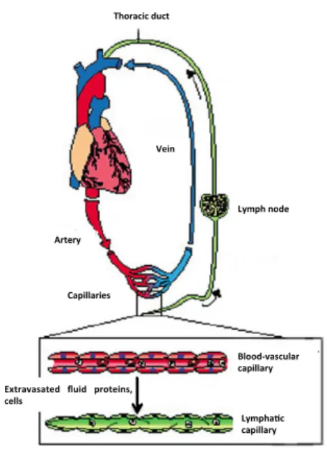
Vertebrates require an efficient circulatory system able to carry out vital functions ensured by two main networks: the blood vascular and the lymphatic system, both formed by endothelial cells (ECs)1,2.
The adult vascular system is composed of an arterial, a venous and a lymphatic compartment. These different compartments respectively provide oxygen and nutrients to peripheral organs and tissues; are able to remove carbon dioxide and other metabolic waste products which have to be transmitted to the excretory organs and maintain an immune barrier to defend the host against foreign organism1.
Blood, which is the carrier of oxygen, carbon dioxide and metabolic products, is pumped from the heart through the arterial system into the tissue capillary bed, where changes occur. The blood is then channeled through the venous system back into the heart. The lymphatic system drains extravasated fluid, the lymph, from the extracellular space and returns it into the venous circulation. The lymphatic vasculature is also essential for the immune defense, as lymph and any foreign material present in it, such as microbial antigens, are filtered through the chain of lymph nodes1
(Figure 1).
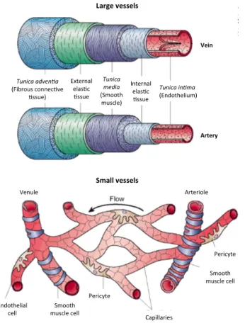
Blood vessels are divided into three main groups: arteries, veins and capillaries. Arteries and veins are divided according to caliber into large, medium and small blood vessels. These vessels are thickest and their walls more complex in the immediate vicinity of the heart, and decrease in caliber when their wall becomes thinner and less complex. Arteries form a high-‐pressure system, enabling the transportation of blood to capillaries, whereas veins face low-‐pressure gradients. The differences in hemodynamic load are reflected in their structures since arteries are supported by layers of vascular smooth muscle cells (SMCs) and a specialized matrix whereas veins are thinner and surrounded by fewer SMCs3.
Histologically, the capillary bed, which comprises the largest surface of the vascular system, is composed only by ECs that support tissue growth and repair, occasionally associated with external pericytes1.
Figure 1. Schematic representation of the circulatory system. Blood is pumped from the heart through arteries, arterioles and capillaries to the tissues, where exchanges occur. Blood is returned to the heart via the venous circulation. Adapted from Eichman et al., 2005 1.
Heterogeneity in vessel wall composition is evident and varies between vessels of different sizes and between arterial and venous vessels (Figure 2). Large vessel vascular walls are composed of multiple layers of cellular and extracellular materials such as the tunica intima, the tunica media and tunica
adventia4. The first one consists of the endothelium, basal membrane and internal elastic layer, the
second one consists of a thick layer of smooth muscle with reticular fibers (collagen III), elastin and proteoglycans and the latter consists of connective tissue with both elastic and collagenous fibers containing vasa vasorum and nerves1. Veins are different from arteries in several aspects. Given that
veins conduct blood back to the heart, the pressure exerted by the heartbeat on them is much less strong than in the arteries. In turn, arteries have strong elastic vessel walls for withstanding the high blood pressure downstream of the heart4. Moreover, veins have semi-‐lunar valves, which prevent
backflow of blood.
Small blood vessels consist of ECs forming the inner lining of the vessel wall and surrounded by a basal lamina covered by pericytes that envelop the surface of the vascular tube5. Pericytes also
referred to vascular SMCs because of their contractile fibers or mural cells, do not serve only as scaffolding, but also communicate with ECs by direct physical contact and paracrine signaling pathways5. Gap junctions provide direct connections between the cytoplasm of pericytes and ECs
enabling the exchange of ions and small molecules. Besides these functions, they have been associated mainly with stabilization and hemodynamic processes of blood vessels, they can sense angiogenic stimuli, guide sprouting tubes, elicit endothelial survival functions, and even exhibit
Thoracic(duct(
Vein(
Lymph(node(
Artery(
Capillaries(
Extravasated( fluid( proteins,( cells(
Blood=vascular( capillary(
Lympha>c( capillary(
macrophages like activities5. When vessels lose pericytes, they become hemorrhagic and
hyperdilated leading to conditions such as edema, diabetic retinophaty and even embryonic lethality6.
Figure 2. Morphology of mural cells associated with large blood vessels (top) and capillaries (bottom). Adapted from Cleaver and Melton, 2003 4.
2. Vasculogenesis and Angiogenesis during embryonic development
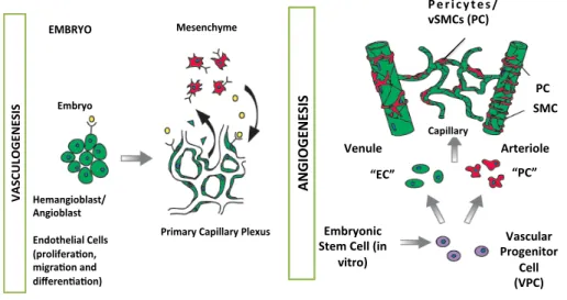
Two distinct molecular mechanisms account for the genesis of blood vessels, vasculogenesis and angiogenesis. Vasculogenesis is the process of de novo blood vessel formation driven by the recruitment and differentiated of mesodermal cells into the endothelial lineage and the de novo assembly of such cells into blood vessels. Angiogenesis is the generation of new blood vessels from pre-‐existing ones, a process driven by EC proliferation.
2.1 Early blood vascular development
From the earliest stages, the embryo develops in the absence blood vessel of vascularization, receiving its nutrition by diffusion. However, in an orderly and sequential manner, the embryo rapidly transforms into a highly vascular organism, survival being dependent on a functional, complex network of capillary plexuses and blood vessels7. The first evidence of blood vessel
Tunica'adven+a' (Fibrous)connec-ve) -ssue)) Tunica' media' (Smooth) muscle)) Tunica'in+ma' (Endothelium)) Internal) elas-c) -ssue) External) elas-c) -ssue) Large&vessels& Small&vessels& Venule) Arteriole) Endothelial) cell) ) Smooth) muscle)cell) Pericyte) Pericyte) Smooth) muscle)cell) Capillaries) Vein& Artery&
development appears on the extra-‐embryonic yolk sac and intraembryonic tissue, where groups of splanchnic mesoderm cells, specified to become hemangioblasts, aggregate and condense, forming what is known as blood islands. Here, hemangioblasts, the precursors of blood cells and ECs, start to differentiate. Cells localized at the perimeter of the blood islands become angioblasts, the precursors of all blood cells7.
In the initial events, angioblasts migrate to distant locations, multiply and differentiate in situ and assemble into solid endothelial cords, forming a primary capillary plexus. This process, where blood vessels are created de novo from endothelial progenitor cells (EPCs), is known as vasculogenesis and it is driven by the recruitment and differentiation of mesodermal cells into the endothelial lineage and the de novo assembly of such cells into blood cells (Figure 3).
This primitive network then differentiates and new blood vessels sprout and branch from pre-‐ existing capillaries. The subsequent growth, expansion and remodeling of these primitive vessels into a mature vascular network is referred to as angiogenesis, a process driven by ECs proliferation. This process is characterized by a combination of sprouting of new vessels from sides and ends of pre-‐existing ones, or by longitudinal division of existing vessels with peri-‐endothelial cells, either of which may then split and branch into precapillary arterioles and capillaries. Depending on the ultimate fate with respect to the type of vessel (artery, vein, capillary) and vascular bed, activated ECs that are migrating and proliferating to form new vessels forming anastomotic connections with each other, become variably surrounded by layers of peri-‐endothelial cells (pericytes for small vessels and smooth muscle cells for large vessels).
Once the peri-‐endothelial cells are recruited, they continue to migrate along sprouting vessels or pre-‐existing vasculature7.
Further functional modifications of larger arteries occur during collateral development also termed as arteriogenesis and thick muscular coat is added, concomitant with acquisition of viscoelastic and vasomotor properties7.
Vascular endothelial growth factor (VEGF) and its receptor-‐2 (VEGFR2) are the most absolute critical drivers of embryonic vessel formation, since the first one regulates endothelial proliferation, permeability and survival and studies with knock-‐out mice for either of both molecules result in embryonic lethality.
Figure 3. Vasculogenesis and angiogenesis in embryo. Endothelial cells (ECs) and pericytes/vSMCs (PC) arise from different precursor cells. ECs develop from angioblasts or hemangioblasts in the embryo, while pericytes/vSMCs are derived from mesenchymal stem cells. In vitro data indicate that exists a common vascular progenitor derived from embryonic stem cells that can give rise to EC and to PC. In the embryo, ECs forts assemble into a simple capillary network. Vessels, then sprout and prune (angiogenesis), become stabilized by pericytes/vSMCs that are recruited by PDGF-‐B secreting ECs, and secrate into different vessel types. Arterioles exhibit a high density of circumferentially oriented SMCs and thicker EC walls to withstand the blood pressure. Venules, like capillaries, have irregularly arranged pericytes with multiple cytoplasmic processes and are composed of thinner EC walls with valves to prevent blood backflow. Adapted from Bergers & Song, 2005 5.
2.2 Maturation of blood vessels
Vascular cells are equipped with a set of molecules that allow them to perform their functions. In quiescent vessels, vascular endothelial cadherin in adherents junctions and claudins, as well as occludin in tight junctions, provide mechanical strength and tightness, and can establish a permeability barrier. These molecules do not only serve as “mechanical zippers”, but also transmit crucial signals for endothelial survival and other functions. When ECs migrate during vessel sprouting, these contacts are transiently dissolved but later re-‐established, once ECs assemble a new sprout8.
The endothelial cell matrix (ECM) provides necessary contacts between ECs and the surrounding tissue, preventing vessels from collapsing. A basement membrane of collagen IV, laminin and other components encases vascular cells allowing pericytes and ECs to be embedded in the same basement membrane8. An interstitial matrix of collagen I and elastin between vascular cells further
provides visco-‐elasticity and strength to the vessel wall. Moreover, ECM also regulates the formation of new vessel sprouts. When vascular cells migrate to form new sprouts, this matrix network is not only proteolytically broken down, but its composition is also altered. A provisional matrix of fibronectin, fibrin and other components provides a support scaffold, guiding ECs to their target.
! ! ! ! ! ! ! ! ! ! ! Mesenchyme) ) Embryo) Hemangioblast/ Angioblast) ) Endothelial)Cells) (prolifera;on,) migra;on)and) differen;a;on)) Primary)Capillary)Plexus) ) ) ) EMBRYO) V AS CU LO G EN ES IS ) ! ! ! ! ! ! ! ! ! ! !Embryonic* Stem*Cell*(in* vitro)* * Vascular* Progenitor* Cell* (VPC)* P e r i c y t e s / vSMCs*(PC)* Venule* Arteriole* VEGF* PDGF?B* “EC”* “PC”* Capillary* PC* SMC* AN G IO G EN ES IS *
Integrins are cell surface receptors of specific ECM molecules that assist vascular cells to build new vessels in coordination with their surroundings by bidirectionally transmitting information between the outside and inside of vascular cells8.
When considering the critical role of ECM in the vessel growth and maintenance, it is conceivable that proteolytic remodeling of the ECM must occur in a balanced manner. Insufficient breakdown prevents vascular cells from leaving their original position, but excessive breakdown removes critical support and guidance for ECs and, in fact, inhibits angiogenesis.
The establishment of a functional vascular network further requires that nascent vessels mature into durable vessels and the association of pericytes and SMCs with newly formed vessels in order to regulate EC proliferation, survival, migration, differentiation, vascular branching, blood flow and vascular permeability8.
3. Postnatal neovascularization
Until 1997, it was generally accepted that vasculogenesis could only occur during embryogenesis. More recently, the existence of a postnatal vasculogenesis has been supported by the evidence that EPCs circulate postnatally in the peripheral blood, and that they may be recruited from the bone marrow and incorporated into sites of active neovascularization. Although vasculogenesis may occur during adulthood, often associated with pathological conditions, new vessels in the adult arise mainly through angiogenesis.
3.1 Physiological angiogenesis
Angiogenesis, as defined before, is the formation of new capillary blood vessels from pre-‐existent vessels and involves differential recruitment of associated supporting cells to different segments of vasculature.
After birth, angiogenesis still contributes to organ growth, although during adulthood most of the blood vessels remain quiescent. However, physiological angiogenesis can occur in response to some particularly situations, since ECs keep their capacity to divide in response to some stimuli. These stimuli may be hypoxia, wound healing and female reproductive organs that are undergoing physiological growth injured tissue.
3.1.1 The angiogenic switch
Angiogenesis is a highly functional process that requires a precise coordination spatially and temporally of multiple steps regulated by an exact balance of inducers (pro-‐angiogenic) and inhibitors (anti-‐angiogenic) factors. Physiologically, the body controls angiogenesis through a series of “on” and “off” regulatory switches. The main “on” switches are known as angiogenesis growth
factors (cytokines) and the main “off” switches are termed as endogenous angiogenesis inhibit9.
When angiogenic growth factors are produced in excess of angiogenesis inhibitors, the balance is tipped in favor of blood vessel growth. In cases where inhibitors are present in excess of stimulators, angiogenesis is “turned off”. Both situations lead to an angiogenic switch and uncontrolled angiogenesis process.
3.1.2 Angiogenic regulators
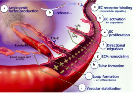
As it was previously referred, angiogenesis requires a precise coordination of multiple steps (Figure 4) regulated by a delicate balance between pro-‐ and antiangiogenic factors.
Figure 4. The process of angiogenesis. Diseased or injured tissues produce and release angiogenic growth factors that diffuse into nearby tissues (a, b); the angiogenic growth factors bind to specific receptors located on the ECs and nearby preexistent blood vessels (c); once growth factors bind to their EC receptors, a cascade of intracellular signaling is activated (c); pericytes detach and blood vessels dilate before the basement membrane and extracellular matrix is degraded by ECs produced enzymes (d); the ECs begin to divide (e) and they migrate out through the dissolved holes of the existing vessel towards the growth factor producer tissue through the action of integrins (αvβ3, αvβ5) that serve as grappling hooks to help pull the sprouting new blood vessel to sprout forward (f); additional enzymes are produced to dissolve and remodel the ECM around the vessel (g); sprouting ECs roll up to form a blood vessel tube (h); individual blood vessel tubes differentiate in arterial-‐venous systems and connect to form blood vessel loops that can circulate blood (i); newly formed blood vessel tubes are stabilized by SMCs and pericytes that provide structural support. Blood flow then begins. Adapted Pandya et al., 2006 9.
3.1.2.1 Vascular endothelial growth factor (VEGF)
VEGF was initially termed vascular permeability factor (VPF) because of its ability to induce vascular leakage. Native VEGF is a heparin-‐binding homodimeric glycoprotein of 45 kDa10. VEGF is a mitogen
for vascular ECs derived from arteries, veins and lymphatics, defined as a unique potent angiogenic factor, that is able to stimulate capillary formation in vivo and has direct mitogenic actions that are restricted to ECs9.
The family currently comprises six members: VEGF-‐A (which denotes the originally VEGF), placenta growth factor (PIGF), VEGF-‐B, VEGF-‐C, VEGF-‐D and the parapox virus VEGF, referred to as VEGF-‐E. The human VEGF gene is located in the short arm of chromosome 6 and organized as eight exons separated by seven introns9,11. Alternative exon splicing of the gene encoding VEGF-‐A was shown to
result in the generation of five isoforms (VEGF-‐A121, VEGF-‐A145, VEGF-‐A165, VEGF-‐A189, VEGF-‐A206)
having respectively 121, 145, 165, 189 and 206 aminoacids, after the signal sequence of cleavage12.
VEGF-‐A165 is the predominant isoform and is over-‐expressed in a variety of solid tumors. VEGF-‐A189 is
thought to be the most potent for vascularization in various cancers.
VEGF expression is transcriptionally regulated by hypoxia, which occurs during tumor expansion and ischemia13. More specifically, oxygen tension has a key role in regulating expression of a variety of
genes. VEGF mRNA expression is induced by exposure to low oxygen tension under a variety of pathophysiological circumstances. Hypoxia is an important stimulus for expansion of the vascular bed that sprouts and matures into a system of stable vessels. Initially, cells are oxygenated by simple diffusion of oxygen, but when tissues grow beyond the limit of oxygen diffusion, hypoxia triggers vessel growth by signaling through hypoxia-‐inducible transcription factors (HIFs). These factors upregulate many angiogenic genes, although the induction of VEGFs is perhaps the most remarkable8.
Other via for VEGF gene expression regulation is through growth factors and oncogenes14. Several
major growth factors, including epidermal growth factor, transforming growth factor-‐α and -‐β (TGF-‐ α and TGF-‐β), keratinocyte growth factor, insulin-‐like growth factor-‐1, fibroblast growth factor (FGF) and platelet-‐derived growth factor (PDGF), all upregulate VEGF mRNA expression, suggesting that the paracrine or autocrine release of such factors cooperates with local hypoxia in the regulation of VEGF release in the microenvironment. Moreover, inflammatory cytokines such as IL-‐α and IL-‐6, induce the VEGF expression in several cell types, including synovial fibroblasts, which is in agreement with the hypothesis that VEGF may be a mediator of angiogenesis and permeability in inflammatory diseases14. Specific transforming events also result in induction of VEGF gene expression. For
mutant Ras-‐dependent VEGF expression is necessary, although insufficient, for progressive tumor growth in vivo14.
Initially, VEGF binding sites were identified on the cell surface of vascular ECs in vitro and in vivo, but subsequently, it became apparent that receptors for VEGF also occur on bone marrow-‐derived cells14.
VEGF binds two related receptor tyrosine kinases (RTKs), VEGFR1 and VEGFR214.
The importance of VEGF in vascular development is highlighted by the fact that the loss of a single VEGF-‐A allele results in abnormal blood vessel development and embryonic death15,16.
A well-‐documented in vitro activity of VEGF is the ability to promote growth of vascular ECs derived from arteries, veins and lymphatics14. VEGF induces a potent angiogenic response in a variety of in vivo models and is a survival factor for ECs, both in vivo and in vitro. As referred before, VEGF is also
known as VPF, based on its ability to induce vascular leakage13,14. Such permeability-‐enhancing
activity underlies significant roles of this molecule in inflammation and other pathological conditions. VEGF induces an increase in hydraulic conductivity of isolated microvessels, which is an effect mediated by increased calcium influx17. Consistent with a role in the regulation of vascular
permeability, VEGF also induces endothelial fenestration in some vascular beds as well as vasodilatation in vitro in a dose-‐dependent fashion as a result of EC-‐derived nitric oxide, and produces transient tachycardia, hypotension and a decrease in cardiac output when injected intravenously in conscious, instrumented rats14.
3.1.2.2 Angiopoietins (Ang1 and Ang2) and Tie receptors
A signaling system involved in vessel maintenance, growth and stabilization is the Tie2 receptor, which binds the angiopoietins (Ang1 and Ang2). The four identified angiopoietins (Ang1, Ang2, Ang3 and Ang4) are a family of secreted proteins that bind tyrosine kinase receptors (TKRs), Tie1 and Tie2. Expression patterns of Tie1 and Tie2 are similar to those of VEGFRs. Tie1 mRNA is highly expressed in embryonic vascular endothelium, angioblasts and endocaridium, whereas it is expressed strongly in lung, capillaries but weakly in endocardium in adult tissues. Tie2 mRNA shows an analogous embryonic localization11.
Ang1 induces EC survival, capillary sprouting and pericytes recruitment. Moreover, it stabilizes blood vessels by increasing the interaction between ECs and pericytes. In vivo studies also suggest that Ang1 is essential for maturation and stabilization of the developing vasculature and for normal remodeling, since mice lacking Ang1 start to develop a primary vasculature which fails to stabilize or remodel leading to embryonic lethality11. Ang2 can either promote or inhibit vessel growth,
ANGPT2 blocked Ang1 mediated Tie2 receptor activation, acting as an anti-‐angiogenic factor capable of promoting in vivo EC apoptosis and regression of blood vessels. In fact, Ang2 antagonizes Ang1 in
vivo in a manner that may act to prevent excessive sprouting and branching of blood vessels by
enhancing the decay of blood vessels. It is now known that these vessels can be prone to two fates as they can suffer regression in the absence of growth factors or they can be predisposed to angiogenic sprouting induced by available angiogenic factors. For example, in the presence of VEGF, ANGPT2 is responsible for an increase in capillary diameter, migration and proliferation of ECs and sprouting of new vessels18.
3.1.2.3 Platelet-‐derived growth factor (PDGF)
PDGF was initially purified from platelets and was then identified in fibroblast, astrocytes, keratinocytes, epithelial cells and other cell types11. PDGFs exist has heterodimers (PDGF-‐AB) or
homodimers (PDGF-‐AA or PDGF-‐BB), composed of chains A and B. PDGF receptors are made up of complexes between α and β subtypes. PDGF has been found to stimulate angiogenesis in vivo, and a role for PDGF in the recruitment of tumor pericytes, required for the development of capillaries, has been demonstrated in knock-‐out mice6. PDGF-‐BB and its receptor, PDGFR-‐B, have essential roles in
the stabilization of nascent blood vessels by recruiting PDGF-‐B positive mesenchymal progenitors. Dropout or insufficient recruitment of mural cells results in EC growth, permeability, fragility, vessel enlargement, bleeding, impaired perfusion and hypoxia in embryos lacking PDGF-‐B.
3.1.2.4 Fibroblast growth factors (FGF)
Acidic and basic FGF are heparin-‐binding protein mitogens that play an important role in angiogenesis. In vitro, both of them induce processes in the ECs that are significant to angiogenesis, such as stimulate EC proliferation and migration as well as production of collagenase11.
FGFs exert their biological activities by binding to high affinity tyrosine kinase FGF receptors (FGFRs) on the surface of target cells. Activation of FGFR1 or FGFR2 by angiogenic FGFs (including FGF1, FGF2 and FGF4) leads to ECs proliferation. The first receptor stimulation leads, also, to ECs migration, protease production and tubular morphogenesis whereas the latter receptor besides ECs proliferation increases only cell motility19.
Alternative splicing of the FGFR mRNA generates receptor variants, which display a range of receptor-‐ligand interactions. Disruption of the genes enconding FGFR1 that is known to be required for the development and maintenance of the vasculature in the embryo or FGFR2 that is important for vascular tone and maintenance of normal blood pressure, leads to embryonic death before
gastrulation. This early lethality has made impossible to define specifically the role of these FGF receptors in the later stages of development and in angiogenesis13,20.
3.1.2.5 Transforming growth factor-‐β (TGF-‐β)
The transforming growth factors –β represent a superfamily of highly conserved cytokines typified by TGF-‐β1. TGF-‐β binds to two different types of serine-‐threonine kinase receptors, known as type I (TβR-‐I) and type II (TβR-‐II). Both receptors are interdependent, meaning that TβR-‐I requires TβR-‐II to bind to TGFβ and TβR-‐II requires TβR-‐I for signaling.
Both pro-‐angiogenic and anti-‐angiogenic properties have been attributed to TGF-‐β1, through effects on ECs and other cell types11. At low doses, TGF-‐β1 contributes to the angiogenic switch by
upregulating angiogenic factors and proteinases, whereas at high doses, TGF-‐β1 inihibits EC growth, promotes basement membrane reformation and stimulates SMCs differentiation and recruitment8,11.
Gene studies in mice demonstrated that the loss of TGF-‐β signaling components results in frail vessels with decreased vessel wall integrity and inactivation of the TGF-‐β1 caused lethality due to defects in the hematopoietic system and yolk sac vasculature21.
In addition, many other factors contribute to the control and influence angiogenesis, although their effects are not as extensive as the previously discussed. Besides growth factors, cytokines and chemokines, a number of membrane-‐bound proteins play important roles in angiogenesis. Integrins, ephrins and cadherins are membrane-‐bound proteins that affect many functions involved in blood vessel assembly (Figure 5).
Induction of the angiogenic switch depends on how heavily that balance tips in favour of pro-‐ angiogenesis or anti-‐angiogenesis.
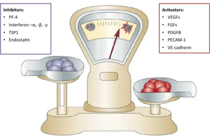
Figure 5. The angiogenic balance. Angiogenesis is orchestrated by a variety of activators and inhibitors. Activators of EC proliferation and migration are mainly receptor tirosine kinase ligands, such as VEGF, fibroblast growth factors (FGFs), platelet-‐derived growth factor (PDGF); and also matrix proteins and adhesion molecules as platelet endothelial cell adhesion molecule-‐1 (PECAM1), which contributes to EC aggregation, tube formation and vessel stabilization and vascular endothelial cadherin (VE-‐cadherin) that besides decreasing EC apoptosis also contributes to vessel stabilization.
On the other hand, some examples of angiogenesis inhibitors are the platelet factor-‐4 (PF-‐4) that decreases FGF mediated angiogenesis; interferon –α, -‐β, -‐γ that reduce FGF and matrix proteins and adhesion molecules as thrombospondin-‐1 (TSP1) which modulates EC proliferation and motility, enhancing EC apoptosis and endoestatin that decreases EC proliferation. Adapted Bergers and Benjamin, 2003 22.
3.2 Pathological angiogenesis
Insufficient vessel growth and regression contribute to numerous disorders, ranging from myocardial infarction and stroke to neurodegeneration. In diseases where an ischemia occurs, angiogenic inhibitors gain weight against stimulators, resulting in EC dysfunction, vessel malformation or regression23. Conversely, uncontrolled vessel growth promotes ocular disorders such as age-‐related
macular degeneration and tumorogenesis, characterized by an excessive angiogenesis24.
3.2.1 Tumor angiogenesis
Angiogenesis is essential for the growth of most primary tumors and their subsequent metastasis. The point at which the “normal processes” differentiate from pathological angiogenesis is in the tightly related balance of pro-‐angiogenic and anti-‐angiogenic signals.
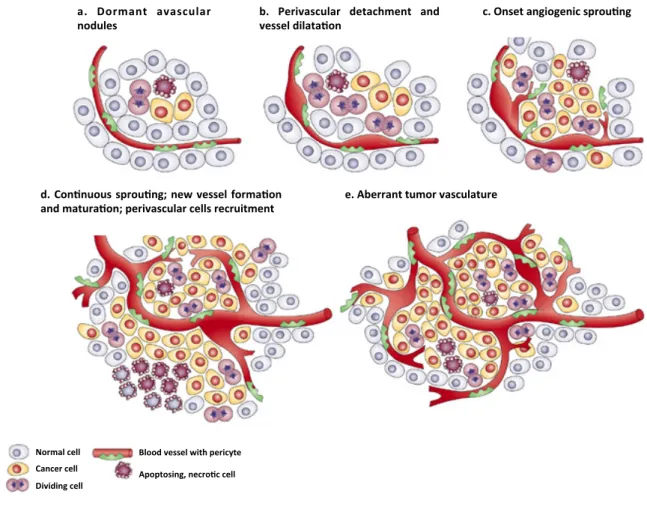
The angiogenic switch can occur at different stages of the tumor-‐progression pathway, depending on the tumor type and environment. It has been shown that dormant lesions and, in some instances, premalignant lesions, also initiate neovascularization, which allows them to progress (Figure 6).
Ac#vators:+ • VEGFs& • FGFs& • PDGFB& • PECAM-1& • VE-cadherin& & & & Inhibitors:+ • PF-4& • Interferon&–α,&-β,&-γ&& • TSP1& • EndostaCn&
Figure 6. The classical angiogenic switch. The angiogenic switch is a step in tumor development that can occur at different stages in the tumor-‐progression pathway, depending on the nature of the tumor and its microenvironment. Most tumors start growing as avascular nodules (a) until they reach a steady-‐state level of proliferating and cells’ apoptosis. The switch begins with perivascular detachment and vessel dilation (b), followed by angiogenic expansion (c), new vessel formation and maturation and the recruitment of perivascular cells (d). Blood vessel formation will continue as long as tumor grows and the blood vessels specifically feed hypoxic and necrotic areas of the tumor proving it the essential nutrients and oxygen (e). Adapted from Bergers & Benjamin, 2003 22.
The classical proof that tumors depend on angiogenesis and thus of blood supply, came from experiments whereby tumor fragments or cultured tumor cells were placed in an avascular site, the cornea of a rabbit eye25. The implants attracted new capillaries that grew in from limbus to vascularize the expanding tumor mass. Although if the capillaries were physically prevented from reaching the implant or were inhibited from undergoing angiogenesis, tumor growth was dramatically impaired, restricting the tumor nodule to a smaller diameter25. Subsequent
experiments have confirmed this result and further revealed that, in the absence of access to an adequate vasculature, tumor cells become necrotic and/or apoptotic, restraining the increase in tumor volume that should result from continuous cell proliferation, the hallmark of cancer26,27.
a.# Dormant# avascular#
nodules# b.# Perivascular# detachment# and#vessel#dilata5on#
d.#Con5nuous#sprou5ng;#new#vessel#forma5on# and#matura5on;#perivascular#cells#recruitment# e.#Aberrant#tumor#vasculature# c.#Onset#angiogenic#sprou5ng## Normal'cell' Cancer'cell' Dividing'cell' Blood'vessel'with'pericyte' ' Apoptosing,'necro:c'cell'
Tumor vessels fail to become quiescent enabling the constant growth of new tumor blood vessels and, consequently, the tumor vasculature develops unique characteristics and becomes quite distinct from the normal supply system. Tumor blood vessels are architecturally different from blood vessels, as they are irregularly shaped, dilated, tortuous and can have dead ends. The vascular network that forms tumors is often leaky and haemorrhagic, partly due to the overproduction of VEGF5. Blood flows irregularly in tumor vessels, moving more slowly and sometimes even oscillating,
leading to dysfunctional capillaries. Also, tumors can be quite heterogeneous in their vascular patterns and are able to overproduce their capillary networks5.
The angiogenic switch seems to result mainly from the action of mutated oncogenes (e.g. Ras family, src, EGFR and erbB-‐2/HER2), or loss or mutational inactivation of a variety of tumor-‐supressor genes (mutant p53)22. Other causes include numerous environmental (epigenetic) factors such as hypoxia,
low pH, inflammatory cytokines and growth factors28.
Factors that stimulate angiogenesis, such as VEGFA, PlGF and Ang1 have been shown to stimulate this process22. High levels of VEGFA expression alone are capable of initiating angiogenesis in a
quiescent vasculature. The earliest stages are defined by vasodilatation and an increased vascular permeability of pre-‐existing capillaries, or post-‐capillary venules, in response to VEGF. This allows extravasation of plasma proteins which lay down a provisional matrix on which activated EC migrate and is accompanied by the loosening of pericyte covering, which is thought to be a function of Ang2.
3.2.2 Peripheral arterial disease
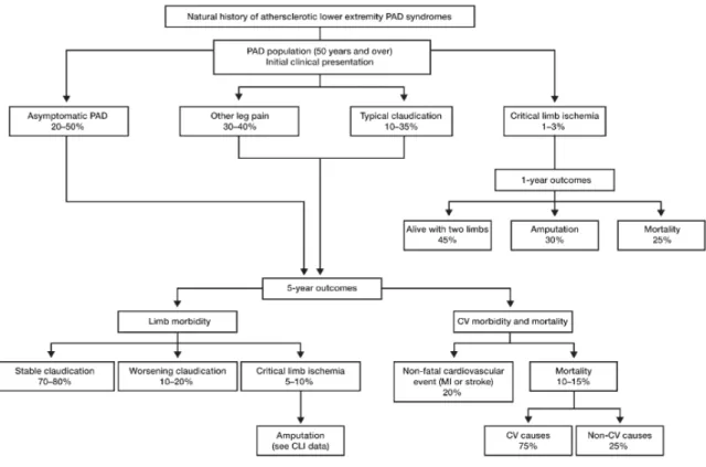
Peripheral arterial disease (PAD) involves the narrowing and obstruction of blood flow in arteries, most commonly affecting the circulation in the arteries of the pelvis and legs due to atherosclerosis related problems. PAD is a progressive illness and the most severe manifestation is termed “critical limb ischemia” (CLI). This disease describes patients with chronic ischemic rest pain or patients with ischemic skin lesions, ischemic ulcers or gangrene and occurs when arterial lesions impair blood flow and distal perfusion pressure to a level insufficient to satisfy the nutritive needs of the limb. The progression of PAD to these acute symptoms may be gradual or progress rapidly reflecting sudden worsening of limb perfusion and often threatening limb viability29,30.
Acute limb ischemia may also occur as the result of an embolic event or a local thrombosis in a previously asymptomatic patient. Moreover, a number of studies have allowed an analysis of the risk factors that seem to be associated with the development of CLI and they appear to be independent, therefore, probably additive29.
There is few information about the incidence of acute leg ischemia, but acute leg ischemia due to emboli has decreased over the years, possibly as a consequence of less cardiac valvular disease from better monitoring and anticoagulant management of atrial fibrillation. Meanwhile, the incidence of
thrombotic acute leg ischemia has increased, even with the extensive use of newer endovascular techniques29.
The only reliable large prospective population studies on the incidence of CLI, showed a figure of approximately between 500 and 1000 new cases per million per year29. In a large proportion of
these patients, the anatomic extent and the distribution of arterial occlusive disease make patients unsuitable for operative or percutaneous revascularization, although, any kind of surgical/endovascular revascularization should be done whenever technically possible. Attempts to manipulate and normalize the microcirculatory flow pharmacologically may enhance the results of revascularization or be one option. In patients with CLI not eligible for arterial revascularization, prostanoids are vasoactive drugs with proven efficacy29,59,60,. However, recent trials do not support
the benefit of prostanoids in promoting amputation-‐free survival58.
Amputation, despite its associated morbidity, mortality rates and functional implications, is often recommended as a solution to the disabling symptoms, in particular excruciating ischemic rest pain. Indeed, a second major amputation is required in nearly 10% of such patients (Figure 7)31.
It is clear that decreasing the level of any risk factor, such as blood pressure and LDL cholesterol, can help improve prognosis. Nevertheless, it is not clear what the optimal values are in the general population and in individual disease states. Modifying several risk factors is at least as beneficial as changing only one. Combination therapy with several drugs will become inevitable; however it must be paid attention to the compliance of the patients who are faced with such therapy.
The prevalence of occlusive vascular disease of peripheral limb circulations underscores the importance of understanding how adaptive remodeling of the vasculature in ischemia is regulated32.
Unlike distal capillaries, which distribute blood flow to individual cells, arteries provide bulk flow to the tissue and are therefore, of utmost importance. When an artery is occluded, its vascular territory becomes ischemic since arterial systems are often inter-‐connected by pre-‐existing collateral vessels. However, the collaterals can enlarge and salvage the ischemic region. The mechanism of angiogenesis and collateral growth differ significantly. Because of the large pressure differences between the perfusion territories, the increased shear stress activates ECs, which then recruit monocytes. These cells produce growth factors, proteinases, which enable SMCs to migrate and divide, explaining why depletion of monocytes impairs, whereas delivery of monocytes enhances collateral growth8. Cytokines that attract monocytes or prolong their life span enhance collateral
growth, whereas anti-‐inflammatory cytokines are inhibitory. PIGF for example enhances collateral growth, not only because it recruits monocytes but also because it stimulates EC and SMCs growth. Delivery of aFGF or bFGF (together with PDGF-‐BB) stimulates collateral growth, in part by upregulating PDGFR expression8.
Preclinical studies have demonstrated that angiogenic growth factors can stimulate the development of collateral arteries in animal models of peripheral and myocardial ischemia. In addition, evidence suggests that new collaterals may form and undergo outward remodeling24. According to literature, flow-‐induced shear stress is an important biomechanical force in the initiation of arteriogenesis. In the most tissues, a small number of preexisting collaterals make interconnections between most arterioles of adjacent vascular beds. Shear stress increases within collaterals when the lumen of the conduit artery supplying one bed becomes narrowed and pressure below it is reduced. Although collateral development, also termed arteriogenesis, has greater potential for restoring blood flow to severely ischemic beds than does angiogenesis alone, less is known about the mechanisms that direct arteriogenesis24,33.
Figure 7. Schematic representation of fate of the claudicant over 5 years (adapted from ACC/AHA guidelines5). PAD – peripheral arterial disease; CLI – critical limb ischemia; CV – cardiovascular; MI – myocardial infarction. Adapted from Norgren et al.,2007 29.
3.3 Therapeutic angiogenesis
The recognition that both vasculogenesis and angiogenesis are fundamental in normal developmental and adult physiological process, requiring the coordinated action of a variety of growth factors and cell-‐adhesion molecules in endothelial and mural cells, has lead to the establishment of numerous in vitro and in vivo research models.