Journal of the American Heart Association
SYSTEMATIC REVIEW AND META- ANALYSIS
Synchronous and Metachronous Thoracic
Aortic Aneurysms in Patients With
Abdominal Aortic Aneurysms: A Systematic
Review and Meta-Analysis
Ryan Gouveia e Melo , MD; Gonçalo Silva Duarte , MD; Alice Lopes , MD; Mariana Alves , MD;
Daniel Caldeira , MD, PhD; Ruy Fernandes e Fernandes , MD; Luís Mendes Pedro , MD, PhD
BACKGROUND: The prevalence of thoracic aortic aneurysms (TAA) in patients with known abdominal aortic aneurysms (AAA) is not well known and understudied. Our aim was to conduct a systematic review and meta-analysis of the overall prevalence of synchronous and metachronous TAA (SM-TAA) in patients with a known AAA and to understand the characteristics of this sub-population.
METHODS AND RESULTS: We searched MEDLINE, EMBASE, and CENTRAL (Cochrane Central Register of Controlled Trials) from inception to November 2019 for all population-based studies reporting on the prevalence of SM-TAAs in a cohort of patients with AAA. Article screening and data extraction were performed by 2 authors and data were pooled using a random-effects model of proportions using Freeman-Tukey double arcsine transformation. The main outcome was the prevalence of SM-TAAs in patients with AAAs. Secondary outcomes were the prevalence of synchronous TAAs, metachronous TAAs, prevalence of TAAs in patients with AAA according to the anatomic location (ascending, arch, and descending) and the differ-ences in prevalence of these aneurysms according to sex and risk factors. Six studies were included. The pooled-prevalence of SM-TAA in AAA patients was 19.2% (95% CI, 12.3–27.3). Results revealed that 15.2% (95% CI, 7.1–25.6) of men and 30.7% (95% CI, 25.2–36.5) of women with AAA had an SM-TAA. Women with AAA had a 2-fold increased risk of having an SM-TAA than men (relative risk [RRs], 2.16; 95% CI, 1.32–3.55). Diabetes mellitus was associated with a 43% decreased risk of having SM-TAA (RRs, 0.57; 95% CI, 0.41–0.80).
CONCLUSIONS: Since a fifth of AAA patients will have an SM-TAA, routine screening of SM-TAA and their clinical impact should be more thoroughly studied in patients with known AAA.
Key Words: abdominal aortic aneurysms ■ meta-analysis ■ metachronous aortic aneurysms ■ synchronous aortic aneurysms
■ thoracic aortic aneurysms
A
neurysmal disease is known nowadays to be asystemic, multifactorial, and unpredictable condi-tion with different causes, behaviors and presen-tations which increase the complexity of its diagnosis,
treatment, and outcomes.1,2
Abdominal aortic aneurysms (AAA) are by far the most common and studied aneurysms and screening
strategies have shown to be effective, reducing the in-cidence in aneurysm rupture rate and improvement of
care in a cost-effective fashion.3
The presence of synchronous and metachronous
aneurysms has been well described in the literature.4
However, in contrast to synchronous peripheral aneu-rysms which are frequently screened and reported,
Correspondence to: Ryan Gouveia e Melo, MD, Hospital de Santa Maria – Centro Hospitalar Universitário Lisboa Norte, EPE, Serviço de Angiologia e Cirurgia Vascular. Avenida Professor Egas Moniz, 1649-028 Lisboa, Portugal. E-mail: [email protected]
Supplementary Material for this article is available at https://www.ahajo urnals.org/doi/suppl/ 10.1161/JAHA.120.017468 For Sources of Funding and Disclosures, see page 10.
© 2020 The Authors. Published on behalf of the American Heart Association, Inc., by Wiley. This is an open access article under the terms of the Creative Commons Attribution-NonCommercial-NoDerivs License, which permits use and distribution in any medium, provided the original work is properly cited, the use is non-commercial and no modifications or adaptations are made.
JAHA is available at: www.ahajournals.org/journal/jaha
the incidence and behavior of synchronous/meta-chronous thoracic aortic aneurysms (SM-TAA) is
underappreciated.4
In a previous study in our center, we found that 18.9% of patients submitted to Thoracic Endovascular Aortic Repair, had a synchronous or
metachronous AAA.5 However, thoracic aortic
an-eurysms (TAA) in general, do not have a screening program to date and the incidence and prevalence of these aneurysms is underappreciated. Since rupture of TAAs in patients with a known AAA or
following an AAA repair has been described,4,6,7
better understanding of the prevalence of these synchronous/metachronous TAAs in patients with a known AAA might be crucial for patients, physi-cians, and policy makers.
Our aim was to estimate the overall prevalence of synchronous and metachronous TAAs in patients with a known AAA.
METHODS
The Preferred Reporting Items for Systematic
Reviews and Meta-Analyses guidelines8 were
fol-lowed for reporting and design of this systematic review. The authors declare that all supporting data are available within the article (and its online supple-mentary files).
Eligibility Criteria
We included all cohort studies, either prospective or retrospective, reporting on the prevalence of tho-racic aortic aneurysms in a population of patients with known abdominal aortic aneurysm, irrespective of the diagnostic method used. We accepted the definition of AAA or TAA provided by the studies. We defined syn-chronous aneurysms as occurring concomitantly and metachronous aneurysms if the diagnosis of a new TAA occurred 2 years after the initial AAA diagnosis and no TAA was present initially.
There were no date or language restrictions. Animal studies were not included.
Papers were excluded if they did not specify the specific number of SM-TAA. If the same population was described in 2 papers 1 of them was excluded to not duplicate events. Papers were also excluded if the studied population (the denominator) included patients without AAA.
Information Sources and Search Method
We searched EMBASE, MEDLINE, and CENTRAL (Cochrane Central Register of Controlled Trials), from inception to November 2019. We also cross-checked references and consulted specialists for additional potential studies. The search strategy is detailed in Table S1.
The search results were cross-checked, and dupli-cates were eliminated.
Study Selection, Data Collection Process,
and Synthesis
Two authors (R.G.M. and A.L.) independently screened the titles and abstracts that yielded from the search. Full-text papers were also independently assessed by both authors and disagreements were resolved by consulting with a third author (G.D.).
After the final search result, data were inde-pendently extracted by 2 authors (R.G.M. and A.L.) using a pre-design report form and uploaded onto a table sheet after cross-checking.
Data retrieved included: study publication data (authors, date, journal), population studied (years of the study and respective centers), study site, number of participants with known AAA, number of patients with SM-TAA and AAA, demographics (age, sex, risk
CLINICAL PERSPECTIVE
What Is New?
• We found that 19.2% of patients with abdomi-nal aortic aneurysm have a synchronous or me-tachronous thoracic aortic aneurysm.
• Women have a 2-fold higher risk of having a synchronous or metachronous abdominal and thoracic aortic aneurysm.
• Diabetes mellitus is associated with a 43% decreased risk of having a synchronous or metachronous abdominal and thoracic aortic aneurysm.
What Are the Clinical Implications?
• Routine screening ofsynchronous/metachro-nous thoracic aortic aneurysms and their clinical impact should be more thoroughly stud-ied in patients with known abdominal aortic aneurysms.
• Differences in prevalence found between men and women might explain, in part, why women have worse outcomes following abdominal aor-tic aneurysm repair.
Nonstandard Abbreviations and Acronyms
CENTRAL Cochrane Central Register ofControlled Trials
SM-TAA synchronous and metachronous thoracic aortic aneurysms TAA thoracic aortic aneurysms
factors), location of the TAA, diagnostic methods used, and definitions of TAA and AAA used.
The main outcome of interest was the overall prev-alence of SM-TAA in patients with known AAA. The prevalence was defined as the number of existing TAAs at the time of the study in the population of pa-tients with known AAA.
The secondary outcomes of interest included the prevalence of synchronous TAAs; the prevalence of metachronous TAAs; the prevalence of SM-TAA ac-cording to the anatomic location and the difference in the prevalence of SM-TAA and AAA according to sex and other risk factors (smoking, hypertension, diabetes
mellitus, family history of AAA/TAA, hyperlipidemia, and chronic obstructive pulmonary disease).
Statistical Analysis
We used the Open-Meta (Analyst) Software9 for
quantitative analysis and to derive forest plots, when appropriate.
The results yielded by the data extraction were ex-pressed in percentages for the prevalence of SM-TAA. The total number of individuals with known AAA was used as the denominator and the number of patients with SM-TAA and AAA as the numerator.
Figure 1. Preferred Reporting Items for Systematic Reviews and Meta-Analyses diagram.
A random-effects model was used to pool the data to account for the heterogeneity of the included
studies.9 The random-effects model of DerSimonian
and Laird was used by default as this approach is the simplest and most commonly used method for fitting the random effects model and is particularly useful for
larger samples.10 Freeman-Tukey transformation
(dou-ble arcsine transformation) was used to adjust the data set to estimate the frequency of the events, limiting the
CI among 0% to 100%.11 For subgroup sex analysis,
we dichotomized the prevalence data and used male sex as the reference group and for subgroup risk fac-tor analysis we used the group with AAA-only as the reference group, results were reported using risk ra-tios [RRs] and 95% CIs. Statistical heterogeneity was
assessed using I2, which was defined as low (25%),
moderate (50%), or high (75%) according to Higgins
and Thompson.12
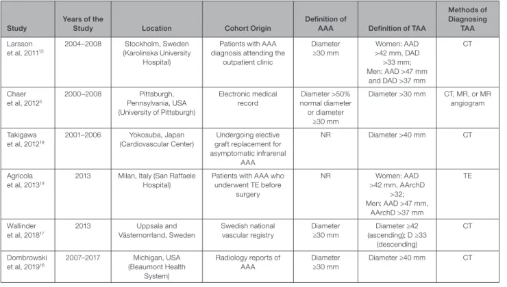
Table 1. Study Details Including Cohort Origin and Definitions
Study
Years of the
Study Location Cohort Origin
Definition of
AAA Definition of TAA
Methods of Diagnosing TAA Larsson et al, 201115 2004–2008 Stockholm, Sweden (Karolinska University Hospital)
Patients with AAA diagnosis attending the
outpatient clinic Diameter ≥30 mm Women: AAD >42 mm, DAD >33 mm; Men: AAD >47 mm and DAD >37 mm CT Chaer et al, 20124 2000–2008 Pittsburgh, Pennsylvania, USA (University of Pittsburgh) Electronic medical record Diameter >50% normal diameter or diameter ≥30 mm Diameter >30 mm CT, MR, or MR angiogram Takigawa et al, 201218 2001–2006 Yokosuba, Japan (Cardiovascular Center) Undergoing elective graft replacement for asymptomatic infrarenal
AAA
NR Diameter >40 mm CT
Agricola et al, 201314
2013 Milan, Italy (San Raffaele Hospital)
Patients with AAA who underwent TE before surgery NR Women: AAD >42 mm, AArchD >32; Men: AAD >47 mm, AArchD >37 mm TE Wallinder et al, 201817 2013 Uppsala and Västernorrland, Sweden Swedish national vascular registry Diameter ≥30 mm Diameter ≥42 (ascending); D ≥33 (descending) CT Dombrowski et al, 201916 2007–2017 Michigan, USA (Beaumont Health System) Radiology reports of AAA Diameter ≥30 mm Diameter ≥40 mm CT
AAA indicates abdominal aortic aneurysms; AAD, ascending aortic diameter; AArchD, aortic arch diameter; CT, computed tomography; DAD, descending aortic diameter; MR, magnetic resonance; NR, not reported; TAA, thoracic aortic aneurysm; and TE, transthoracic echocardiography.
Table 2. Study Data
Study No. of Participants Mean Age, y (SD) Men/Women, n (%) Patients With TAA (n) Synchronous; Metachronous TAA (n) Location of the Aneurysm—Asc; Desc; Arch (n) TAAs—Men/ Women, n (%) Larsson et al, 201115 354 74 (NR) 274 (77.4)/80 (22.6) 100 100; NR 12; 94; 6 62 (62)/38 (38) Chaer et al, 20124 1082 74.6 (9) 724 (66.9)/358 (33.1) 253 117; 136 NR 143 (68.5)/105 (41.5) Takigawa et al, 201218 157 72.7 (7.5) 128 (82)/29 (18) 13 13; NR 2; 8; 3 NR Agricola et al, 201314 1305 NR 1034 (79.2)/271 (20.8) 137 137; NR 52; NR; 85 66 (48)/71 (52) Wallinder et al, 201817 217 75 (NR) 0 (0)/217 (100) 67 NR; NR 8; 53; 2 NR/67 (100) Dombrowski et al, 201916 218 74 (NR) 136 (62.4)/82 (37.6) 40 40; NR 19; 13; 8 20 (50)/20 (50)
AAA indicates abdominal aortic aneurysms; Arch, aortic arch; Asc, ascending thoracic aorta; CT, computed tomography; Desc, descending thoracic aorta; NR, not reported; and TAA, thoracic aortic aneurysm.
Sensitivity analysis was performed according to the diagnostic method used (computed tomography [CT] scan or magnetic resonance/magnetic resonance-an-giogram versus transthoracic echocardiography), to the definition for AAA used (only patients admitted for AAA repair versus all patients with an abdominal aortic diameter ≥3 cm) and type of patients included (both men and women versus only women).
Risk of Bias
We adapted and used the Critical Appraisal Skills Program cohort study checklist to assess for risk of bias, in which we categorized 9 items as having high,
unclear, or low risk of bias.13 Two authors (R.G.M. and
A.L.) independently assessed each included paper. The overall risk of bias for each study was divided as high- or low-risk, with high-risk studies considered those in which at least 2 items were assessed at a high risk of bias, or where >3 items were rated as unclear.
RESULTS
Included Studies
The search yielded 3563 papers which resulted in 3197 articles after duplicates were removed. After title and
abstract screening, 44 papers were included in the full-text assessment. Of these, 6 articles were included in
the review.4,14–18 The reasons for exclusion of the
remain-der 38 full-text articles assessed are detailed in Figure 1.
Study Characteristics and Demographic
Data
Overall, 3333 patients with known AAA were included. Of these, 610 were found to have an SM-TAA.
Definitions of both AAA and TAA varied across
stud-ies (Table 1). Four papers4,15–17 defined AAA as an
in-crease in the aortic diameter ≥30 mm and 2 papers14,18
did not give precise definition of AAA. However, both of these later studies included patients who were undergo-ing elective AAA repair, so we can assume the definition
to be a diameter of, at least 50 mm.14,18,19 The
diagnos-tic method of TAA also varied: CT in 4 studies15–18; CT,
magnetic resonance, or magnetic
resonance-angio-gram in one,4 and transthoracic echocardiography in
another.14 Specific demographic data, including risk
factors, for all participants in both groups (synchro-nous/metachronous and only AAA patients) were only
detailed in 2 studies.4,16 Mean age in the total cohort
varied between 72.5 and 75 years. Most patients in the cohort were men, except for the paper by Wallinder et
al17 which only analyzed female patients (Table 2).
Figure 2. Forest plot analyzing the prevalence of synchronous/metachronous thoracic aortic aneurysm in patients with known abdominal aortic aneurysm.
AAA indicates abdominal aortic aneurysm; and SM-TAA, synchronous/metachronous thoracic aortic aneurysm.
Figure 3. Forest plot analyzing the prevalence of synchronous thoracic aortic aneurysm in patients with known abdominal aortic aneurysm.
AAA indicates abdominal aortic aneurysm; and S-TAA, synchronous thoracic aortic aneurysm.
Only 1 paper4 provided with individualized data for both synchronous and metachronous TAAs, 4 pa-pers14–16,18 only reported on synchronous TAAs and another included both synchronous and metachronous
TAAs but did not report the specific number of each.17
Three papers provided the indications for chest
imaging: in the Agricola et al14 study the reason was
to measure the ascending aorta and aortic arch diameter and in the other 2 studies the majority of patients performed a chest CT for non-aortic re-lated problems, mostly for pulmonary indications
(74% in Chaer et al4 and 68% in Dombrowski et al16).
Information on specific aortic diameter of the TAAs
was only available in 2 studies.4,17 In the Wallinder
et al17 study, one ascending aortic aneurysm had
≥60 mm and 14 descending aortic aneurysms had ≥55 mm leading to 13 of the patients having
under-gone repair. In the Chaer et al4 study they reported
that 61 of 253 patients underwent repair, ruptured, or had a TAA >55 mm, and 13 patients died from a ruptured TAA. Mortality from TAA was not described in any other paper.
Risk of Bias
Overall, the risk of bias was considered to be high. The main source of risk of bias was the absence of adjust-ing for key risk factors, which occurred in all studies,
such as age, smoking, and hypertension. Additional sources of risk of bias were: the selective recruitment of patients undergoing AAA repair in the studies by
Agricola et al14 and Takigawa et al,18 rather than
in-cluding every patient with AAA (ie, diameter ≥30 mm); and in the way the outcome (prevalence of TAA) was
measured in the study by Agricola et al,14 which used
transthoracic echocardiography only (Figure S1).
Prevalence of Synchronous/
Metachronous TAA in Patients With
Known AAA
Overall, we found that 19.2% of patients with AAA had
an SM-TAA (95% CI, 12.3–27.3; I2=96%; 6 studies;
3333 participants)—Figure 2.
Individualized Prevalence of Synchronous
and Metachronous TAA in Patients With
Known AAA
The prevalence of synchronous TAA in patients with
known AAA was 14.6% (95% CI, 0.09–20.9; I2=94%;
5 studies;, 3116 participants)—Figure 3. Only the study
from Chaer et al4 specified the number of
metachro-nous TAAs in patients with known AAA and found a prevalence of 12.7%.
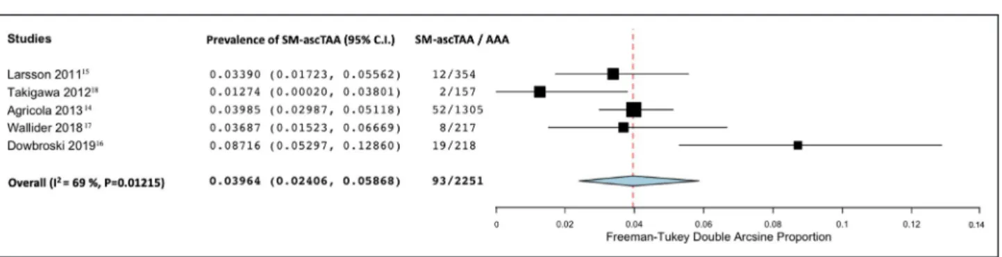
Figure 4. Forest plot analyzing the prevalence of synchronous/metachronous ascending thoracic aortic aneurysm in patients with known abdominal aortic aneurysm.
AAA indicates abdominal aortic aneurysm; and SM-ascTAA: synchronous/metachronous ascending thoracic aortic aneurysm.
Figure 5. Forest plot analyzing the prevalence of synchronous/metachronous descending thoracic aortic aneurysm in patients with known abdominal aortic aneurysm.
AAA indicates abdominal aortic aneurysm; and SM-descTAA, synchronous/metachronous descending thoracic aortic aneurysm.
Prevalence of Synchronous/
Metachronous TAA in Patients With
Known AAA According to Anatomic
Location
The prevalence of SM-TAA in patients with known AAA according to the anatomic location of the TAA was 4% in
the ascending thoracic aorta (95% CI, 2.4–5.7; I2=69%;
5 studies; 2251 participants)—Figure 4; 14.1% in the
descending thoracic aorta (95% CI, 4.7–27.3; I2=96%;
4 studies; 946 participants)—Figure 5 and 2.8%
involv-ing the aortic arch (95% CI, 0.9–5.5; I2=87%; 5 studies;
2251 participants)—Figure 6.
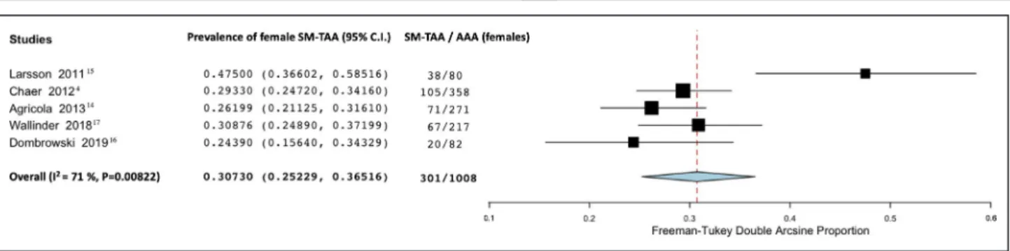
Prevalence and Risk of Synchronous/
Metachronous TAA in Patients With
Known AAA According to Sex
We found that 15.2% of male patients with AAA had
an SM-TAA (95% CI, 7.1–25.6; I2=97%; 4 studies; 2168
participants)—Figure 7. In female patients, the preva-lence was higher: 30.7% of female patients with AAA
had an SM-TAA (95% CI; 25.2–36.5; I2=71%; 5 studies;
1008 participants)—Figure 8.
Comparing both groups, women with AAA had a 2-fold increased risk of having SM-TAA compared with
men with AAA (RRs, 2.16; 95% CI, 1.32–3.55; I2=90%;
4 studies; 2168 participants)—Figure 9.
Risk of Synchronous/Metachronous TAA
in Patients With Known AAA According to
Other Risk Factors
Information on risk factors in both groups (SM-TAA and only AAA patients) was only available in 2
stud-ies.4,16 Overall, diabetes mellitus was associated with a
43% decreased risk of having SM-TAA and AAA (RRs,
0.57; 95% CI, 0.41–0.80; I2=0%; 2 studies; 1007
par-ticipants)—Figure 10. All other identified risk factors: smoking; hypertension; chronic obstructive pulmonary disease; family history of aortic aneurysm; and hyper-lipidemia were not associated with an increased or de-creased risk of having an SM-TAA and AAA (Table 3).
Sensitivity Analysis
To analyze the impact of using different diagnostic methods (namely transthoracic echocardiography), dif-ferent definitions for AAA and only including female pa-tients in the study, we performed a sensitivity analysis evaluating these effects (Table 4). Excluding the paper
from Agricola et al,14 which used transthoracic
echocar-diography as the diagnostic method to screen for TAAs and screened only for ascending and arch aneurysms, showed an increase in the prevalence (19.2% versus 21.5%). However, when analyzing according to the ana-tomic location the prevalence remained the same on ascending TAAs and was lower on arch TAAs (2.8%
Figure 7. Forest plot analyzing the prevalence in men of synchronous/metachronous thoracic aortic aneurysm in patients with known abdominal aortic aneurysm.
AAA indicates abdominal aortic aneurysm; and SM-TAA, synchronous/metachronous thoracic aortic aneurysm.
Figure 6. Forest plot analyzing the prevalence of synchronous/metachronous thoracic aortic arch aneurysm in patients with known abdominal aortic aneurysm.
AAA indicates abdominal aortic aneurysm; and SM-archTAA: synchronous/metachronous arch thoracic aortic aneurysm.
versus 1.9%). Also, when we only included papers using
a definition of ≥30 mm4,15–17 for AAAs the prevalence of
SM-TAA also increased (19.2% versus 25%).
DISCUSSION
The main findings of this review were: (1) the overall prevalence of SM-TAA in patients with a known AAA was 19.2% (95% CI, 12.3–27.3) and (2) the prevalence of SM-TAA was higher in women—30.7% (95% CI, 25.2–36.5) versus 15.2% (95% CI, 7.1–25.6)—with a 2-fold increase in risk.
These findings are surprising as almost a fifth of every patient with an AAA will have a TAA and that number increases to almost a third in women.
Screening strategies for AAA with an abdominal ultrasound have shown to be effective, reducing the incidence in aneurysm rupture rate and improvement
of care in a cost-effective fashion.3 Contrary to AAAs,
implementing a screening strategy for TAAs is difficult, since for an accurate diagnosis, a chest CT is usually necessary. Transthoracic echocardiography might find some but certainly not all TAAs, especially in the descending thoracic aorta. We found this in our sen-sitivity analysis: the prevalence increased (19.2% ver-sus 21.5%) when we excluded the study from Agricola
et al.14 In fact, in this latter study,14 which used only
transthoracic echocardiography to screen for TAAs,
the prevalence described for synchronous TAAs in pa-tients with known AAA was only 10.5%, which is prob-ably because of the fact that descending TAAs were not screened (Table 4).
The absence of a timely diagnosis has led to a lot of TAAs passing undiagnosed until a complication
oc-curs, such as rupture, which is usually fatal.20,21 A TAA
diagnosed before rupture is a potential life saved and this is usually made accidentally. We found that 19.2% of AAAs have an SM-TAA, this means that screening every patient with a known AAA with a chest CT might be useful. Moreover, death attributable to rupture of other aneurysms, such as TAAs, is already a problem
recognized in long-term follow-up of AAA repair.4,6,7,22,23
Currently, there are no clear indications in the current
Society of Vascular Surgery guidelines19 about full
aor-tic imaging when an AAA is diagnosed. Although more research is needed to demonstrate cost-effectiveness and the real impact of these SM-TAA, we believe TAA screening in patients with a known AAA should probably become common practice since 19.2% is not negligible.
The difference found between male and female sex is striking. It has been known that women have a lower threshold for aneurysm rupture and have worse outcomes after AAA repair, even when operated at
smaller diameters.24,25 No one has yet clearly
under-stood why this occurs. The higher prevalence of syn-chronous/metachronous thoracic and abdominal aortic
Figure 9. Forest plot analyzing relative risk of female sex comparing with male sex on the prevalence of synchronous thoracic aortic aneurysm in patients with known abdominal aortic aneurysm.
AAA indicates abdominal aortic aneurysm; and SM-TAA, synchronous/metachronous thoracic aortic aneurysm.
Figure 8. Forest plot analyzing the prevalence in women of synchronous/metachronous thoracic aortic aneurysm in patients with known abdominal aortic aneurysm.
AAA indicates abdominal aortic aneurysm; and SM-TAA, synchronous/metachronous thoracic aortic aneurysm.
aneurysms in women, might indicate that aneurysm disease has a different pathophysiology in the female sex, with a different and probably more aggressive and systemic behavior than in men. This might be one of the causes for these worse long-term outcomes found in women. Understanding this fact might lead us to in-crease our diagnostic suspicion for synchronous and metachronous TAAs and lead us to better surveillance and ultimately improved care.
Because of lack of data, we were not able to an-alyze in-depth other risk factors which might be pre-dictive of higher prevalence of SM-TAA. We found diabetes mellitus to be negatively associated with the risk of having SM-TAA and AAA, however, this data
were only available in 2 studies,4,16 which limits our
findings. In the paper from Chaer et al4 positive
pre-dictors of SM-TAA were Black race; family history of TAA; hypertension; and obesity; and negative predic-tors were diabetes mellitus, infra-renal location of the AAA; and smoking.
This review has some limitations, the difference in study designs on diagnostic method and definitions of both AAA and TAAs brings some clinical and possibly statistical heterogeneity to our results in general. The fact that the data retrieved was not age-standardized
also limits our findings, and explains, at least, par-tially, the statistical heterogeneity. The lack of data about risk factors in both groups (synchronous and AAA-only patients), such as smoking or hyperten-sion, limited our analysis on possible confounding or predictive factors for the presence of SM-TAA. Other important data that were not available across all stud-ies was the size of the TAAs found, the number of TAAs that ruptured, and their mortality before repair, which limits our analysis about risk and the prognosis of these aneurysms.
Further studies are needed to understand the clini-cal behavior of synchronous and metachronous TAAs in AAA patients (including their morbidity and mortal-ity), what is the true impact of screening patients with an AAA with a chest CT, to assess the cost-effective-ness of such a screening program and to understand their relationship with the female sex.
A feasible observational study would be to compare 2 time periods: pre- and post-SM-TAA screening with a chest CT in all patients with a known AAA to address the impact of screening in the number of treatable/near treatable TAAs and the number of preventable ruptured TAAs and deaths. Also, using large registry data would be useful to identify other clinical characteristics that might be more common in patients with synchronous or metachronous aortic aneurysms.
CONCLUSIONS
This meta-analysis increases the evidence about the presence of synchronous/metachronous TAA in pa-tients with known AAA. The higher prevalence of these aneurysms in women is striking and might explain one of the aspects why worse outcomes on follow-up of AAA are observed in women and shows the impor-tance of aortic screening in women.
To improve short- and long-term outcomes after AAA repair, the authors recommend that routine screening of synchronous and metachronous TAAs and their clinical impact should be more thoroughly studied, since 19.2% of AAAs with an SM-TAA is not a negligible number.
Table 3. Relative Risk of Having a Synchronous TAA and AAA Compared With Patients With AAA Only Across the Different Risk Factors
Risk Factor Relative Risk
Diabetes mellitus 0.57 (95% CI, 0.41–0.80; I2=0%;
2 studies; 1007 participants) Smoking 0.97 (95% CI, 0.85–1.10; I2=62%; 2 studies; 1007 participants) Hypertension 1.01 (95% CI, 0.94–1.01; I2=70%; 2 studies; 1007 participants) Hyperlipidemia 1.10 (95% CI, 0.89–1.3; I2=58%; 2 studies; 1007 participants) COPD 1.09 (95% CI, 0.93–1.28; I2=0%; 2 studies; 1007 participants) Family history of AAA/TAA 2.37 (95% CI, 0.97–5.77; I2=35%;
2 studies; 1007 participants) AAA indicates abdominal aortic aneurysm; COPD, chronic obstructive pulmonary disease; and TAAs, thoracic aortic aneurysms.
Figure 10. Forest plot analyzing relative risk of diabetes mellitus on the prevalence of synchronous thoracic aortic aneurysm in patients with known abdominal aortic aneurysm.
AAA indicates abdominal aortic aneurysm; DM, diabetes mellitus; RR, relative risk; and SM-TAA, synchronous/metachronous thoracic aortic aneurysm.
ARTICLE INFORMATION
Received May 7, 2020; accepted September 18, 2020.
Affiliations
From the Vascular Surgery Department, Hospital Santa Maria, Centro Hospitalar Universitário Lisboa Norte (CHULN), Lisboa, Portugal (R.G.e.M., A.L., R.F.e.F., L.M.P.); Faculty of Medicine, University of Lisbon, Lisboa, Portugal (R.G.e.M., G.S.D., M.A., D.C., R.F.e.F., L.M.P.); Cardiovascular Center of the University of Lisbon (CCUL), Lisboa, Portugal (R.G.e.M., A.L., D.C., R.F.e.F., L.M.P.); Laboratory of Clinical Pharmacology and Therapeutics, Faculty of Medicine (G.S.D., M.A., D.C.) and Instituto de Medicina Molecular, Faculty of Medicine (G.S.D., M.A., D.C.), University of Lisbon, Lisboa, Portugal; Serviço de Medicina III, Hospital Pulido Valente (CHULN), Lisboa, Portugal (M.A.); and Serviço de Cardiologia, Hospital Universitário de Santa Maria (CHULN), Lisboa, Portugal (D.C.).
Acknowledgments
Gouveia e Melo and Silva Duarte conceived the idea for the protocol and made the main contribution to planning and preparation of timelines for completion. Gouveia e Melo and Lopes analyzed all papers, extracted the data, and analyzed the risk of bias. Gouveia e Melo performed the statistical analysis. Caldeira and Alves analyzed the data and confirmed the statistical analysis. Gouveia e Melo designed the tables and wrote the first draft of the manuscript, which was then reviewed and amended by Alves, Caldeira, Fernandes e Fernandes, and Pedro. All authors then approved the final writ-ten manuscript. Gouveia e Melo is the guarantor for the work.
Sources of Funding None. Disclosures None. Supplementary Material Table S1 Figure S1 REFERENCES
1. Kuivaniemi H, Ryer EJ, Elmore JR, Tromp G. Understanding the patho-genesis of abdominal aortic aneurysms. Expert Rev Cardiovasc Ther. 2015;13:975–987.
2. Ruddy JM, Jones JA, Spinale FG, Ikonomidis JS. Regional hetero-geneity within the aorta: relevance to aneurysm disease. J Thorac
Cardiovasc Surg. 2008;136:1123–1130.
3. Guirguis-Blake JM, Beil TL, Senger CA, Coppola EL. Primary care screening for abdominal aortic aneurysm updated evidence report and systematic review for the US Preventive Services Task Force. JAMA. 2019;322:2219–2238.
4. Chaer RA, Vasoncelos R, Marone LK, Al-Khoury G, Rhee RY, Cho JS, Makaroun MS. Synchronous and metachronous thoracic an-eurysms in patients with abdominal aortic anan-eurysms. J Vasc Surg. 2012;56:1261–1265.
5. Garrido P, Pedro LM, Fernandes RF, Silvestre L, Sousa G, Martins C, Fernandes JF. Endovascular treatment of synchronous and metachro-nous aneurysms of the thoracic aorta. Is there an increase risk in the procedural risk? Angiol Cir Vasc. 2016;12:226–233.
6. Clouse WD, Marone LK, Davison JK, Dorer DJ, Brewster DC, LaMuraglia GM, Cambria RP. Late aortic and graft-related events after thoracoab-dominal aneurysm repair. J Vasc Surg. 2003;37:254–261.
7. Plate G, Hollier LA, O’Brien P, Pairolero PC, Cherry KJ, Kazmier FJ. Recurrent aneurysms and late vascular complications following repair of abdominal aortic aneurysms. Arch Surg. 1985;120:590–594. 8. Liberati A, Altman DG, Tetzlaff J, Mulrow C, Gotzche PC, Ioannidis
JPA, Clarke M, Devereaux PJ, Jos K, Moher D. The PRISMA statement for reporting systematic reviews and meta-analyses of studies that evaluate healthcare interventions: explanation and elaboration. BMJ. 2009;339:2700.
9. Wallace BC, Dahabreh IJ, Trikalinos TA, Lau J, Trow P, Schmid CH. Closing the gap between methodologists and end-users: R as a com-putational back-end. J Stat Softw. 2012;49:1–15.
10. Dersimonian R, Laird N. Meta-analysis in clinical trials. Stat Med. 1986;188:177–188.
11. Barendregt JJ, Doi SA, Lee YY, Norman RE, Vos T. Meta-analysis of prevalence. J Epidemiol Community Health. 2013;67:974–978. 12. Higgins JPT, Thompson SG. Quantifying heterogeneity in a
meta-anal-ysis. Stat Med. 2002;21:1539–1558.
13. Critical Appraisal Skills Programme. CASP (cohort study) checklist. [on-line]. 2018. Available at: https://casp-uk.net/wp-conte nt/uploa ds/2018/01/ CASP-Cohor t-Study -Check list_2018.pdf. Accessed April 25, 2020. 14. Agricola E, Slavich M, Tufaro V, Fisicaro A, Oppizzi M, Melissano G,
Bertoglio L, Marone E, Civilini E, Margonato A, et al. Prevalence of thoracic ascending aortic aneurysms in adult patients with known ab-dominal aortic an aneurysm: an echocardiographic study. Int J Cardiol. 2013;168:3147–3148.
15. Larsson E, Vishnevskaya L, Kalin B, Granath F, Swedenbor J, Hultgren R. High frequency of thoracic aneurysms in patients with abdominal aortic aneurysms. Ann Surg. 2011;253:180–184.
Table 4. Sensitivity Analysis
Synchronous/Metachronous TAA in Patients With Known AAA Prevalence (%)
Overall Prevalence of SM-TAA in Patients With Known AAA 19.2 (95% CI, 12.3‒27.3; I2=96%; 6 studies; 3333 participants)
Studies using CT and/or MR for the diagnosis of TAA 21.5 (95% CI, 15.4–28.2; I2=90%; 5 studies; 2028 participants)
Studies using TE for the diagnosis of TAA 10.5 (95% CI, 8.9–12.2, 1 study; 1305 participants) Using AAA definition of a diameter ≥30 mm 25.0 (95% CI, 20.6–29.7; I2=76%; 4 studies; 1871 participants)
Including only AAA patients admitted for surgery 10.2 (95% CI, 8.6–11.8; I2=0%; 2 studies; 1462 participants)
Including both male and female patients 17.2 (95% CI, 10.2–25.5; I2=96%, 4 studies, 3116 participants)
Studies only analyzing female patients 30.9 (95% CI, 24.9–37.2)
Overall Prevalence of Snchronous/Metachronous Ascending
TAAs in Patients With Known AAA 4% (95% CI, 2.4–5.7; I2=69%; 5 studies; 2251 participants)
Studies using CT and/or MR for the diagnosis of ascending TAA 4% (95% CI, 1.7–7.0; I2=76%; 4 studies; 946 participants)
Overall Prevalence of Synchronous/Metachronous Arch
TAAs in Patients With Known AAA 2.8% (95% CI, 0.9‒5.5; I2=87%; 5 studies; 2251 participants)
Studies using CT and/or MR for the diagnosis of arch TAA 1.9% (95% CI, 1.0–3.1; I2=23%; 4 studies; 946 participants)
AAA indicates abdominal aortic aneurysm; CT, computed tomography; MR, magnetic resonance; SM-TAA, synchronous/metachronous thoracic aortic aneurysms; TAAs, thoracic aortic aneurysms; and TE, transthoracic echocardiography.
16. Dombrowski D, Long G, Chan J, Brown O. Screening chest computed tomography is indicated in all patients with abdominal aortic aneurysm.
Ann Vasc Surg. 2019;65:190–195.
17. Wallinder J, Georgiou A, Wanhainen A, Björck M. Prevalence of syn-chronous and metasyn-chronous aneurysms in women with abdominal aortic aneurysm. Eur J Vasc Endovasc Surg. 2018;56:435–440. 18. Takigawa M, Yoshimuta T, Akutsu K, Takeshita S, Yokoyama N.
Prevalence and predictors of coexistent silent atherosclerotic car-diovascular disease in patients with abdominal aortic aneurysm without previous symptomatic cardiovascular diseases. Angiology. 2012;63:380–385.
19. Chaikof EL, Dalman RL, Eskandari MK, Jackson BM, Lee WA, Mansour MA, Mastracci TM, Mell M, Murad MH, Nguyen LL, et al. The Society for Vascular Surgery practice guidelines on the care of patients with an abdominal aortic aneurysm. J Vasc Surg. 2018;67:2–77.
20. Bickerstaff LK, Pairolero PC, Hollier LH, Melton J, Van Pennen HJ, Cherry KJ, Joyce JW, Lie JT. Thoracic aortic aneurysms: a popula-tion-based study. Surgery. 1982;92:1103–1108.
21. Olsson C, Thelin S, Stahle E, Ekbom A, Granath F. Thoracic aortic an-eurysm and dissection: increasing prevalence and improved outcomes reported in a nationwide population-based study of more than 14,000 cases from 1987 to 2002. Circulation. 2006;114:2611–2618.
22. Goodney PP, Tavris D, Lee L, Gross T, Fisher ES, Finlayson SRG. Causes of late mortality after endovascular and open surgical repair of infrarenal abdominal aortic aneurysms. J Vasc Surg. 2010;51:1340–1347. 23. Van Schaik TG, Yeung KK, Verhagen HJ, Bruin JL, Van Sambeek
MRHM, Bam R, Zeebregts CJ, Van Herwaarden JA, Blankensteijn JD; DREAM trial participants. Long-term survival and secondary proce-dures after open or endovascular repair of abdominal aortic aneurysms.
J Vasc Surg. 2017;66:1379–1389.
24. Deery SE, Soden PA, Zettervall SL, Shean KE, Bodewes TCF, Pothof AB, Lo RC, Schermerhorn ML. Sex differences in mortality and morbid-ity following repair of intact abdominal aortic aneurysms. J Vasc Surg. 2017;65:1006–1013.
25. Lo CR, Schermerhorn ML. Abdominal aortic aneurysms in women. J
Vasc Surg. 2016;63:839–844.
SUPPLEMENTAL MATERIAL
1. Exp Aortic Aneurysm, Thoracic/
2. Exp Aneurysm, Ruptured/
3.
((thoracoabdominal or thoraco-abdominal or thorax or thoracic) adj3 (aneurysm or
dilated or ectasia) adj3 (aorta or aortic)).ti,ab,kw.
4.
1 or 2 or 3
5.
Incidence/ or incidence.ti.ab.kw.
6.
Epidemiologic studies/
7.
Cohort studies/ or cohort.ti.ab.kw.
8.
Prevalence/ or prevalence.ti.ab.kw
9.
5 or 6 or 7 or 8
10.
4 and 9
11.
Exp animals/ not humans.sh.
12.
10 not 11
Original CASP items Adapted CASP items Reasons for downgrading
1.Did the study address a clearly focused issue?
1.Did the study address a clearly focused issue? 2.Was the cohort recruited in an
acceptable way? 2.Was the cohort recruited in an acceptable way? Not including every AAA with a diameter equal or higher than 30mm 3.Was the exposure accurately
measured to minimise bias?
3.Was the exposure accurately measured to minimise bias?
No clear explanation of how the diagnosis of AAA was obtained
4.Was the outcome accurately
measured to minimise bias? 4.Was the outcome accurately measured to minimise bias? Did not use either chest MR or CT to diagnose a TAA 5. (a) Have the authors identified all
important confounding factors?
5. (a) Have the authors identified all important confounding factors – age, smoking, hypertension
These risk factors have not been identified and quantified in the overall cohort and outcome group
5. (b) Have they taken account of the confounding factors in the design and/or analysis?
5. (b) Have they taken account of the confounding factors in the design and/or analysis?
No adjustment has been made for confounding factors (age, smoking, hypertension)
6. (a) Was the follow up of subjects complete enough?
Not used – not aplicable 6. (b) Was the follow up of subjects
long enough?
Not used – not aplicable
7. What are the results of this study? Not used – not applicable (open question format) 8. How precise are the results? Not used – not applicable (open question format)
9. Do you believe the results? 9. Do you believe the results? No clear definition of TAA and methods used for diagnosis are provided
10. Can the results be applied to the local population?
10. Can the results be applied to the local population?
The population is not a valid cohort for AAA patients in a hospital/community setting 11. Do the results of this study fit
with other available evidence? 11. Do the results of this study fit with other available evidence? The results are not in concordance with the overall available data regarding synchronous TAA and AAA
12. What are the implications of this study for practice?
Not used – not applicable (open question format)