UNIVERSIDADE DE LISBOA
FACULDADE DE MEDICINA DE LISBOA
Synthetic Pathogens for Integrated Biophysical and
Genetic Dissection of Antigen Cross-Presentation
RUI PEDRO DA SILVA ALBUQUERQUE E FREITAS
Doutoramento em Ciências Biomédicas
Especialidade em Ciências Morfológicas
UNIVERSIDADE DE LISBOA
FACULDADE DE MEDICINA DE LISBOA
Synthetic Pathogens for Integrated Biophysical and
Genetic Dissection of Antigen Cross-Presentation
RUI PEDRO DA SILVA ALBUQUERQUE E FREITAS
Tese orientada por:
Professor Doutor Luís Filipe Ferreira Moita
Professor Doutor Darrell J. Irvine
Doutoramento em Ciências Biomédicas
Especialidade em Ciências Morfológicas
2010
Todas as afirmações efectuadas no presente documento são da exclusiva
responsabilidade do seu autor, não cabendo qualquer responsabilidade
à Faculdade de Medicina de Lisboa pelos conteúdos nele apresentados.
A impressão desta dissertação foi aprovada pela Comissão
Coordenadora do Conselho Científico da Faculdade de
Medicina de Lisboa em reunião de 27 de Janeiro de 2010
O desenvolvimento e execução gráfica da presente dissertação foram
financiados pela Fundação para a Ciência e Tecnologia
“Para ser grande, sê inteiro: nada Teu exagera ou exclui. Sê todo em cada coisa. Põe quanto és No mínimo que fazes. Assim em cada lago a lua toda
Brilha, porque alta vive” Fernando Pessoa
iii
One of the most important and difficult steps in a scientist short life is to decide start writing the PhD thesis, but even before, to be brave or naive enough to start a scientific career and to run the unknown and tricky road until the end. It is a kind of matrimonial relation, where almost all scientists begin to love science, adore and enjoy the idea of discover something new and interesting with the propose of finding a solution or eliminating something that was not so bright. Therefore, everything starts with conceptual dreamers without frontiers.
This thesis describes the work carried between January 2006 and July 2009 mainly at the Instituto de Medicina Molecular (Lisbon, Portugal). During this period, part of the research was done at Massachusetts General Hospital (Boston, USA), at MIT (Cambridge, USA) and at the Institute Currie (Paris, France). The main goal was to study how the biochemical and biophysical properties of specific particulate antigens influence the cross-presentation pathway(s) and try to dissect and indentify the mechanism(s) behind it.
This thesis was divided in 6 Chapters:
The introduction comprises a general overview of specific key immunology concepts, such as Innate Immunity, Dendritic Cell biology, and antigen presentation mechanism with emphasis on antigen cross-presentation.
The second chapter focuses on particulate antigen design and the goal of specific properties introduced in the particles; the shRNA lentiviral library production and its application in high-throughput approaches. It includes a summary description of my participation in the work done within this period and the resulting publications.
The third chapter is composed by the materials and methods used throughout my work, including the different particulate platforms design and biochemical and cellular techniques for antigen presentation studies.
Results of my main project are described on Chapter 4, where different platforms of particulate antigen were used to study antigen cross-presentation mechanism(s).
Discussion is presented on Chapter 5 and concluding remarks on Chapter 6.
The results presented in this thesis, in collaboration with Darrell Irvine’s lab at MIT, are under preparation for publication.
v
“A person who never made a mistake never tried anything new”
vii
The study of host-pathogen interactions is crucial to unveil the diversity of the immune response outcome. Dendritic Cells (DCs) play a central role in the initiation and regulation of T-Cell immunity, functioning as master switches that
control whether the outcome of antigen presentation results in tolerance, or immunity. Antigen cross-presentation is a necessary mechanism to generate
immunity against tumors, bacteria and viruses. In addition, it is extremely important to induce cytotoxic immunity by vaccination with antigens. Moreover,
particulate antigens have been used in vaccine design tools as a platform to deliver different types of signals and in the modulation of DC-dependent immune responses. DCs express a series of different receptors that mediate the transfer of signals from the environment. Among them, Toll-Like Receptors (TLRs) play a critical role in the early innate immune response to invading pathogens. These receptors have the ability to recognize a broad range of pathogen-associated molecular patterns (PAMPs), turning them, key receptors in distinguishing between self/non-self antigens. The precise mechanisms underlying the crosstalk between TLRs and antigen presentation are not entirely understood. Therefore, the main goal of this project is to understand how TLR agonists coupled to particulate antigens influence antigen cross-presentation.
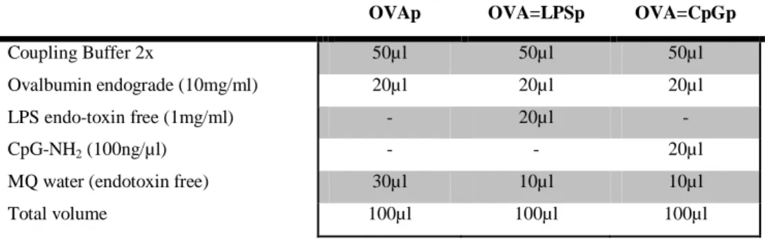
In our studies, we have used newly synthesized particle antigens, denominated as 'synthetic pathogens', coupled with a model antigen (Ovalbumin - OVA), and/or a model ligand (TLR agonist). These particle platforms have distinct, well-defined physical and biochemical properties, and function as a novel approach to elucidate the intrinsic mechanism(s) of antigen cross-presentation. In addition, they represent a valuable and powerful tool, which might be explored for therapy applications.
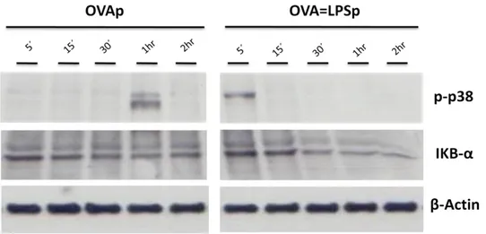
TLR4 is unique among TLRs as it can signal through both MyD88 and TRIF adaptors upon LPS stimulation, but mainly by the TRIF pathway when LipidA is the agonist. Our results revealed that when LPS is in the same cargo as the particle antigen, it impairs antigen cross-presentation and dictates a shift to MHC class- II presentation. This antigen cross-presentation abolishment is recovered on TLR4 deficient DCs and in the presence of the p38 MAPK pathway inhibitor, but not in the absence of the MyD88 adaptor. Moreover, LipidA reproduces the same phenotype as LPS, implicating the TLR4/TRIF-mediated signaling on particulate antigen cross-presentation impairment. Thus, here we describe a new mechanism of antigen selection in DCs for antigen cross-presentation that is dependent on the antigen based-environment. We show that the efficiency of presenting antigens from phagocytosed cargo is dependent on the presence of TLR ligands within the cargo. The influence of the compartmentalization on the crosstalk between the TLR-signaling and the antigen cross-presentation pathway(s), may constitute a tool used by DCs in order to discriminate the contents of phagosomes and present an appropriate immune response to specific stimuli. Therefore, DCs may have the “capacity” to decide which kind of destiny an antigen should have depending on the type and origin of the stimuli.
In order to dissect the mechanisms behind the cross-presentation phenotype, we have addressed the role of particle LPS on several antigen presentation key steps. Our data show that LPS-containing phagosomes enhance phagosome maturation (higher levels of colocalization with lysosomes) characterized by higher rates of phagosomal acidification and a decrease of phagosomal reactive oxygen species (ROS) production. The induction of phagosome maturation mediated by LPS signaling seems to shut down the machinery for antigen release into the cytosol, where the epitopes for MHC class-I are predominantly generated by the proteasome. Moreover, lower levels of antigen degradation occur when LPS is in
ix
the same cargo as antigen, mainly in a proteasome-dependent manner. This phenotype mediated by particulate LPS stimulus seems to be related with lower levels of particle antigen cross-presentation.
Therefore, we propose that antigen cross-presentation is enhanced during the brief period of time when phagosomal acidification is “sustained” and an immature phenotype is predominant, where endoplasmic reticulum machinery important for MHC class-I presentation probably is available. In addition this phenotype allows antigen escape into cytosol and the generation of epitopes for MHC class-I by the proteasome. On the other hand, antigen cross-presentation is impaired when a stimulus that induces phagosome maturation/acidification is in the same cargo as the antigen, producing a mature phenotype, allowing the generation of epitopes on the endocytic pathway that is compromised for MHC class-II antigen presentation. In order to address if the abolishment on antigen cross-presentation phenotype is transversal to others TLRs, studies were extended using different TLR specific agonists. When particle antigen contains TLR agonists that preferentially signal through MAPK/NF-kB pathways, antigen cross-presentation is induced. In contrast, in the presence of TLR agonists that preferentially signals through IFN-Type I pathway, particle antigen cross-presentation is inhibited. Therefore, a signaling pathway correlation may exist in the outcome of antigen presentation pathway(s) mediated by TLR agonist-containing particle antigens.
In sum, this work shows for the first time the inhibitory effect of TLR4 signaling on cross-presentation when agonists are delivered in the same cargo as particulate antigen. This phenotype is likely to be mediated by TRIF-dependent signaling, mainly by p38 MAPK activation. This knowledge could have a major impact in the dissection of the antigen cross-presentation mechanism, which will be highly valuable for novel vaccine design inducing T-Cell responses of the desired type and specificity.
xi
O estudo das interações patogénio-hospedeiro é fundamental para a compreensão da diversidade da resposta imunitária e para o desenvolvimento de novas estratégias terapêuticas. As células dendríticas (DCs) desempenham um papel central na iniciação e regulação da imunidade mediada por linfócitos T, funcionando como “interruptores”, que podem originar uma resposta de tolerância ou imunidade em relação a um determinado antigénio. O mecanismo de cross-presentation de antigénios tem sido descrito como necessário para gerar imunidade contra tumores, bactérias e vírus, e fudamental na indução de imunidade citotóxica mediada por vacinação. Por outro lado, os antigénios particulados têm sido utilizados como ferramentas no design de vacinas, possibilitando uma plataforma na qual se podem integrar diferentes tipos de estímulos.
As DCs expressam uma diversidade de receptores à superficie, o que permite uma detecção e transmissão eficazes dos vários tipos de “sinais” do meio ambiente. Entre eles, os Toll-Like Receptors (TLRs) desempenham um papel crucial, na resposta imune inata contra patogénios invasores. Estes receptores têm a capacidade de reconhecer uma ampla gama de padrões moleculares associados a patogénios (PAMPs), implicando-os como receptores-chave na distinção entre antigénios próprios e não-próprios. O mecanismo subjacente à crosstalk entre TLRs e a apresentação de antigénios não é totalmente conhecido. Por isso, um dos principais objetivos do meu trabalho foi compreender como é que os agonistas dos TLRs no mesmo contexto que antigénios particulados, influenciam a sua cross-presentation.
Neste projecto foram utilizadas partículas sintéticas – designadas por synthetic pathogens - na presença de um antigénio modelo (Ovalbumina) e/ou de um ligando
(agonista dos TLRs). Estas plataformas têm propriedades físicas e bioquímicas distintas e bem definidas, pelo que funcionam como uma nova abordagem para dissecar o mecanismo de cross-presentation de antigénios, bem como explorar o seu potencial para utilizaçao nas mais diversas terapias.
Os resultados demonstram que quando o Lipopolissacarídeo (LPS - agonista do TLR4) está presente no mesmo contexto que as partículas contendo o antigénio, ocorre uma redução nos níveis de cross-presentation de antigénios. Este fenótipo é acompanhado por uma mudança na via de apresentação de antigénios para MHC classe- II, que é induzida quando comparada com as partículas só com o antigénio. Este mecanismo foi demonstrado como sendo mediado pelo TLR4, onde a cross-presentation de antigénios é restabelecida em DCs deficientes nesse receptor .
A origem física dos estímulos (partícula vs solúvel) parece ser crucial para a regulação da via de cross-presentation de antigénios. Quando o LPS solúvel é co-incubado com partículas contendo o antigénio (dois estímulos físicos diferentes), verifica-se um aumento da activação/proliferação de células T em ambos os contextos de apresentação de antigénios - MHC classe-I e MHC classe -II. No entanto, quando o LPS é utilizado numa partícula diferente daquela que contém o antigénio, não se verificam diferenças significativas na eficiência das duas vias.
A influência da compartimentação no crosstalk entre a sinalização mediada pelos TLR e a via de cross-presentation de antigénios, pode constituir uma ferramenta importante que as DCs utilizam para discriminar o conteúdo dos fagossomas e iniciar uma resposta imune apropriada aos estímulos específicos. Esta observação é de extrema importância para compreender o papel de estímulos "patogénicos" no destino da apresentação de antigénios. Com o objectivo de compreender o mecanismo adjacente ao fenótipo observado da via da cross-presentation de antigénios particulados, o papel da activação do TLR4 foi estudado em vários processos importantes na apresentação de antigénios. Os resultados
xiii
obtidos indicam que fagossomas que contêm LPS têm uma indução na maturação (níveis mais elevados de colocalização com lisossomas), caracterizada por taxas mais elevadas de acidificação e uma diminuição da produção de espécies reactivas de oxigénio (ROS). A indução da maturação dos fagossomas mediada pela sinalização por LPS parece bloquear o mecanismo de libertação do antigénio dos fagossomas para o citosol, onde os epítopos para a apresentação em MHC classe -I são predominantemente gerados pelo proteassoma. Para além disso, níveis mais baixos de degradação do antigénio, mediada principalmente pelo proteassoma, ocorrem quando o LPS está no mesmo contexto. Este fenótipo é devido à sinalização mediada pelo LPS e parece estar relacionado com níveis baixos de cross-presentation do antigénio particulado.
Posto isto, sugerimos que a cross-presentation de antigénios é reforçada durante um breve período de tempo quando o pH dos fagossomas é mantido em valores próximos do estado basal, onde um fenótipo imaturo é predominante. Este estado imaturo é caracterizado pela existência de componentes do retículo endoplasmático (ER) importantes para a apresentação em MHC classe-I, permitindo o escape do antigénio para o citoplasma, onde os epítopos podem então ser gerados pelo proteassoma e apresentados no contexto MHC classe-I à superfície. Por outro lado, a cross-presentation de antigénios é diminuída quando um estímulo que induz a maturação dos fagossomas que contem o antigénio está no mesmo contexto. A formação de fagolisossomas leva à rápida acidificação e produz um fenótipo maduro, permitindo a geração de epítopos na via endocítica que é direccionada para a via MHC classe-II de apresentação de antigénios.
O TLR4 é singular entre os TLRs, uma vez que pode sinalizar tanto pelo adaptador MyD88 ou pelo TRIF quando estimulado por LPS, mas preferencialmente pelo adaptador TRIF quando LipidA é o agonista. Com o objectivo de estudar o impacto da sinalização do TLR4 na via de
cross-presentation dos antigénios particulados, o LipidA foi utilizado no mesmo contexto que o antigénio particulado. Observou-se uma reprodução do fenótipo de supressão da via de cross-presentation de antigénios obtido na presença do LPS. Além disso, a cross-presentation de antigénios particulados na presença de LPS não foi recuperada usando DCs deficientes no adaptador MyD88, ao contrário do que acontece quando se usa DCs deficientes no TLR4 e na presença de inibidores de activação da via das MAPK, principalmente a p38 MAPK. Estes resultados implicam a via TLR4/TRIF na inibição da cross-presentation de antigénios.
Com o objectivo de verificar se o efeito inibitório na via de cross-presentation de antigénios é transversal aos outros TLRs, os estudos foram alargados usando agonistas dos diversos TLRs no mesmo contexto que as partículas de antigénio. Verificou-se que, os agonistas dos TLR que sinalizam preferencialmente através da via MAPK/NF-kB induzem a cross-presentation de antigénios particulados. Em oposição, os agonistas dos TLR que sinalizam preferencialmente através da via do IFN Tipo-I, levam à inibição da cross-presentation destes antigénios. O TLR4 pode sinalizar através dos dois adaptadores (MyD88 e TRIF) em diferentes localizações, sendo preferencialmente via TRIF quando o TLR4 é internalizado nos endossomas. A inibição da via de cross-preserntation de antigénios mediada pelo LPS quando no mesmo contexto que o antigénio particulado, poderia indicar que o LPS em partículas sinaliza preferencialmente através da via TRIF, quando estas são internalizadas. Esta observação corrobora os dados obtidos com os outros agonistas de TLR que preferencialmente sinalizam através da via do IFN Tipo-I, como é o caso do TLR3, do TLR7 e do TLR9, que estão localizados em endossomas. Conclui-se assim que os vários TLRs estão envolvidos em mecanismos diferentes que levam a efeitos distintos nas vias de apresentação de antigénios. Além disso, um padrão da via de cross-presentation de antigénios
xv
parece existir mediado por partículas contendo os vários agonistas dos diferentes TLRs.
Em colaboração com o grupo do Prof. Darrell Irvine do MIT, pretendemos alargar estes estudos para outras plataformas de antigénios. Partículas de poli(ácido lático-co-ácido glicólico), PLGA, e hidrogel têm sido usadas como plataforma para administrar drogas, assim como em aplicações de biomateriais e concepção de vacinas. De acordo com os dados obtidos para a plataforma “fixa” de antigénios (partículas utilizadas nos ensaios anteriores), o LipidA no mesmo contexto que o antigénio particulado inviabiliza a via de cross-presentation de antigénios, comprovando a sua acção como um inibidor de sinalização mediada pelo TLR4, por um mecanismo dependente do adaptador TRIF. Assim, podemos concluir que mesmo na presença de partículas com propriedades distintas, a via mediada pelo adaptador TRIF tem um papel importante na inibição da via de cross-presentation de antigénios quando estimulada pelos agonistas do TLR4.
Este trabalho mostrou pela primeira vez, o efeito inibitório da crosstalk entre a sinalização pelo TLR4 e a cross-presentation de antigénios quando os agonistas estão no mesmo contexto do antigénio particulado. Este fenótipo é susceptível de ser mediado pela via TLR4/TRIF, principalmente através da activação da via p38 MAPK. Estes resultados podem ter assim um impacto deveras importante na dissecção do mecanismo de cross-presentation de antigénios, assim como na concepção de novos protocolos de vacinação e na indução de respostas específicas mediadas por linfócitos T.
xvii
Em primeiro lugar queria agradecer a todos que de alguma maneira ao longo destes anos sempre tiveram disponibilidade para ensinar, apoiar, dar, partilhar, aconselhar mas acima de tudo acreditar e fazer acreditar.
O primeiro agradecimento ao Professor Doutor Luís Ferreira Moita, pela orientação e por me ter proporcionado as condições necessárias para desenvolver o meu trabalho.
I would also like to thank my co-supervisor, Professor Darrell Irvine, to have received me at MIT and to support important technology used on my work and for helpful brainstorms. Professor Nir Hacohen (Harvard Medical School) and Professor Sebastian Amigorena (Institute Curie) for their hospitality and availability to help me to train and learn important techniques and for helpful science improvement, discussion and supervision. To Anna Bershteyn for the important collaboration, helpful work discussion and particles design.
Aos membros do comité de tese, Professor Doutor João Gonçalves, Professor Doutor Tiago Outeiro e Professora Doutora Margarida Gama-Carvalho pela sua paciência e conselhos durante todo este processo.
Ao Sérgio, ao “Malino”, ao “Mister”, à Ju, à Sofia, ao Daniel e ao “Zé” pela ajuda preciosa na elaboração da tese e pela “energia” partilhada. Ao José Rino pela ajuda na aquisição e análise de imagens por microscopia confocal.
Um agradecimento muito especial aos colegas de laboratório e dos quais nasceram verdadeiros amigos... principalmente aos companheiros de longas tertúlias científicas e que marcaram a diferença: “Malino”, “Mister” e Bruno. À Catarina, à Teresa, à Helena Raquel, à Raquel, à Ana e ao Nuno um muito obrigado.
Finalmente, gostaria de deixar um último agradecimento e abraço, aqueles que ao longo do meu percurso académico e principalmente no IMM se interessam por ciência e tentam fazer a diferença, aos verdadeiros amigos que nasceram entre as pipetas, jogos de futebol e rambóia ... pela continua amizade e conselhos. Àqueles que passaram pela minha vida e que a marcaram de alguma forma ... fazendo sentir, sofrer, chorar, rir, pensar, reflectir, amar, parar, andar, vibrar, gostar, odiar, transpirar, viajar, respirar, querer ... viver. Obrigado pela essência.
Aos meus Pais por serem únicos, à minha irmã e sobrinho por serem especiais, aos meus avós onde quer que estejam ... este “momento” é vosso.
This work was funded by Human Frontier Science Program (HFSP - RGY0058/2006-C), FCT (PTDC/SAU-MII/69280/2006) and FCT PhD fellowship - SFRH/BD/14316/2003
xix
Ag Antigen
APCs Antigen Presenting Cells
BMDCs Bone Marrow-derived Dendritic Cells
Cat.S Cathepsin S
cDCs Concentional Dendritic Cells
CFSE Carboxyfluorescein diacetate Succinimidyl Ester
CpG Cytosine-guanosine oligonucleotide
CTL Cytotoxic T Lymphocyte
Cyt c Cytochrome c
Cytcp Particles loaded with Cyt c
Cytc=LPSp Particles loaded with Cyt c and LPS
DCs Dendritic Cells
DHR123 Dihydrorhodamine 123
DQ-OVAp Particles loaded with DQ-OVA
DQ-OVA=LPSp Particles loaded with DQ-OVA and LPS
ELISA Enzyme-linked Immunosorbent Assay
ER Endoplasmatic Reticulum
ERAD ER-associated Degradation
FACS Fluorescence Activated Cell Sorter
GM-CSF Granulocyte Macrophage- Colony Stimulating Factor Hydrogel=OVAp Hydrogel particles loaded with OVA
Hydrogel=OVA=MPLAp Hydrogel particles loaded with OVA and MPLA
IFN-β Interferon beta
IFN-γ Interferon gamma
IL-… Interleukin-…
LPS Lipopolysaccharide
MPAKs Mitogen-activated protein kinases
MHC Major Histocompatibility Complex
MPLA (LipidA) Monophosphoryl Lipid A
MyD88 Myeloid differentiation primary response gene 88
NF-κB Nuclear Factor kappa B
OVA Ovalbumin
OVA488 Ovalbumin–Alexa488
OVAp Particles loaded with OVA
OVA=CpGp Particles loaded with OVA and CpG
OVA=Flagellinp Particles loaded with OVA and Flagellin
OVA=LPS Particles loaded with OVA and LPS
OVA=LipidAp Particles loaded with OVA and LipidA OVA=Pam2p Particles loaded with OVA and Pam2
LIST OF
OVA=Pam3p Particles loaded with OVA and Pam3 OVA=PolyI:Cp Particles loaded with OVA and PolyI:C OVA=ssRNA40p Particles loaded with OVA and ssRNAS40
OVA488p Particles loaded with OVA488
OVA488=LPSp Particles loaded with OVA488 and LPS OVA=DHR123p Particles loaded with OVA and DHR123 OVA=DHR123=LPSp Particles loaded with OVA, DHR123 and LPS PAMPs Pathogen-Associated Molecular Patterns
pDCs Plasmacytoid Dendritic Cells
PLGA poly(lactic-co-glycolic acid)
PLGA=OVAp poly(lactic-co-glycolic acid) particles loaded with OVA
PLGA=OVA=MPLAp poly(lactic-co-glycolic acid) particles loaded with OVA and MPLA
PRRs Pattern Recognition Receptors
RNAi RNA interference
ROS Reactive Oxygen Species
sDCs Splenic Dendritic Cells
shRNA short hairpin RNA
SIINFEKLp Particles loaded with SIINFEKL
SIINFKL=LPSp Particles loaded with SIINFEKL and LPS
TRIF TIR-domain-containing adapter-inducing interferon-β TAP Transporter Associated with Antigen Processing
TCR T-Cell Receptor
TLR Toll-Like Receptor
u.v. Ultraviolet radiation
xxi Preface____________________________________________________________i Abstract _________________________________________________________vii Resumo__________________________________________________________xi Acknowledgements________________________________________________xiv List of Abbreviations_______________________________________________xix Table of Contents _________________________________________________xxi
CHAPTER 1 –Introduction___________________________________________1
CHAPTER 2 - Objectives and Integrated Research Plans: Synthetic Pathogen Platform & shRNA genetic tools ______________________________________59
Part I: Synthetic Pathogen Platform design and characterization ______66
Part II: shRNA Library: New tools for the genetic dissection of cross-presentation pathway(s) ______________________________________ 87
CHAPTER 3 - Materials and Methods _________________________________97
CHAPTER 4 - Results_____________________________________________129
CHAPTER 5- Discussion __________________________________________179
CHAPTER 6- Concluding Remarks __________________________________203
REFERENCES__________________________________________________211
TABLE OF
CONTENTS
Introduction
Chapter 1_________________________________________________________________
_______________________________________________________________Introduction
3
1. Immune System_____________________________________________
The immune system is one of nature's most fascinating creations. It is composed of different cell types and uses almost all known proteins, with different ―jobs‖ in fighting foreign invaders and to recognize and tolerate self. It protects against bacteria, viruses, and other parasites, but unfortunately some pathogens can bypass and establish diseases. In some cases, immune cells fail their function and progress to recognize self-antigens (autoimmune response). For these reasons, immunology is one of the most studied subjects, and its knowledge seems to be interminable and fascinating. The immune system has been ―conceptually‖ subdivided in 2 sub-systems, innate and adaptive, each one with a different function and role. The main distinction relates to the mechanisms and receptors used for the immune recognition, which will be described bellow (Medzhitov and Janeway, 2000).1.1. Innate Immune System
The idea of a generic host defence was introduced over 100 years ago by Eli Metchnikoff, who revealed the role of ―phagocytes‖ in destroying invading microbes. In 1957, important components of innate immunity, named interferons, were discovered by Alick Isaacs. The interferons were recognized as important molecules for host defence mechanism activation with broad specificity. In 1973, Steinman and Cohn made a big step in immunology, by identification of Dendritic Cells (DCs) as a subtype of phagocytes, which were specialized for the capture and presentation of antigens. Nonetheless, the ―big bang‖ in innate immunity was introduced by Charles Janeway Jr. in his seminal 1989 commentary. There he introduced the ―immunologist´s dirty little secret‖, i.e. that most antigens would only elicit an adequate immune response when mixed with adjuvants containing microbial products.
The Innate Immune system is an evolutionarily ancient part of the host defense mechanisms and is found in all metazoans (the same molecular modules are found
Chapter 1_________________________________________________________________
4
in plants and animals, meaning that it arose before the split into these two kingdoms) (Hoffmann et al., 1999). Innate immunity major functions are: a) inflammatory responses through the production of chemical factors (cytokines and chemokines); b) activation of the complement cascade that promote the clearance of pathogens, dead cells or antibody complexes; c) identification and removal of cellular debris, foreign particles or microorganism by phagocytosis; d) activation of the adaptive immune system through a process known as antigen presentation. Innate immune cells originate from pluripotent hematopoietic stem cells present in the bone marrow and include: Natural killer cells, Mast Cells, Eosinophils, Basophils and the phagocytic cells (Macrophages, Neutrophils and DCs) (Janeway and Medzhitov, 2002). There are hundreds of receptors involved in innate immune recognition and approximately 1014 and 1018 different somatically generated immunoglobulin receptors and T-Cell receptors respectively. However, microbes are extremely heterogenous and can mutate at a much higher rate than any of their hosts (Medzhitov and Janeway, 2000).
Therefore, the strategy of the innate immune response may not be to recognize every possible antigen, but rather to focus on a few, highly conserved structures present in large groups of microorganisms, that are structurally distinct from the host. These structures are referred to as Pathogen-Associated Molecular Patterns (PAMPs), and the receptors of the innate immune system that evolved to recognize them are called Pattern-Recognition Receptors (PRRs) (Janeway, 1989). These receptors allow the first line of defence: discrimination between self and non-self (Janeway, 1989). When Charles Janeway Jr. answered the questions: “How does the immune system determine the origin of the antigen, and how does it decide whether to induce and immune response or not? Does innate immune system has a major role on that?”, the PAMPs and their recognition by PRRs, emerged as essential components for the innate immune system to respond or not to a specific antigen (Janeway and Medzhitov, 2002).
_______________________________________________________________Introduction
5
1.2 Adaptive Immune System
Adaptive Immunity is present only in vertebrates and it was the ―big bang‖ in immunity. It is composed of highly specialized, systemic cells: B Lymphocytes (B cells), T Lymphocytes (T-Cells) - CD8+ T-Cell or Cytotoxic T-Cell, CD4+ T-Cell or Helper T-Cells (Th1 or Th2) and gamma/delta T-Cells (Medzhitov and Janeway, 2000; Reis e Sousa, 2004a). Adaptive immunity is a relative newcomer on the evolutionary landscape. Because the mechanism of generating receptors in the adaptive immune system involves great variability and rearrangement of receptor gene segments, the adaptive immune system can provide specific recognition of foreign antigens, immunological memory of infection, and pathogen-specific adaptor proteins. However, the adaptive immune response is also responsible for allergy, autoimmunity, and the rejection of tissue grafts (Janeway and Medzhitov, 2002). This variability of receptors is due to Somatic hypermutation on two types of antigen receptors: T-Cell receptors (TCRs) on T-Cells and immunoglobulin receptors (IgR) on B cells. These antigen receptors are generated by random somatic gene rearrangement and are expressed in a clonal fashion on lymphocytes. Since each lymphocyte displays a single kind of structurally unique receptor, the repertoire of antigen receptors in the entire population of lymphocyte is extremely unique (each lymphocyte with a structurally unique receptor). Thus, these receptors are able to recognize almost all antigens that exist in nature. However, these receptors recognize not only pathogenic, but also environmental or self-antigens (Hoffmann et al., 1999; Medzhitov and Janeway, 1998). Rarely, responses from these receptors to environmental or self-antigens could lead to allergies or autoimmune diseases. What adaptive immunity adds to the underlying innate immune system is specific recognition of proteins, carbohydrates, lipids, nucleic acids (Janeway, 1989). The signals induced on recognition by the innate immune system, in turn, control the activation of adaptive immune responses that respond to a pathogen only after it has been recognizing by the innate immune system
Chapter 1_________________________________________________________________
6
(Medzhitov and Janeway, 2000). DCs make the link between innate and adaptive immune system by presenting antigens to naive T-Cells and expressing molecules such as cytokines, chemokines, costimulatory molecules and proteases to initiate an immune response (Steinman, 1991). To generate an efficient immune response to a specific pathogen, it is critical that the recognition of a specific antigen by lymphocyte receptors could trigger its activation and proliferation. This process termed clonal selection is the basic property of the adaptive immune system. Unfortunately, these receptors cannot be passed on to the next generation, even if they give a survival advantage. Antigen Receptors have to be reinvented by every generation (Medzhitov and Janeway, 2000). This mechanism takes 3-5 days to produce enough number of clones and to differentiate into effector cells. For most pathogens, this period could be enough to damage the host and establish disease (Medzhitov and Janeway, 2000). However, the effector mechanism of innate immune system (antimicrobial peptides, phagocytosis and alternative complement pathway), is activated immediately after infection, which normally leads to a rapid control of the infection pathogen. ―Retarding‖ the infection until the adaptive immune system is ready to deal with it, is one of the main functions of innate immunity (Janeway and Medzhitov, 2002).
_______________________________________________________________Introduction
7
2. Pathogen and Antigen Recognition
________________________________2.1. Pattern Recognition Receptors
The Pattern Recognition Receptors (PRRs) concept was introduced by Janeway 20 years ago, with the idea that immune system senses microbial infection by these receptors that are predominantly expressed on sentinels cells. Pathogen-associated molecular patterns (PAMPs) are the molecular signature recognized by PRRs, which are broadly expressed in pathogens but not in host cells. Therefore, PRRs are able to discriminate between self from non-self (Janeway, 1989). During the past years, different families of PRRs were identified. The most important are: Toll-like receptors (TLR), RIG-I-like receptors (RLR), NOD-like receptors (NLR) and C-Type Lectin Receptor (CLR).
Studies have shown the existence of a cytosolic detection system for intracellular PAMPs. These cytosolic PRRs include retinoic acid-inducible gene-I (RIG-I)-like receptors (RLRs) and nucleotide-binding oligomerization domain (NOD)-like receptors (NLRs). RLRs belong to the RNA helicases family that specifically detects RNA species derived from viruses in the cytoplasm and coordinate anti-viral programs via type I IFN induction (Yoneyama and Fujita, 2008). NLRs constitute a large family of intracellular PRRs, where the NOD1, NOD2 and NALP3 are the most relevant (Ting et al., 2008). NOD1 and NOD2 recognize intracellular bacterial cell products, and NALP3 responds to multiple stimuli to form a multi-protein complex termed the NALP3 inflammasome, which promotes the release of the IL-1 family of cytokines (Fritz et al., 2006; Inohara et al., 2005; Kanneganti et al., 2007; Meylan et al., 2006; Schweichel et al., 2006; Yu and Finlay, 2008). CLRs, are diverse families of receptors containing one or more C-type lectin domains, identified as a carbohydrate-binding structure. The different groups vary widely in ligand recognition and function and only a few acts as innate PRR mediating the recognition of PAMPs by DCs (Robinson et al., 2006). CLR
Chapter 1_________________________________________________________________
8
expressed by DCs included dectin-1, DEC-205 and DC-SIGN, which all belong to a separate group of CLR (Zelensky and Gready, 2005). Dectin-1 has been shown to act as PRR resulting in DC activation (Rogers et al., 2005). It recognizes carbohydrate structures in the form of fungal β 1,3-glucans (Palma et al., 2006). DEC-205 and DC-SIGN play a role in uptake, processing and presentation of antigens from pathogens, but does not induce DC activation (Jiang et al., 1995; Mahnke et al., 2000).
In addition to PAMPs, innate immunity has the potential to respond to endogenous molecules that are released by host cells as a result of necrosis, pathogen infection, damage, injury and certain pathological conditions, which are directly or indirectly recognized by TLRs, NLRs, RLRs or as-yet undefined sensors. The recognition of endogenous molecules by PRRs is tightly linked to the pathogenesis of autoimmune and inflammatory diseases (Kawai and Akira, 2009).
TLR family detects PAMPs either on the cell surface or the lumen of intracellular vesicles such as endosomes or lysosomes (Kawai and Akira, 2009). This work was focused on TLRs function in antigen cross-presentation; therefore they will be described more extensively as follows.
2.1.1. Toll-like Receptors: Overview
The first member of the Toll family, Drosophila Toll, was discovered as one of 12 maternal effect genes that function in a pathway required for dorso-ventral axis formation in fly embryos (Hashimoto et al., 1988). Analysis of the sequence of the toll gene revealed that it encodes a transmembrane protein with a large extracellular domain containing leucine-rich repeats. The sequence of the cytoplasmic domain of the toll turned out to be similar to the cytoplasmic domain of the human interleukin-1 receptor (hIL-1R), and it became apparent that they had possible functional similarities (Gay and Keith, 1991). Both hIL1-R and toll in drosophila have homologous cytoplasmic TIR domains and signal through
_______________________________________________________________Introduction
9
homologous protein kinases (Pelle and IRAK). Their signal-transduction leads to activation of transcription factors of the nuclear factor-kB (NF-kB) family pathway (Anderson, 2000; Belvin and Anderson, 1996). Earlier studies done by Ruslan Medzhitov in 1997, lead to the identification of a family of membrane-bound receptors in mammalian similar to drosophila toll (Medzhitov and Janeway, 1997). Therefore, a family of pattern-recognition receptors homologues of drosophila toll have been identified in mammals and are referred as toll-like receptors (TLRs) (Medzhitov and Janeway, 1997; Rock et al., 1998). Until now, there are described 13 members of mammalian TLR family (Akira et al., 2006). TLR1-9 are conserved between humans and mice, TLR10 is not functional in mice because of a retrovirus insertion, and TLR11, TLR12 and TLR13 are lost in human genomes.
TLRs differ from each other in: (1) ligand specificities, (2) expression patterns and in (3) target genes they can induce (Akira et al., 2006). The TLR family members can be conveniently divided into two subpopulations with regard to their cellular localization. TLR1, TLR2, TLR4, TLR5, TLR6 and TLR11 are expressed exclusively on the cell surface and recognize microbial membrane components such as lipids, lipoproteins and proteins. TLR3, TLR7, TLR8 and TLR9 are localized in intracellular vesicles such as the endosomes or lysosome and the endoplasmic reticulum (ER) and predominantly recognize microbial nucleic acid species (Akira and Takeda, 2004).
Therefore, TLR were initially implicated in the recognition of bacterial, fungal patterns and viruses (Pasare and Medzhitov, 2005; Takeda and Akira, 2004) but the exact mechanism of PAMPs recognition has not yet been well characterized. Recent studies have revealed the crystal structure of TLR1, TLR2, TLR3 and TLR4 and suggest their mechanism of ligand recognition (Jin and Lee, 2008). The ligands for most TLRs were identified trough generation of mice deficient for each TLR. Genetic studies revealed that TLR are able to recognize a wide range of PAMPs including lipids, lipoproteins, proteins, glycans and nucleic acids and play
Chapter 1_________________________________________________________________
10
a central role in initiating innate immune responses (Akira et al., 2006). Bacterial lipopeptide, a ligand for the TLR-1-TLR2 heterodimer, interacts with internal protein pockets, and hydrophobic interactions are responsible for ligand recognitions (Jin et al. 2007). On the other end, viral double-stranded RNA (dsRNA), a TLR3 ligand, interacts with both the N-terminal and the C-terminal sites on the lateral side of the convex surface of TLR3 by ionic and hydrogen bonds with the sugar-phosphate backbones of dsRNA (Choe et al., 2005). TLR4 is responsible for bacterial LPS recognition but there are no direct interactions. TLR4 form a complex with another LRR protein known as MD-2 (LPS-binding component) by ionic and hydrogen bonds in two oppositely charged patches (Kim et al., 2007; Ohto et al., 2007). All these TLR ligands induce a homodimer or heterodimer of TLRs (TLR1-TLR2, TLR3-TLR3, TLR4-TLR4) showing an ―m‖-shaped complexes. This dimerization is necessary for triggering downstream signaling by recruitment the TIR domain-containing adapter protein (fig.1).
_______________________________________________________________Introduction
11 Fig.1: TLR-mediated immune responses. The TLR family can be divided into subfamilies: the TLR at the cell surface (TLR1, TLR2, TLR4, TLR5 and TLR11) primarily detected bacterial, fungal and protozoan cell components, while intracellular TLR (TLR3, TLR7/8 and TLR9) recognize nucleic acid ligands in specific endosomal compartments. TLR2 in concert with TLR1 or TLR6 discriminates between the molecular patterns of triacyl and diacyl lipopeptide, respectively. TLR3 recognizes dsRNA. TLR4 recognizes bacterial LPS. TLR7/8 mediates recognition of imidazoquinolines and ssRNA. TLR9 recognizes CpG DNA of bacteria and viruses. TLR5 recognizes bacterial flagellin and mouse TLR11 recognizes components of uropathogenic bacteria and profilin like molecule of the protozoan parasite Toxoplasma gondii. TLR1/2 and TLR2/6 utilize MyD88 and TIRAP as essential adapters. TLR3 utilizes TRIF. TLR4 utilizes four adapters, including MyD88, TIRAP, TRIF and TRAM. TLR7/8, TLR9, TLR5 and TLR11 use only MyD88. The MyD88-dependent pathway controls inflammatory responses, while TRIF mainly mediates type I IFN responses. In addition, TLR7/8 and TLR9 induce type I IFN in a MyD88-dependent manner. Adapted from (Kawai and Akira, 2006).
2.1.2. Toll-like Receptors at cell surface
Toll-like Receptor 4: The first human TLR (hTLR), that was identified by Ruslan Medzhitov in 1997 is now referred to as toll-like receptor 4, and it was shown to activate, like its drosophila homolog, NF-kB signaling pathway (Medzhitov et al., 1997). In 1998, further four TLR were reported (Rock et al., 1998). Through NF-kB pathway, activation of TLR4 induces the expression of a variety of inflammatory cytokines and co-stimulatory molecules that are crucial to adaptive immune response (Medzhitov et al., 1997). This evidence implies TLRs as receptors of the immune system (Medzhitov and Janeway, 1997). The first link arises when it was shown that TLR4 is the receptor for lipopolysaccharide (LPS) in mice. Mice with either spontaneous mutation or a target disruption of the tlr4 gene, have no response to LPS and are thus resistant to endotoxin shock (Poltorak et al., 1998; Qureshi et al., 1999).Together, these studies demonstrated the essential role for TLR4 in recognition of LPS, a major component of gram-negative bacteria, which is a potent immunostimulatory molecule and cause septic shock.
TLR4 is not directly involved in LPS recognition. Soluble LPS molecules first interact with a serum protein called lipopolysaccharide-binding protein (LBP)that is present as a soluble protein or as a plasma membrane protein (Ulevitch and Tobias, 1995). At the plasma membrane, LBP binds CD14, a receptor that is
Chapter 1_________________________________________________________________
12
anchored to cell surface by a glycosylphosphoinositol tail (GPI-linked cell surface protein) and delivers LPS-LBP to the TLR4 (Miyake, 2007; Wright et al., 1990; Wright et al., 1989).
A small protein, MD-2, is also required for TLR4-mediated recognition of LPS, making the three components of LPS-recognition complex (TLR4, CD14 and MD-2). MD-2 lacks a transmembrane anchor but is constitutively associated with the extracellular region of TLR4, whereas CD14 is presumably recruited to the complex after binding LPS (Shimazu et al., 1999). The cell-surface events that lead to LPS recognition are not clear, but important data indicate that a complex of TLR4/MD-2/CD14 directly binds LPS (da Silva Correia et al., 2001; Lien et al., 2000; Poltorak et al., 2000). ‗Smooth‘ LPS is composed of a polysaccharide O-antigen side chain and has complete core oligosaccharides, whereas ‗rough‘ LPS lacks O-antigen and has shorter core oligosaccharides; both forms contain lipid A, a biologically active component of LPS. Cells lacking CD14 are unresponsive to smooth LPS; however, they still respond to rough LPS or lipid A (Dybdahl et al., 2002; Vabulas et al., 2002). TLR4 is known to activate two signaling pathways - the myeloid differentiation primary response gene 88 (MyD88)-dependent pathway and the TIR-containing adapter inducing IFN-β (TRIF)-dependent pathway. Monophosphoryl lipid A (MPLA) is a low-toxicity derivative of LPS with useful
immunostimulatory properties. MPLA is inefficient with respect to stimulation of
TLR4/MyD88-induced gene expression, it has fortuitously retained TLR4/TRIF-associated activities, such as induction of type I interferon (Mata-Haro et al., 2007).
These results suggest that the diversity of the structures of LPS among bacterial species may influence selective activation of these pathways. In addition to the detection of components of Gram-negative bacteria, TLR4 has been implicated as well, in the recognition of liptocheic acid (LTA), the heat shock protein hsp60, and the fusion protein of the respiratory syncytial virus (RSV) and mouse mammary
_______________________________________________________________Introduction
13
tumors virus (Kurt-Jones et al., 2000; Ohashi et al., 2000; Takeuchi et al., 1999; Vabulas et al., 2001). The physiological relevance of these putative TLR4 ligands remains to be demonstrated. However it is clear that mammalian TLRs do not discriminate between classes of pathogens (Janeway and Medzhitov, 2002).
TLR4 has recently been shown to signal from two locations (Kagan et al., 2008). At the plasma membrane, TLR4 recruits the second TIR domain adapter to be described, termed MyD88-adapter-like protein (MAL), and MyD88, leading to NF-kB activation. It then appears to traffic to the endosome, where it recruits two other TIR domain adapters, translocating chain-associating membrane protein (TRAM) and TIR-domain-containing adapter-inducing interferon-β (TRIF). Consequently leading to activation of the protein kinase TANK-binding kinase-1 (TBK-1) and activation of interferon regulatory factor 3 (IRF3), the transcription factor required for induction of Type I interferons and many other genes that contain the interferon responsive response element. This capacity for signaling from two locations appears to be unique to TLR4 among the TIR domain-containing receptors (O'Neill, 2008).
Toll-like Receptor 2 and heterodimers: TLR2 recognizes a wide range of PAMPs derived from various pathogens, ranging from bacteria, fungi, parasites and viruses (Akira et al., 2006). These ligands include triacyl lipopeptides from bacteria and mycobacteria, diacyl lipopeptides from mycoplasma, peptidoglycan (PGN) and lipoteichoic acid (LTA) from Gram-positive bacteria, porin from Neisseria, lipoarabinomannan from mycobacteria, zymosan (containing b-glucan, mannans, chitin, lipid and protein) from fungi, Trypanosoma GPI-mucin (tGPI-mucin) and hemagglutinin protein from measles virus. TLR2 generally forms a heterodimer with TLR1, TLR6 or non-TLR molecules such as CD36, CD14 and dectin-1 to discriminate the molecular structure of the ligands. TLR2–TLR6
Chapter 1_________________________________________________________________
14
recognizes the mycobacterial diacylated lipopeptide, LTA and zymosan, whereas TLR2–TLR1 recognizes the bacterial triacylated lipopeptide.
The role of extracellular TLRs may be as sensors for danger signals. TLR2 and TLR4 are also implicated in the recognition of endogenous molecules. These include heat shock proteins (HSP60, HSP70, gp96 and HSP22), fibrinogen, the extra domain A of fibronectins, hyaluronic acid, heparan sulfate, fatty acids, high-mobility group box 1 (HMGB1), modified low-density lipoprotein and β-defensin 2, most of which are released during inflammation or tissue damages or by necrotic cells (Akira and Takeda, 2004).These endogenous ligands trigger production of TNF-α, IL-12 and nitric oxide (Miyake, 2007).
Toll-like Receptor 5: TLR5 recognizes a highly conserved central site of flagelin, a protein that is a component of bacterial flagella, which is required for protofilament formation and bacterial motility (Hayashi et al., 2001). TLR5 is highly expressed on the basolateral surface of intestinal epithelial cells and moreover preferentially in CD11c+ CD11b+ lamina propria of DCs (LPDCs) in the small intestine (Uematsu et al., 2006), which suggests a role of TLR5 in the detection of invasive flagellated bacteria in the gut. TLR5 on LPDCs plays a critical role in regulating both innate and adaptive immune response in the intestine (Uematsu et al., 2008).
Toll-like Receptor 11: Mouse TLR11, which is a relative to TLR5, is highly expressed in the kidney and bladder. Because TLR11-deficient mice are susceptible to uropathogenic bacteria infection, is likely to sense its products, however a specific ligand has not been identified yet (Zhang et al., 2004). TLR11 also recognizes a parasite component from Toxoplasma gondii tachyzoites known as soluble Toxoplasma antigen that is a potent inducer for IL-12. The active component is a profiling-like molecule that functions as an actin-binding protein
_______________________________________________________________Introduction
15
and involved in parasite motility and invasion (Plattner et al., 2008) and is recognized by mouse TLR11 (Yarovinsky et al., 2005).
2.1.3. Intracellular Toll-like Receptors
The intracellular TLRs, TLR3, TLR7, TLR8 and TLR9, are expressed in intracellular compartments. They are on the ER in resting cells and trafficked to the endosomal compartments such as endosomes and lysosomes, in response to PAMP-mediated stimulation (Latz et al., 2004; Nishiya et al., 2005). This intracellular localization is important for the recognition of viral nucleic acids that are delivered to TLR expressing intracellular vesicles through the endosomal pathway. Moreover, this is also important for discrimination of self from non-self nucleic acids since ectopic expression of TLR9 on the macrophage cell surface causes it to respond to DNA derived from self (Barton et al., 2006). Intracellular TLRs appear to be sensors of foreign nucleic acids and trigger anti-viral innate immune responses by producing type I IFN and inflammatory cytokines.
Toll-like Receptor 3: TLR3 is a receptor for dsRNA. It recognizes a synthethic analogue of dsRNA polyinosinic-polycytidylic acid (poly:IC), genomic RNA purified by dsRNA viruses (such as reovirus) and dsRNA produced during the course of single-stranded RNA (ssRNA) viruses replication (such as RSV, encephalomyocarditis virus (EMCV) and West Nile virus (WNV) (Alexopoulou et al., 2001; Wang et al., 2004). TLR3 is also implicated in the recognition of small interfering RNA (siRNA) in a sequence independent manner (Kleinman et al., 2008). TLR3 mRNA is expressed in conventional Dendritic Cells (cDCs) but mostly in CD8α+ DCs, which have high phagocytic activity for apoptotic bodies of virus-infected or dsRNA-loaded cells. This allows dsRNA to gain access to TLR3 compartments and signaling to produce IL-12p40 and IFN-β (Schulz et al., 2005).
Chapter 1_________________________________________________________________
16
Toll-like Receptor 7: TLR7 is a natural receptor for ssRNA. It was originally identified to recognize imidazoquinoline derivatives (imiquimod and resiquimod (R-848)), and guanine analogues such as loxoribine. These compounds have anti-viral and anti-tumor properties (Hemmi et al., 2002). Synthethic polyuridine ssRNA and some siRNAs as well as natural compounds such guanosine-rich and uridine-rich ssRNA from HIV or influenza virus were identified as ligands for TLR7 (Diebold et al., 2004; Heil et al., 2004). TLR7 is mostly expressed in a subset of DCs, plasmacytoid Dendritic Cells (pDCs), which are unique in their capacity to rapidly secrete vast amounts of type I IFN in response to viral infections. These response to influenza or VSV was impaired in TLR7-deficient pDCs and it is independent of envelope virus replication such influenza or herpes viruses. It seems that these virus are endocyted to intracellular compartments, were viral particles are degraded, allowing the viral RNA to engage TLR7 (Gilliet et al., 2008).
Toll-like Receptor 8: TLR8 is related phylogenetically with TLR7. TLR8 is mostly expressed in monocytes, and is up-regulated upon bacterial infection. Human TLR8 recognizes R-848, ssRNA from virus (HIV, VSV and influenza A) and bacterial RNA, however, TLR8-deficient mice responds normally to these molecules suggesting a species-specific function of TLR8 (Heil et al., 2004).
Toll-like Receptor 9: TLR9 was identified to recognize DNA motifs that are frequently present in bacteria, but are rare in mammalians - unmethylated 2´-deoxyribo cytidine-phosphate-guanosine (CpG) (Hemmi et al., 2000). Synthethic CpG oligodeoxynuvleotides (ODNs) are TLR9 ligands. This recognition is independent of the base sequence, and the sugar backbone 2´-deoxyribose of DNA is sufficient for signaling (Haas et al., 2008). TLR9 is highly expressed in pDCs as TLR3 and TLR7, which serve as sensor for virus infection. pDCs respond to DNA
_______________________________________________________________Introduction
17
virus infection (such as MCMV,HSV-1 and HSV-2) and to CpG ODNs by production high amounts of type I IFN and this response was totally dependent on TLR9 (Krug et al., 2004a; Krug et al., 2004b; Lund et al., 2003). Recently, a compound derived from Plasmodium falciparum - hemozoin (Hz)– potentially activates, trough TLR9, macrophages and DCs to produce inflammatory cytokines and chemokines (Coban et al., 2005; Parroche et al., 2007; Pichyangkul et al., 2004).
2.1.4. Toll-like Receptors signaling pathway(s)
TLRs are type I transmembrane proteins (N-terminal is outside de membrane) composed of three major domains. The ectodomain is responsible for PAMPs recognition and is characterized by Leucin-Rich Repeats (LRRs). There is a transmembrane domain and an intracellular domain homologous to that of the IL1-R known as Toll/IL1IL1-R (TIIL1-R) domain which is required for initiation of the downstream signaling pathways. The LRR domain is composed of 19-25 tandem copies of LRR motifs, 20-30 amino acids in length, that contain the ‗xLxxLxLxx‘ motif as well as ‗xUxxUxxxxUxxLx (U: hydrophobic)‘ sequences. LRR domain contains a beta-strand and an alpha-helix linked by loops, which leads to the prediction that the LRR has a horseshoe-like structure.
Differences in the TIR-domain-containing adaptors used for downstream signaling by TLRs have a crucial influence on the cytokine patterns induced in response to ligand recognition (fig.2). Except for TLR3 which exclusively signals via the TIR domain-containing adaptor inducing IFN-β (TRIF), all TLR share the adaptor molecule MyD88 (O'Neill and Bowie, 2007). While the TIR domain of MyD88 is recruited directly to the cytoplasmic TIR domain of most TLR, TLR2 and TLR4 require the MyD88-adaptor-like adaptor (MAL), also called TIR-domain-containing adaptor protein (TIRAP), as a bridge for MyD88 recruitment (Fitzgerald et al., 2001; Horng et al., 2002; Kagan and Medzhitov, 2006;
Chapter 1_________________________________________________________________
18
Yamamoto et al., 2002). In addition to these two TIR-containing adaptor molecules, TLR4 also signals via TRIF and TRIF-related adaptor molecule (TRAM) (Fitzgerald et al., 2003; Yamamoto et al., 2003). These differences in adaptor molecule usage between the TLR leads to activation of various transcription factors in the downstream signaling cascade. Triggering of TLR2 homo and heterodimers and TLR5 lead exclusively to NF-kB activation, whereas TLR4 additionally induces interferon regulatory factor 3 (IRF3) in a TRIF/TRAM-dependent manner (Fitzgerald et al., 2003). While IRF3 is crucial for the induction of interferon-β (IFN-β) in response to TLR4, NF-kB-mediated immune activation leads to the rapid induction of pro-inflammatory cytokines such IL-6, IL-12 and TNF-α. IRF5 was shown to play a crucial role in the induction of pro-inflammatory cytokines and IFN type-I in response to TLR activation in general (Takaoka et al., 2005).
As a summary, the general consequence of TLR activation results in induction of mitogen activated protein kinases (MAPK) p38, ERK and JNK and transcription of nuclear factor kB (NF-kB) and interferon regulatory factor (IRF)-responsive genes pivotal to immunity (fig.2).
_______________________________________________________________Introduction
19 Fig.2: TLR signaling pathway(s). Lipopolysaccharide recognition by TLR4 initiates both MyD88-dependent and TRIF-MyD88-dependent pathways. The TLR4–MD-2 complex engages with LPS on the cell surface via LBP and CD14 and then recruits a TIR domain-containing adapter complex including TIRAP and MyD88. The TLR4–MD-2–LPS complex is subsequently trafficked to the endosome, where it recruits TRAM and TRIF adapters (not shown (Kagan et al., 2008)). TIRAP–MyD88 recruits IRAK family members and TRAF6 to activate TAK1. The TAK1 complex activates the IKK complex composed of IKKa, IKKb and NEMO (IKKc), which catalyze phosphorylation of IkB proteins. Phosphorylated IkB proteins are degraded, allowing NF-kB to translocate to the nucleus. TAK1 simultaneously activates the MAPK pathway that induces the activator protein-1 (AP-1). The activation of NF-kB and AP-1 results in induction of inflammatory cytokine genes (MyD88- dependent pathway). TRAM–TRIF recruits TRAF6 and RIP-1 for activation of TAK1 as well as TRAF3 for activation of TBK1– IKKi that phosphorylates and activates IRF3, in addition to NF-κB and AP-1. Whereas NF-kB and MAPK regulate expression of inflammatory cytokine genes in both pathways, IRF3 regulates expression of type I IFN in the TRIF-dependent pathway only. TLR7 and TLR9 reside in the ER and interact with UNC93B and traffic to the endosome to recognize viral ssRNA and DNA, respectively. These TLRs recruit MyD88, IRAK4 and TRAF6, which in turn activates TAK1, IRF5 and TRAF3. TAK1 mediates activation of NF-kB and MAPK, which leads to the induction of inflammatory cytokine genes. TRAF3 activates IRAK1 and IKKα, which catalyze
Chapter 1_________________________________________________________________
20
the phosphorylation of IRF7 and induce type I IFN genes. TLR3 signaling basically goes through the same pathway as the TLR4-TRIF-dependent pathway, without TRAM. TLR7 and -9 initiate only the MyD88-dependent pathway, but they induce IFNα expression by activating IRF7 via TNF receptor-associated factor 3 (TRAF3). Adapted from (Kawai and Akira, 2009; Lee and Kim, 2007).
Upon PAMPs recognition, TLRs induce inflammatory responses and a variety of antimicrobial effector responses. In particular, TLR ligation on specialized antigen-presenting cells called Dendritic Cells (DCs) directly induces a differentiation program, called DC maturation, which is characterized by the induction of co-stimulatory molecules on the cell surface. The co-stimulatory signal "flags" the antigenic peptides as foreign and is required (along with the TCR ligand–MHC/peptide complex) for the activation of T lymphocytes. Thus, by recognizing microbial molecular patterns, TLRs couple recognition of infection with the induction of pathogen-specific adaptive immune responses. TLR have also been implicated in autoimmunity and their manipulation has been seen to be extremely important in immunotherapies (Janeway and Medzhitov, 2002).
The activation of DCs determines the ability to deliver three signals to naive T lymphocytes (fig.3). The three signal model is composed by: Signal 1: delivered to the T-Cell receptor by the engagement of peptide-MHC complex of DCs. Antigens internalized that are delivered to late endosomes compartments, could be processed and loaded onto MHC class-II molecules (Turley et al., 2000). However, Blander and Medzhitov have shown that the response to peptide loading after phagocytosis by MHC class-II only occurs if TLR signals are triggered (Blander and Medzhitov, 2006b), however little is known about the implication of TLR signaling on MHC class-I antigen cross-presentation. Signal 2: delivered to T-Cells through co-stimulatory molecules such, CD40, CD80, CD83 and CD86. T-Cell activation is determined by the expression of these co-stimulatory molecules on DCs (Banchereau and Steinman, 1998; Iwasaki and Medzhitov, 2004; Reis e Sousa, 2006) that is induced by triggering TLR signaling pathways (Iwasaki and Medzhitov, 2004). In the absence of co-stimulatory signal naive T-Cells are
_______________________________________________________________Introduction
21
tolerized (when receive signal 1 alone) and primed when signal 1 and signal 2 are both present. Signal 3: refers to DC-derived signals, such as cytokines, and determine the T-Cell differentiation fate and for consequence the outcome of immune response (Reis e Sousa, 2006). As initially postulated, many of these signals are controlled by TLR (Akira et al., 2006; Amsen et al., 2004; Reis e Sousa, 2006).
Fig.3: Interactions between Dendritic Cell and naive T-Cell. Signal 1 is the antigen-specific signal that is mediated through T-Cell receptor (TCR) triggering by MHC class molecules peptides processed from pathogens after internalization through specialized pattern recognition receptors (PRRs). Signal 2 is the co-stimulatory signal, mainly mediated by triggering of CD28 by CD80 and CD86 that are expressed by DCs after ligation of PRRs, such as Toll-like receptors (TLRs) that are specialized to sense infection through recognition of pathogen-associated molecular patterns (PAMPs) or inflammatory tissue factors. Signal 3 is the polarizing signal that is mediated by various soluble or membrane-bound factors, such as interleukins and chemokines. The nature of signal 3 depends on the activation of particular PRRs, such as TLRs by PAMPs. Optimal activation of DCs requires feedback stimulation by CD40 ligand (CD40L) expressed by T-Cells after activation by signals 1 and 2. Adapted from (Kapsenberg, 2003).
Chapter 1_________________________________________________________________
22
3. Antigen Presenting Cells (APCs)_______________________________
3.1. The importance of being professionalVirtually any cell type expressing cell surface MHC class-I and II α/β heterodimers was able to engage T-Cells in an antigen-specific manner (Malissen et al., 1984). However, different cell types can process and present antigens with different efficiencies (Mellman et al., 1998; Robadey et al., 1996; Schneider and Sercarz, 1997). This finding has led investigators to consider certain cells as antigen presenting cells (APCs), a group that typically includes B Lymphocytes (B Cells), Macrophages (MØ), and especially Dendritic Cells (DCs).
3.2. Dendritic Cells (DCs): The key players in antigen presentation
The historical, functional, and morphological definition of a DC is a veiled cell characterized by the presence of numerous membrane processes, that extended for up to hundreds micrometers in the form of dendrites, pseudopods or veils. Additional, morphological features of DCs included high concentrations of intracellular structures related to antigen processing such as endosomes and lysosomes. The fundamental characteristic is the unique ability to stimulate a naive T-Cell into cycle (Steinman, 1991). Phenotypically, DCs expressed in their surface large amounts of MHC class-II molecules, but are excluded of lineage markers including CD14 (monocyte), CD3 (T-Cell) CD19, 20, 24 (B cell), DCD56 (natural killer cell) and CD66b (Granulocyte) (Hart, 1997). Concerning their antigen presenting function, DCs also express various adhesion molecules: CD11a (LFA-1), CD11c, CD50 (ICAM-2), CD54 (ICAM-(LFA-1), CD58 (LFA-3) and CD102 (ICAM-3), as well expressed in monocytes and macrophages (Hart and Prickett, 1993); and co-stimulatory molecules: such as CD80 (B7.1), CD86 (B7.2) and CD40. The adhesion molecules, co-stimulatory molecules and MHC class-II